NEUROPHYSIOLOGY Dr. Steven Resnick, D.O. Neurology Resident Jackson Memorial Hospital
LECTURE OUTLINE I  INTRO - OVERVIEW OF THE  NERVOUS SYSTEM II  NEURON III  ACTION POTENTIAL IV  SYNAPSE V  BRAIN VI  SPINAL CORD VII  AUTONOMIC NERVOUS SYSTEM VIII  CRANIAL NERVES IX  CONCLUSION
Basic Functions of the Nervous System Sensory Function Detection of both internal and external stimuli Interpretive Function Analyzes, stores, and makes decisions based on sensory information Motor Function (Response) Response to interpretive data
Nervous System Afferent  (sensory) Neurons – Carry impulses toward the CNS Efferent  (Motor) Neurons – Carry impulses from the CNS Interneurons  – conduct impulses within the spinal cord (between afferent and efferent) (Syn. Association, Internuncial) Ganglia  are small masses of nervous tissue located outside the brain and spinal cord.
Fine Anatomy of the Nervous System Three kinds of neurons: Sensory  Motor Interneurons 1) Sensory: Sensory organs to CNS “ Receptors” 2) Motor: CNS to muscles and organs “ Effectors” 3) Interneurons:  Connections within CNS “ Processors” Neurons Sensory Motor Interneurons
Sensory Neurons INPUT  From   sensory organs to the brain and spinal cord. Somatosensory neuron - spinal Vision,  hearing,  taste and smell - cranial Spinal Cord Brain Sensory Neuron Touch receptors in skin
Motor Neurons OUTPUT   From the brain and spinal cord to muscles and glands Motor neurons in spinal cord Spinal Cord Brain Motor Neuron
Interneurons PROCESSING  Relay information between other neurons Spinal Cord Brain Inter- Neurons Interneurons in brain
General Organisation of the Nervous System The Nervous System Central Nervous System Peripheral Nervous System Brain Spinal Cord Somatic Autonomic Sensory Motor Parasympathetic Sympathetic
The Nervous System Two subsystems Central nervous system (CNS) Brain and Spinal Cord Integrate, correlate, and respond to many different kinds of sensory information Source of thoughts, emotions, and memories Peripheral nervous system (PNS) Includes all nervous tissue outside of the CNS Cranial nerves, spinal nerves, etc.
Divisions of the  PNS Somatic Nervous System (SNS) Sensory neurons that convey information from sensory receptors in the head, body wall and limbs to the CNS Motor neurons from the CNS that conduct impulses to the skeletal (voluntary) muscles only.
Divisions of the PNS Autonomic Nervous System (ANS) The motor portion of the ANS consists of two divisions Sympathetic (thoracolumbar region) Fight/flight (prepares the body to cope with stressful situations) Parasympathetic (craniosacral region) Rest/Repose (generally an opposite response to the sympathetic nervous system)
Divisions of the PNS Autonomic Nervous System (ANS) Sensory neurons convey information from receptors in the viscera (internal organs), to the CNS. Motor neurons then convey information from the CNS to smooth muscle, cardiac muscle, glands, etc.  Motor functions in the ANS are not normally under conscious control; they are  involuntary .
Major Divisions of the Nervous System { sensory motor autonomic nervous system { somatic nervous system { somatic nervous system { brain spinal cord { afferent nerves efferent nerves { parasympathetic nervous system sympathetic nervous system peripheral nervous system (PNS) nervous system central nervous system (CNS)
Peripheral  Nervous System: Afferent and  Efferent Neurons
Neurons “Functional unit” of nervous system Several Types Conduct nerve impulses Electrical excitability  Structures Cell body Axon Dendrites
 
The Nerve Cell Body An enlarged part of the nerve cell containing abundant cytoplasm and cell organelles. It is sometimes called the soma. Receives information from dendrites  and sends messages out through the axon. The primary site for maintaining the life of the nerve cell which support the dendrites and axon.
Nervous Tissue Cell body Nucleus surrounded by cytoplasm Contain organelles such as lysosomes, mitochondria, Golgi complexes, and rough ER (Nissl bodies) for protein production. No mitotic apparatus
The Dendrite  An incoming nerve cell process that can act as a receptor or connect to separate specialized receptors. Conducts stimulus information to the nerve cell body. Produces voltage changes in response to various stimuli and assists in nerve impulse formation.
Nervous Tissue Dendrites  (little trees) Receiving end of the neuron Conduct impulses toward the cell body Short and highly branched Also contain organelles
The Axon Hillock The junction site between the nerve cell body and the axon. Processes voltage changes , or generator potentials (GP’s) from the cell body and dendrites, and assists the formation of a transmittable nerve impulse.
The Axon Conducts nerve impulses away from the nerve cell to the axon terminals. Is very small in diameter, but can be very long (e.g. the length of a leg). Each nerve cell has only one axon. If an axon is cut, the distal portion degenerates due a disruption of the cytoplasm extending from the cell body.
Axon Conducts impulses away from the cell body toward another neuron, muscle fiber or gland cell. Also contains organelles “ Axon hillock – where the axon joins the cell body. The “initial segment” is the beginning of the axon. The “trigger zone” is the junction between the axon hillock and the initial segment.
Axon Terminals Axon terminals are bulbous distal endings of the many branches that extend from the end of an axon. These bulb-like structures can also be called synaptic knobs, boutons or even “end feet”. The axon terminal serves as a secretory component that releases neurotransmitters in response to nerve impulses.
Nervous Tissue Axon Terminals Site of communication between two neurons or between a neuron and an effector cell.
Representative neuron in out Soma “ cell body”
Neuron that Delivers signal To the synapse Region where an Axon termimal Meets its target Cell The cell that Receives the signal
Nervous Tissue (2 Types) Neuroglia = “glue”  Smaller than neurons but more numerous Do not generate or conduct nerve impulses Support neurons  Form myelin sheath Participate in phagocytosis
Neuroglia CNS Astrocytes-maintain chemical environment (Ca & K) - Blood Brain Barrier Oligodendrocytes-produce myelin in CNS  Microglia-participate in phagocytosis  Epedymal cells-form and circulate CSF PNS Schwann cells-produces myelin in PNS  Satellite cells-support neurons in PNS
Astrocytes: Nutrient Take up K+  And Neuro- Transmitters From ECF GLIAL CELLS OF THE CNS
GLIAL CELLS OF THE CNS Ependymal Cells: Epithelial Cells Create Selectively Permeable Barrier Between Compartments Of the Brain
GLIAL CELLS OF THE CNS Oligodendrocytes: Support and Insulate axons By creating myelin In CNS, one Oligodentrocyte Forms myelin Around portions of Several Axons
Glial Cells Peripheral nervous system: Schwann cells:   produce myelin sheath of peripheral nerve fibers; wrap around the axon **spaces between adjacent sections of myelin where the axon’s plasma membrane is exposed to extracellular frluid are the  Nodes of Ranvier . Each Schwann cell associates with a single axon. Satellite cells: nonmyelinating Schwann cell:  Form supportive capsules around nerve cell bodies that are located in the ganglia, outside the CNS
The Schwann Cell A specialized cell that supports and maintains the fibers (axons and dendrites) of nerve cells in the peripheral nervous system (PNS). Contains myelin material. Assists the in repair and regeneration of fibers. Wraps around a section of a nerve fiber and creates a protective myelin sheath.
Nodes of Ranvier A Node of Ranvier is a space or gap found on a nerve cell process (axon or dendrite) and is located between the myelin sheaths formed by cells such as the Schwann Cell. The exposed cell membrane located in the node can facilitate the formation and transmission of nerve impulses.
The Myelin Sheath The Schwann Cell wraps around a section of nerve cell fiber in “jellyroll”fashion resulting in a tight coil of concentric membranes called the Myelin Sheath. The whitish, fatty myelin material acts as an excellent insulator and protector of the nerve cell fiber.
Myelin Sheath Cover for a nerve fiber Lipid (white matter) and Protein Insulates and increases impulse speed Formed by Schwann cell membranes (PNS) Oligodendrocytes (CNS) Multiple Sclerosis Autoimmune destruction of myelin sheaths
The Neurilemma The most external portion of the plasma or cell membrane of the Schwann Cell. This specialized membrane surrounds the myelin sheath. The neurilemma is sometimes called the sheath of the Schwann Cell or a neuron “husk”.
Myelination 1 mm in length Up to 100 layers Neurolemma (sheath of Schwann) assists with regeneration of damaged neurons
Nerve Tissue Regeneration At birth, the cell bodies of neurons lose their mitotic features  (Can not be replaced by daughter cells) To regenerate, neurons must: Be myelinated Have intact cell body Have functional Schwann cells
Regeneration Wallerian Degeneration is anterograde degeneration characterized by the disappearance of axons and myelin sheaths and secondary proliferation of Schwann cells (occurs in CNS and PNS) Chromatolysis- the result of retrograde degeneratin in the neurons of the CNS and PNS.
Regeneration CNS- Effective regeneration does not occur in the CNS. For examaple, there is no regeneration of the optic nerve or no basement membrane/endoneural surrounding the axons of the CNS PNS- Regeneration does not occur in the PNS. The proximal tip of a severed axon grows into the endoneural  tube which consists of Schwann basement membrane and endonerium.
Nerve Tissue  Regeneration The axons sprout grows at the  rate of 3 mm/day
Nerve Tissue Regeneration Axons in the CNS do not form neurilemmas, so they do not survive axonal damage.  Damaged Neurons in the CNS are also rapidly converted to scar tissue (proliferation of astrocytes)
ACTION POTENTIAL Nerve signals are transmitted by action potentials that are abrupt, pulse-like changes in the membrane potential that last a few ten thousandths of a second. Action potentials can be divided into three phases: the resting or polarized state, depolarization, and repolarization The amplitude of an action potential is nearly constant and is not related to the size of the stimulus, so action potentials are all-or-nothing events.
ACTION POTENTIAL Cell membranes of excitable tissue, including neurons, contain ion channels that are responsible for generating action potentials These membrane channels are guarded by voltage-dependent gates that open and close with change in the membrane potential There are separate voltage-gated channels for the sodium., potassium, and calcium channels.
Membrane potential 2 factors influence membrane potential: 1.)  Concentration gradients of different ions across the membrane 2.) The permeability of the membrane to these ions
Ionic Concentration Gradients Cell Membrane in resting state K+ Na+ Cl- K+ A- Outside of Cell Inside of Cell Na + Cl- 0 mV
RESTING MEMBRANE POTENTIAL Approximately -70mV, cell negative is the result of the high resting conductance to K+, which drives the membrane potential toward the K+ equilibrium potential At rest, the Na+, channels are closed and Na+ conductance is low
The Resting Potential: Ionic Gradients  & a Semi-Permeable Membrane Cell Membrane at rest Na+ Cl- K+ Na+ Cl- K+ A- Outside of Cell Inside of Cell Potassium (K+) can pass through open channels to equilibrate its concentration Sodium and Chlorine cannot pass through Result - inside is negative relative to outside - 70 mV
Resting Potential
ACTION POTENTIAL •  K +  is high  inside  so increased K +  permeability causes more K +  to leave cell (inside becomes negative). Na +  is high  outside  so increased Na +  permeability causes more Na +  to enter cell (inside becomes positive). Membrane potential is a compromise based on which ion is more permanent
UPSTROKE OF A.P. 1) Inward current depolarizes the membrane potential to threshold 2) Depolarization causes rapid opening of the activation gates of the Na channel, and the Na+ conductance of the membrane promptly increases. 3) The Na+ conductance becomes higher than the K conductance and so the membrane potential is driven toward the Na+ equilibrium potential of +65 mV ** The rapid depolarization during the upstroke is caused by an inward NA+ current
REPOLARIZATION OF A.P. Depolarization also closes the inactivation gates of the Na+ channel (more slowly than it opens the activation gates). Closure of the inactivation gates means that the Na+ channels close, and so Na+ conductance returns toward zero Depolarization slowly opens K+ channels and increases K+ conductance to even higher levels that at rest. The combined effect of closing the Na channels and greater opening of the K channels makes the K conductance higher than the Na conductance and the membrane potential is repolarized. ** Repolarization is caused by an outward K current
Action potential initiation S.I.Z.
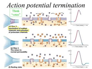
Action potential termination Think “votes”
 
Positive feedback loop Na+ enters (depolarization) V-gate Na+  channels open graded Na+ potential Reach “ threshold”? If YES, then...
 
 
Action Potential
Absolute Refractory Period. Is the period during which another action potential cannot be elicited no matter how large the stimulus coincides with almost the entire duration of the action potential Explanation: the inactivation gates of the Na channel are closed and will remain closed until repolarization occurs. No action potential can occur until the inactivation gates open.
Relative refractory period Begins at the end of the absolute refractory period and continues until the membrane potential returns to the resting level An action potential can be elicited during this period if a stronger than usual current is provided Explanation: The K+ conductance is higher than at rest, the membrane potential is closer to the K equilibrium potential and farther away for threshold; more current is required to bring the membrane to threshold.
Refractory periods
Saltatory Conduction Propagation of action potentials occurs by spread of local currents to adjacent areas of membrane, which are then depolarized to threshold and generate action potentials. Conduction velocity is increased by: 1) increasing diameter of a verve fiber resuts in decreased internal resistance and so conduction velocity down the nerve is faster 2) Myelination. Myelin acts as an insulator around nerve axons and increases conduction velocity. Myelinated nerve exhibits saltatory conduction because action potentials can be generated only at the nodes of Ranvier, where there are gaps in the myelin sheath.
Saltatory Conduction
 
Terminology Synapse Region at which neurons come nearly together to communicate.  (neuron or effector organ) Synaptic Cleft Gap between neurons (at a synapse) Impulses can not propagate across a cleft Synaptic Vesicle Packets of neurotransmitter in presynaptic neuron
Terminology Presynaptic Neuron Neuron sending a signal (before the synapse) Postsynaptic Neuron Neuron receiving a signal (after the synapse) Neurotransmitter Substance that tends to cause excitement Required to transmit impulses across a synaptic cleft
Direction of chemical synapse One Direction Synaptic vesicles are only located in the pre-synaptic nerve ending.  Only the post-synaptic neuron contains receptors for the neurotransmitters


Chemical Synapses An action potential in the presynaptic cell causes depolarization of the presynaptic terminal As a result of the depolarization, Ca enters the presynaptic terminal Ca entry causes release of neurotransmitter in the presynaptic cleft Neurotransmitter diffuse across the synaptic cleft and combines with receptors on the postsynaptic cell membrane, causing a change in its permeablilty to ions and its membrane potential.
 
Synaptic Physiology
Locks and Keys Neurotransmitter molecules have specific shapes positive ions (NA+ ) depolarize the neuron  negative ions (CL-) hyperpolarize When NT binds to  receptor, ions enter Receptor molecules have binding sites
Synaptic transmission Excitatory postsynaptic potentials(EPSP)/ Inhibitory postsynaptic potentials (IPSP) are inputs that depolarize/heperpolarize the postsynaptic cell bringing it closer/away from firing an action potential EPSP are cause by opening of channels that are permeable to Na and K  Neurotransmitters :Ach, NE, Epinephrine,  dopamine, serotonin IPSP are cause by opening Cl channels  Neurotransmitters: GABA and glycine
Postsynaptic Inhibition IPSP EPSP
Types of Neurotransmitters Acetylcholine Serotonin Norepinephrine Dopamine Endorphins GABA  Glutamate
Acetylcholine Found at neuro-muscular junction Involved in muscle movements (nicotine, curare) In CNS: recticular activating system Slow excitation of cerebral neurons (muscarine, atropine) Memory
Neuromuscular Junction Is the synapse between axons of motorneurons and skeletal muscle. The neurotransmitter released from the presynaptic terminal Ach, and the receptor on the postsynaptic membrane is nicotinic
Neuromuscular Junction and Ach 1. Synthesis and storage of Ach in the presynpatic terminal  choline acetylransferase catalyzes the formation from acetyl coA and choline in the presynaptic terminal.  2. Depolarization of the presynaptic terminal and Ca uptake 3. Calcium uptake causes release of Ach into the synaptic cleft 4. Diffusion of Ach to the poststnaptic membrane (muscle end-plate) and binding to specific receptors 5.  depolarization of adjacent membrane to threshold 6. Degradation of Ach to acetyl CoA and choline by acetylcholinesterase (acheE) on the muscle end plate
Myasthenia gravis Is characterized by skeletal muscle weakness and fatigability resulting from a reduced number of Ach receptors on the muscle end plate ( due to autoimmune antibodies against acetylcholine receptors at the neuromusclar juntion Diagnosis and treatment involves Acetylchlinesterase inhibitors (neostigmine)- prolong the action of Ach at the muscle end plate.
Curare - ACh antagonist  paralysis Nicotine - ACh agonist stimulates skeletal muscles, trembling movements Nerve gases/Black Widow spider venom -release of ACh  Severe muscle spasms and death Atropine - ACh antagonist Stupefying agent - delirium & coma Disorders of Cholinergic Transmission
Synaptic Function - Drug Effects 1) Release Inhibitors - ex:  Botulinum  toxin (in botulism) 2) Acetylcholinesterase Inhibitors - ex: Nerve Gas 3) Postsynaptic Blockers - ex: Curare and most Snake venoms 4) Sodium Channel Blockers - ex: Pufferfish venom and red tide toxins
Serotonin Involved in mood, depression Prozac works by blocking reuptake Ecstasy (MDMA) kills 5-HT terminals in forebrain, releasing massive amounts  Pain regulation (descending brainstem) Involved in regulation of cortical activity (sleep?)
Dopamine Involved in movement, attention, learning, motivation, reward Overactive dopamine: Schizophrenia  Underactive (loss of) dopamine: Parkinson’s Disease
Loss of dopamine neurons in the substantia nigra Symptoms: difficulty starting/stopping voluntary movements tremors at rest stooped posture rigidity poor balance Parkinson’s Disease
Main inhibitory neurotransmitter in CNS Benzodiazepines (Valium) and alcohol agonise GABA receptor complexes Also some anti-epileptic drugs  Gamma-Aminobutyric Acid (GABA)
Huntington’s Chorea Involves loss of neurons in striatum that utilize GABA Symptoms: jerky involuntary movements mental deterioration
 
Central Nervous System Cerebral ganglia (brain) Spinal Cord
Basic Directions dorsal caudal or posterior rostral or anterior ventral dorsal neuraxis dorsal ventral lateral medial medial lateral dorsal lateral medial medial lateral ventral caudal or posterior caudal or posterior dorsal ventral rostral or anterior neuraxis
Basic Directions
Basic Directions FIGURES 4.3 & 4.4 on pgs. 90-91 X dorsal ventral
CNS - The REAL Forebrain
This is Your Brain 1.5kg of water, lipids and protein contained in your cranium The most important and complex organ in your body 100 billion neurons 100 million billion connections between neurons Top Bottom Side Middle
6 Major Regions of the Brain 1.)  Cerebrum 2.)  Diencephalon 3.)  Midbrain 4.)  Pons 5.)  Cerebellum 6.)  Medulla Oblongata