Joints Goniometry
Shoulder Goniometry
Introduction
1. It is the measuring of angles created by the bones
of the body at the joints.
2. These joints are measured by a goniometer.
3. It has a moving arm, stationary arm, and the
fulcrum.
4. The fulcrum or body is placed over the joint being
measured and on it is a scale from 0 to 180.
5. The stationary arm will be aligned with the inactive
part of the joint measured, while the moving arm is
placed on the part of the limb which is moved in
the joint’s motion.
6. For example, when measuring knee flexion, the
stationary arm will be aligned over the thigh in line
with the greater trochanter of the femur.
Introduction - continue
7. The fulcrum is aligned over the knee joint or lateral epicondyle of
the femur, and the moving arm with the midline of the leg or
lateral malleolus.
8. Performing these tests is important for many reasons.
• The mobility of joints is important for diagnosis and
determining the presence or absence of dysfunction.
9. In a chronic condition, goniometry can measure the progression
of the disorder.
• An example of this is the progression of rheumatoid
arthritis.
10. Furthermore, joint motion measurement can evaluate
improvements or lack of progression during rehabilitation.
11. This not only provides motivation for the patient when there are
improvements, but also can decipher if modifications need to
be made if treatment is not effective.
Flexion
Patient Instructions:
• Once the goniometer is aligned
properly ask the patient to lift the arm
up just as if they were raising their
hand to ask a question.
• Be sure that the patient keeps the palm
of their hand facing in toward their
body.
Starting Position
• Patient is supine with arm
at side and the palm of the
hand facing the body.
• The fulcrum of the
goniometer is placed over
the acromion process.
• The stationary and moving
arms are aligned with the
midline of the humerus
and lateral epicondyle.
Ending Position
• The moving arm remains in line with the lateral
epicondyle and midline of the humerus.
• The examiner supporting the patient’s extremity.
• The stationary arm should remain in its starting position,
only now it should be in line with the lateral midline of
the thorax.
• Normal ROM for glenohumeral flexion is 160 to 180º; in
the picture the patient is in 180º of flexion.
Extension
• Patient Instructions:
• Ask the patient to simply lift their arm off
the table as far as they can.
Starting Position
• Patient is prone with arm
at side; make sure the
head is facing away from
the shoulder being tested.
• Elbow bent slightly and the
palm facing in toward the
body.
• The fulcrum is placed over
the acromion process.
• The stationary and moving
arms are aligned with the
lateral midline of the
humerus and the lateral
epicondyle.
Ending Position
• The moving arm remains in line with the lateral
epicondyle and the examiner should support the
patient’s extremity.
• The stationary arm in line with the midline of the thorax.
• Normal ROM for glenohumeral extension is 40 to 60º; in
the picture the patient is in 61º of extension.
Abduction
• Patient Instructions:
• Have the patient bring their arm out to
their side and as close to their head as
they can.
• Make sure that their palm faces upward
throughout the motion.
Starting Position
• The patient is supine
with arm at side; the
palm should be facing
interiorly.
• The fulcrum is placed at
the acromion process.
• The stationary and
moving arms are
aligned with the
anterior midline of the
humerus.
Ending Position
• The stationary arm should remain still and
parallel to the sternum.
• The moving arm should still be resting at the
anterior midline of the humerus.
• Normal ROM between 160 and 180º; the patient
in the picture is in 174º of abduction
Medial (Internal) Rotation
• Patient Instructions:
• Ask the patient to rotate their arm
down as far as they can.
Starting Position
• Supine with 90º of
shoulder abduction and
the elbow is in 90º of
flexion.
• The table should not
support the elbow.
• The fulcrum centered over
the olecranon process.
• The moving arm is aligned
with the ulnar styloid and
the stationary arm should
be perpendicular to the
floor.
Ending Position
• Same as above
• Normal ROM is 60-70˚; the patient is in 68º
of internal rotation.
Lateral (External) Rotation
• Patient Instructions:
• Ask the patient to rotate their arm up
toward their head as far as they can.
Starting and Ending Position
• Supine with 90º of shoulder
abduction and 90º of elbow
flexion.
• The table should not support
the elbow. (Refer to above
picture)
• Fulcrum on the olecranon
process.
• The moving arm should be
aligned with the ulnar styloid
and the stationary arm
should be perpendicular to
the floor.
• Ending Position:
• Same as before
Normal ROM Reference Values
Typical ROM
Shoulder
160 - 180˚
Flexion
40 - 60˚
Extension
160 - 180˚
Abduction
60-70˚
Internal Rotation
80 - 100˚
External Rotation
Elbow Goniometry
Flexion
• Patient Instructions:
• Ask the patient to bend their elbow as
far as they can, try and touch their
shoulder.
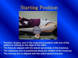
Starting Position
• Position: Supine, arm in the anatomical position with arm of the
patient is resting on the edge of the table.
• The fulcrum aligned with the lateral epicondyle of the humerus.
• The stationary arm is positioned along the midline of the humerus
• The moving arm is aligned with the radial styloid process.
Ending Position
• The arm is now flexed
at the elbow, the
goniometer should still
be aligned with the
correct anatomical
landmarks as described
below.
• Normal ROM is between
150-160º, the patient
has 155º of elbow
flexion.
Pronation
• Patient Instructions:
• Have the patient turn their
wrist down toward the ground.
• Starting Position:
• Patient sitting up with elbow
bent 90 degrees and at
patient’s side, wrist in a
handshake position.
• The fulcrum is placed just
behind the ulnar styloid
process.
• The moving arm and
stationary arm are parallel with
the anterior midline of the
humerus.
Ending Position
• The fulcrum should remain
in the same position as
above.
• The stationary arm will still
be aligned parallel to the
midline of the humerus,
the moving arm will lie
across the dorsum of the
forearm just behind the
ulnar and radial styloid
processes.
• Normal ROM is 90-96º, the
patient has 95º of
pronation.
Supination
• Patient Instructions:
• Have the patient turn their
palm up as if they are
holding something in the
palm of their hand.
• Starting Position:
• Patient position is the same
as for pronation.
• The goniometer is placed on
the medial aspect of the
forearm with the fulcrum at
the radioulnar joint.
• The arms are both aligned
with the anterior midline of
the humerus.
Ending Position
• The moving arm will
be resting on the
medial forearm at the
radioulnar joint.
• The moving arm
should remain parallel
to the midline of the
humerus.
• Normal ROM is 81-93º,
the patient has 90º of
Supination.
Normal ROM Reference Values
Typical ROM
Elbow
150-160º
Flexion
0
Extension
90-96º
Pronation
81-93º
Supination
Wrist Flexion
Flexion
• Position: Sitting next to supporting surface. Shoulder abducted 90º,
Elbow flexed 90º, Forearm in 0º supination-pronation, resting on
supporting surface, hand free to move. Avoid wrist radial-ulnar flexion
& finger flexion
• Stabilization: Stabilize radius & ulna to prevent supination or pronation
• Center: Lateral aspect of wrist over triquetrum.
• Proximal Arm: Lateral midline of ulna. Reference olecranon & ulnar
styloid process.
• Distal Arm: Lateral midline of 5th metacarpal
END POSITION
Wrist Extension
Extension
• Position: Sitting next to supporting surface. G-H abd 90º, Elbow flexed
90º, Forearm in 0º supination-pronation, resting on supporting surface,
hand free to move. Avoid wrist radial-ulnar flexion & finger flexion .
• Stabilization: Stabilize radius & ulna to prevent supination or
pronation.
• Center: Lateral aspect of wrist over triquetrum
• Proximal Arm: Lateral midline of ulna. Olecranon & ulnar styloid
process
• Distal Arm: Lateral midline of 5th metacarpal
END POSITION
Radial Flexion
• Position: Sitting next to supporting surface. G-H abd 90º, Elbow flexed
90º, Forearm in 0º supination-pronation, resting on supporting surface,
hand free to move. Avoid wrist radial-ulnar flexion & finger flexion
• Stabilization: Stabilize distal ends of radius & ulna to prevent pronation
& supination of forearm & elbow flexion beyond 90º
• Center: Middle of dorsal aspect of wrist over capitate
• Proximal Arm: Dorsal midline of forearm. lateral epicondyle of humerus
• Distal Arm: Dorsal midline of 3rd metacarpal. Reference 3rd phalanx
END POSITION
Ulnar Flexion
• Position: Sitting next to supporting surface. G-H abd 90º, Elbow flexed
90º, Forearm in 0º supination-pronation, resting on supporting surface,
hand free to move. Avoid wrist radial-ulnar flexion & finger flexion
• Stabilization: Stabilize distal ends of radius & ulna to prevent pronation
& supination of forearm & elbow flexion beyond 90º
• Center: Middle of dorsal aspect of wrist over capitate
• Proximal Arm: Dorsal midline of forearm. lateral epicondyle of humerus
• Distal Arm: Dorsal midline of 3rd metacarpal. Reference 3rd phalanx
END POSITION
Hip Goniometry
Flexion
• Patient Instructions:
– Ask the patient to bend their knee and bend
their leg and bring it as close to their chest as
is comfortable for them.
Starting and Ending Position
• The patient should be lying
supine in the anatomical
position.
• The fulcrum is aligned with
the greater trochanter of the
femur.
• The stationary arm is
positioned along the lateral
midline of the abdomen,
using the pelvis for
reference, the moving arm
along the lateral midline of
the femur.
• Normal ROM is between 115
and 125º.
• The patient is in 115º of knee
flexion.
Ending Position
Extension
• Patient Instructions:
• Have the patient lift their leg
off the table as far as they
can.
• Starting and Ending Position:
• Patient is lying prone with
legs together and arms at
sides.
• Goniometer positioning is the
same as for hip flexion.
• Normal ROM for hip extension
is 10 to 30º, patient has 25º of
hip extension.
Ending Position
Abduction
• Patient Instructions:
• The patient should be asked to
move their leg out to the side as far
as they can.
Starting Position
• Patient is supine in anatomical
position.
• Fulcrum is placed in line with
the anterior superior iliac spine.
• The moving arm of the
goniometer is aligned with the
midline of the patella, the
stationary arm with the ASIS of
the opposite side.
• Note: This places the
goniometer reading at 90
degrees, to determine the actual
reading make sure to read 90 as
0 and determine the
measurement in that manner.
Ending Position
Same as before
• Normal ROM for hip abduction is 40 to 50º; the
patient has 48º of hip abduction.
Adduction
Patient Instructions:
• Ask the patient to move their leg to
the inside toward their opposite leg.
Starting Position
• Patient is supine the leg not
being measured is abducted to
allow full adduction of the
opposite leg.
• The goniometer positioning is
the same as for abduction,
fulcrum at the ASIS, moving arm
aligned with the midline of the
patella, and the stationary arm
with the ASIS of the opposite
side.
• Also, remember to adjust due to
the goniometer starting at a
reading of 90º.
Ending Position
• Same as before
• The normal ROM is 15 to 25º.
• The patient has 18º of hip adduction.
Medial (Internal) Rotation
• Patient Instructions:
• Have the patient bring their leg out to the side.
• Starting Position:
• The patient will be sitting off of the edge of the table,
knees against the table, with their legs dangling down off
the table.
• The fulcrum is aligned with the patella and both arms of
the goniometer with the midline of the tibia.
Ending Position
• The fulcrum and moving arm remain in the same
position as before.
• The stationary arm should now be hanging freely
but should be perpendicular to the floor.
• The normal ROM for internal rotation is 35 to 50º.
Patient has 49º of internal rotation at the hip.
Normal ROM Reference Values
Typical ROM
Hip
115 - 125º
Flexion
10 - 30º
Extension
40 - 50º
Abduction
15 - 25º
Adduction
35 - 50º
internal Rotation
35 - 50º
External Rotation
Knee Goniometry
Flexion – Start Position
• Position: Supine, knee in ext.
Initially hip in 0º ext, abd, add,
but as knee flexes, hip also
flexes
• Stabilization: Stabilize femur
to prevent rotation, abduction
& adduction
• Center: Over lateral
epicondyle of femur
• Proximal arm: Lateral midline
of femur, referencing greater
trochanter
• Distal arm: Lateral midline of
fibula, reference lateral
malleolus & fibular head
Normal ROM is 135-150º.
The patient in the picture is in 146ºof knee flexion.
Flexion – End Position
Patient Instructions:
• Ask the patient to bend their knee as far as they can.
• If the patient is able, he/she should also flex at the hip to get complete knee
flexion.
• If the patient cannot flex at the hip this measurement can also be done in the
prone position.
Extension
Start & End Position
• Position: Supine, knee in ext.
Initially hip in 0º ext, abd, add,
but as knee flexes, hip also
flexes
• Stabilization: Stabilize femur
to prevent rotation, abduction
& adduction
• Center: Over lateral
epicondyle of femur
• Proximal arm: Lateral midline
of femur, referencing greater
trochanter
• Distal arm: Lateral midline of
fibula, reference lateral
malleolus & fibular head
ANKLE GONIOMETRY
Dorsiflexion
Starting Position:
• Patient is sitting with legs off the
table.
• The fulcrum is aligned with the
lateral malleolus.
• The stationary arm is in line with
the midline of the lower leg; use
the head of the fibula for
reference.
• The moving arm is parallel to the
fifth metatarsal.
Patient Instructions:
Ask the patient to bend the ankle and point their toes up
towards the ceiling
Note:
Remember that the
goniometer will be at 90º and
to adjust for that when
recording the angles.
Ending Position
• Normal ROM for Dorsiflexion is between 0º and 10º.
• The patient has 3º of Dorsiflexion.
Plantar flexion
Starting Position and Ending
Position:
• Patient is sitting with legs
off table.
• Goniometer alignment is
the same as for
Dorsiflexion.
• Normal ROM is 20º to 30º,
the patient is at 27
degrees.
Patient Instructions:
Ask the patient to point their foot down toward the
ground.
Inversion at the Tarsal joints
• Starting and Ending Position:
• Patient sitting with legs off the
table, or patient can be supine
on the table with the foot resting
on the table.
• The fulcrum is positioned
between the two malleoli.
• The stationary arm with the
midline of the tibia and the
moving arm with the second
metatarsal.
• Normal ROM is 35º to 45º
patient has 40 degrees of
inversion.
Patient Instructions: Have the patient turn their foot in.
Eversion at the Tarsal joints
• Starting and Ending Position:
• Patient sitting with legs off
the table, or patient can be
supine on the table with the
foot resting on the table.
• The fulcrum is positioned
between the two malleoli.
• The stationary arm with the
midline of the tibia and the
moving arm with the second
metatarsal.
Patient Instructions: Ask the patient to turn their foot out.
Normal ROM is 15º to 25º; patient is in 30º of Eversion