Presented by : Radhika Ahuja
BPT 3rd year
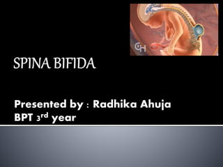
 Latin : “ Split spine”
Developmental congenital disorder
 caused by incomplete closure of embryonic
neural tube
 Some vertebrae overlying the spinal cord
are not fully formed and remain unfused
and open
 Opening is large enough that this allows spinal cord to
protrude through it
 there may or may not be a fluid filled sac around spinal
cord
 Worldwide incidence of one or two causes
per 1000 birth
 Myelomeningocele, most significant
common cause , this leads to disability in
most affected indivdual
 more likely to affect females , idiopathic
cause
 Unknown etiology
 Genetic predisposition triggered by
environmental factors- drugs “Valpronic
Acid” during pregnancy
 Women age more than 35 years
 Women already have 1 child affected
 Maternal Folic acid deficiency , 0.4 mg/day
during 1st trimester
 Arrest in the orderly formation of vertebral
arches and spinal column between 4-6th wk
of pregnancy
 Incomplete closure of neural tube during
4th week of embryonic age
 The neural tube forms adequately and then
ruptures
Spina bifida malformation lies in three
categories:
A. spina bifida occult ( 10% of the population)
B. spina bifida cystica with meningocele
C. spina bifida cystica with
myelomeningocele
( most common site are sacral and lumbar)
 Occult means “Hidden” (Mildest form)
 Here , outer part of some vertebrae is not
completely closed
 Split is small thus S.C do not protrude
 Site of lesion is normal but may have some
growing hairs , a dimple in the skin , or a
birthmark
 Least common form of spina bifida is
posterior meningocele
 Here, vertebrae develops normally but
meninges are forced into the gap between
the vertebraes.
 This gives more severe complications
 Here , unfuse portion of spinal column allows
spinal cord to protrude through an opening
 Meningeal memebrane encloses spinal cord
form a sac enclosing spinal elements
 Spina bifida with myeloschisis is most severe
form of myelomeningocele ,
 Involved area is represented by flattened ,
plate-like mass of nervous tissue with no
overlapping membranes
 Exposure of these nerves and tissues make
the baby more prone to life –threatening
infections such as meningitis
 Protrudes nerves are damaged or not
properly formed
 As a result , there is usually some degree of
paralysis and loss of sensation below level
of spinal cord defect.
 PHYSICALSIGNS ;
 Orthopedic abnormalities ( i.e Club foot ,
Hip dislocation )
 Bowel and Bladder incontinence
 Urinary tract infections
 Poor renal function
 Pressure sores and skin irritation
 Abnormal eye movement
 68% affected people have allergy to
latex
 Paralysis
 Scoliosis
 Back pain
 Partial or complete loss of sensation
 Weakness of hips, legs , feet of a
newborn
 OTHER SYMPTOMS ARE ;
 Growing hairs at the back part of the pelvis
 Dimpling of sacral area
 Difficulty in swallowing , lead to chocking
 Hoarseness
 Below average IQ
 Many individual with spina bifida have an
associated abnormality of cerebellum
called ARNALD CHIARI II MALFORMATION
 Here , back portion of the brain is displaced
from the back of the skull down into the
upper neck
 Specific area of difficulty in some individual
include; Planning ;
Organization ;
Initiation ;
Memory ;
Problem-solving ;
Abstraction ;
andVisual Planning .
 Children with spina bifida and shunted
hydrocephalus have higher rate of ADHD
 Individual with spina bifida may struggle
academically especially in the subjects like
Mathematics and Reading .
 In one study ,it is found that 60% of the
children with spina bifida were diagnosed
with learning disability.
 AFP SCREENING
Neural tube defect can usually be detected
during pregnancy by testing the mother’s
blood
 Increased level of MATERNAL SERUM
ALPHA FETOPROTEIN (MSAFP) should be
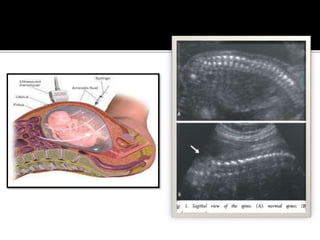
follow up to two test : ULTRASOUND OF
FETAL SPINE
& AMNIOCENTESIS of the mother amniotic
fluid for MSAFP and acetylcholinesterase
 FOLIC ACID , dietary supplements has
been shown to be helpful in reducing
incidence of spina bifida.
 pregnant women is recommended to take
0.4 mg of folic acid a day
 There is no known cure for neural damage
caused by spina bifida
 Spinal cord and its nerve roots are put back
inside the spine and covered with meninges
 A shunt may be surgically installed to
provide continuous drain excessive CSF
produce in brain
 This shunt most commonly drain into chest
 Monitor growth and development of bones
, muscles and joints
 Physical therapy
 Speech therapy
 Place the child in prone position
 Cover the site with the sterile guaze piece
dipped in normal saline
 Maintain hydration
 Monitor for associated defect
 Catheters
 Braces
 High fiber diet
 Antibiotics for meningitis and urinary
infections
• Difficult delivery with problem resulting
from a traumatic birth , including
CEREBRAL PALSY and decreased oxygen
to brain
• Frequently UTI
• Hydrocephalus
• Loss of bowel and bladder control
• Meningitis
• Permanent weakness or paralysis of brain
Spina bifida ppt
Spina bifida ppt