Amr Hassan MD,FEBN
Associate Professor of Neurology - Cairo
University
FLOPPY INFANT
 Definition
 Clinical examination
 Differential diagnosis
 Investigations
 Definition
 Clinical examination
 Differential diagnosis
 Investigations
Floppy infant refers to those children
presenting with generalized hypotonia, most
often arising out of an insult incurred during
fetal or neonatal period.
 Definition
 Clinical examination
 Differential diagnosis
 Investigations
 Definition
 Clinical examination
 Differential diagnosis
 Investigations
Prenatal risk factors:
 • History of drug or teratogen exposure
 • Presence of polyhydramnios
 • Maternal diseases (diabetes, epilepsy)
 • Parental age
 • Consanguinity
 • Family history of neuromuscular disease
 • Other affected siblings
 History
 History since delivery
▪ Respiratory effort
▪ Ability to feed
▪ Level of alertness
▪ Level of spontaneous activity
▪ Character of cry
▪ Apgar scores
▪ Resuscitation requirements
 History
 Any significant family history
▪ Affected parents
▪ Siblings
▪ Consanguinity
▪ Stillbirths
▪ Childhood deaths
NeurologyChapter of IAP
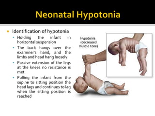
 Identification of hypotonia
 Holding the infant in
horizontal suspension
 The back hangs over the
examiner's hand, and the
limbs and head hang loosely
 Passive extension of the legs
at the knees no resistance is
met
 Pulling the infant from the
supine to sitting position the
head lags and continues to lag
when the sitting position is
reached
 Identification of
hypotonia
 Holding the infant under
the arms
 The legs will be
extended
 Decreased tone of the
shoulder girdle allows
the infant to slip through
the examiner's hands
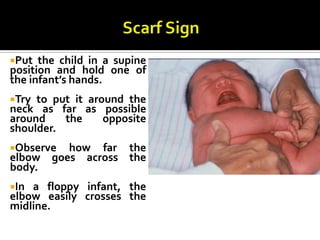
Put the child in a supine
position and hold one of
the infant’s hands.
Try to put it around the
neck as far as possible
around the opposite
shoulder.
Observe how far the
elbow goes across the
body.
In a floppy infant, the
elbow easily crosses the
midline.
The same infant in horizontal
suspension. Note the inverted U posture.
A 12-week-old male infant with excessive head-
lag evident on ‘pull-to-sit’. Note the hypotonic
posture of the legs with external rotation.
Classification( Location)
Brain
Spinal cord
Peripheral nerves
NeuromuscularTransmission
Muscles
Systemic disorders
Bodensteiner JB. The evaluation of the hypotonic infant. Semin Pediatr Neurol. 2008
 Physical Examination
 Clues and Pitfalls
▪ Profound central hypotonia may have absent DTR
▪ Absent DTR in the first few DOL would not rule out a
central cause for the hypotonia
 Physical Examination
 Clues and Pitfalls
▪ Presence of profound weakness and hypotonia suggest:
▪ Disorder of the lower motor neuron
 A sign of this may be a weak cry
▪ Weakness is uncommon in central hypotonia except in
the acute stages
 Physical Examination
 Clues and Pitfalls
▪ Arthrogryposis (the fixation of joints at birth)
▪ Associated with:
 Neonatal hypotonia
 More commonly with lower motor neuron unit
 Multisystem abnormalities
Clues in cerebral hypotonia
Cerebral Hypotonia in newborns usually does not pose
diagnostic difficulty.The history and physical examination
identify the problem.
Normal or Brisk reflexes
Other abnormal brain functions: delay, seizures
Fisting
Movement through postural reflexes
Scissoring on vertical suspension
Dysmorphic features
Extra-cranial organ malformations
Clues in motor unit hypotonia
Disorders of the motor unit are not associated with
malformations of other organs except for joint
deformities and the mal development of bone
structures.
Absent or Depressed reflexes
Intact brain function
Muscle atrophy
Fasciculations
Failure of movement through postural reflexes
No extra-cranial organ malformations
 Physical Examination
 Anterior horn cells
▪ Generalized weakness
▪ Decreased/ absent DTRs
▪ Fasciculations
▪ Often described as alert
 Physical Examination
 Nerve
▪ Weakness, distal>proximal
▪ Decreased/ Absent DTRs
▪ +/- fasciculations
 Physical Examination
 Neuromuscular Junction
▪ Weakness, face/ eyes/ bulbar
▪ Normal DTRs
▪ No fasciculations
Ptosis and external ophthalmoplegia in a floppy weak child.
Suggestive of myasthenia gravis.
 Physical Examination
 Muscles
▪ Weakness, proximal>distal
▪ Decreased DTRs
 Definition
 Clinical examination
 Differential diagnosis
 Investigations
 Definition
 Clinical examination
 Differential diagnosis
 Investigations
 Physical Examination
 Clues
▪ Abnormal odor
▪ Metabolic disorders
▪ Hypopigmentation, undesceded testes
▪ Prader Willi
▪ Hepatomegaly
▪ Retinitis pigmentosa
▪ Neonatal adrenoleukodystrophy
 Physical Examination
 Clues
▪ Abnormal odor
▪ Metabolic disorders
▪ Hypopigmentation, undesceded testes
▪ Prader Willi
▪ Hepatomegaly
▪ Retinitis pigmentosa
▪ Neonatal adrenoleukodystrophy
Central nervous system
 Perinatal asphyxia, neonatal, encephalopathy, kernicterus,
cerebral palsy (atonic type).
 Intracranial hemorrhage.
 Cerebral malformations
 Chromosomal abnormalities (e.g.Trisomy 21, Prader-Willi
syndrome)
 Congenital infectionTORCH
 Acquired infections
 Peroxisomal disorders
 Drug effects (e.g. benzodiazepines)
 Inborn errors of metabolism e.g., aminocidurias,
mucopolysaccharidosis and cerebral lipidosis.
Many hypotonic children due to causes in central
nervous system are mentally retarded.
In atonic or hypotonic cerebral palsy, reflexes are
brisk in spite of generalized flaccidity.
Floppy infant due to cerebral causes is associated
with lethargy, poor feeding, and lack of alertness,
poor Moro’s reflex, and seizures during the neonatal
period.
Spinal cord
 Spinal Cord Injury – Broken Neck
 Infections
Enterovirus - POLIO
Transverse Myelitis
 Mass lesions
Injuries in Breech Presentation
 Injuries to the cervical spinal cord occur almost
exclusively during vaginal delivery;
 approximately 75% are associated with breech
presentation and 25% with cephalic presentation.
 Because the injuries are always associated with a
difficult and prolonged delivery, decreased
consciousness is common, and hypotonia is falsely
attributed to asphyxia or cerebral trauma.
 MRI of the spine shows intraspinal edema and
hemorrhage
Injuries in Cephalic Presentation
 Twisting of the neck during midforceps
rotation causes high cervical cord injuries in
cephalic presentation.
 The trunk fails to rotate with the head.
 The risk is greatest when amniotic fluid is
absent because of delay from the time of
membrane rupture to the application of
forceps.
Poliovirus Infection
 Small RNA virus : Neurotropic
 Seasonal epidemics
 Prodromal illness
 Pain -->Asymmetric Paralysis
 Rare but still occurs
 vaccine related : 1 in 12 million
Mass lesions of spinal cord
 Rare
 Intra-AbdominalTumors
 Neuroblastoma
 Early in Infancy
12/21/2018 44
1. The anterior (ventral) horn cell
2.The radicle (root).
3.The peripheral nerve.
4.The neuromuscular junction.
5. The muscle.
5
4
2
1
2
2
3
Anterior Horn cell (neuronal) degeneration
ProgressiveWeakness: Proximal > Distal
Hypotonia
Areflexia
Atrophy / Fasciculations
Intact Brain Development
NeurologyChapter of IAP
 SMA is the second most common autosomal recessive
disease in the US after cystic fibrosis.
 Incidence:
 Type 1: 1 per 10,000 live births
 Types II and III: 1 per 24,000 births
 Worldwide 7.8-10 cases per 100,000 live births
 ? M:F predominance or M>F
 No ethnic predominance.
 The genetic defects associated with SMA
types I-III are localized on chromosome
5q11.2-13.3.
 Mutations in the SMN gene result in a loss of
function of the SMN protein.
 Many classification systems based on
inheritance, clinical, and genetic criteria.
SMA type I, (Werdnig-Hoffmann acute infantile),
occur birth – 6 months (95% by 3 months)
Severe, progressive muscle weakness and flaccid or
reduced muscle tone (hypotonia).
Bulbar dysfunction includes poor suck ability, reduced
swallowing, and respiratory failure.
Patients have no involvement of the extraocular
muscles, and facial weakness is often minimal or
absent.
They have no evidence of cerebral involvement, and
infants appear alert.
 Impaired fetal movements are observed in 30% of
cases
 60% of infants with SMA type I are floppy babies at
birth. Prolonged cyanosis may be noted at delivery.
 In some instances, the disease can cause fulminant
weakness in the first few days of life. Such severe
weakness and early bulbar dysfunction -> mean
survival of 5.9 months.
 Affected children never sit or stand.
 In 95% of cases, infants die from complications of
the disease by 18 months.
 SMA type II (chronic infantile, sitters) usually
begin between 6 - 18 months.
 Most common form of SMA
 Most common manifestation is developmental
motor delay. Infants with SMA type II often have
difficulties with sitting independently or failure
to stand by 1 year of age.
 These children may learn to sit but will never be
able to stand or walk.
 An unusual feature of the disease is a postural
tremor affecting the fingers. This is thought to be
related to fasciculations in the skeletal muscles
 Pseudohypertrophy of the gastrocnemius muscle,
musculoskeletal deformities, and respiratory failure
can occur.
 The lifespan of patients with SMA type II varies from
2 years to the third decade of life. Respiratory
infections account for most deaths.
 SMA type III (Kugelberg-Welander, chronic juvenile,
walkers) appear 18 months – adult.
 Slowly progressive proximal weakness. Most can stand
and walk but have trouble with motor skills, such as
going up and down stairs.
 Bulbar dysfunction occurs late in the disease.
 Patients may show evidence of pseudohypertrophy.
 The disease progresses slowly, and the overall course is
mild. Many patients have normal life expectancies.
 Congenital SMA with arthrogryposis (persistent
contracture of joints with fixed abnormal posture
of the limb) is a rare disorder. Manifestations
include
1. severe contractures,
2. curvature of the spine,
3. chest deformity,
4. respiratory problems,
5. an unusually small jaw, and
6. drooping upper eyelids.
12/21/2018 62
1. The anterior (ventral) horn cell
2.The radicle (root).
3.The peripheral nerve.
4.The neuromuscular junction.
5. The muscle.
5
4
2
1
2
2
3
 Polyneuropathy
 Dysmyelination
▪ Autoimmune
▪ Congenital / genetic
 Dysautonomia
 Peripheral nerves
 Hereditary sensory motor neuropathies
▪ Charcot-Marie-Tooth disease
12/21/2018 65
1. The anterior (ventral) horn cell
2.The radicle (root).
3.The peripheral nerve.
4.The neuromuscular junction.
5. The muscle.
5
4
2
1
2
2
3
 Toxins
▪ Botulism
 Myasthenia
▪ Congenital
▪ Neonatal transitory
Infantile myasthenia
 FAMILIAL-INFANTILE
Multiple Genetic Defects: AR + AD
Pre & Post Synaptic AChR abnormalities
Respiratory or feeding problems at birth
 CONGENITAL
Usually Bilateral Ptosis & Ophthalmoplegia
Multiple Genetic Defects: AR
 NEONATAL-TRANSITORY
10 - 15% of myasthenic mothers
The CMS conditions represent genetic defects of neuromuscular transmission
Infantile botulism
Infants usually 2 - 26 weeks old
Clostridium Botulinum --> Exotoxin
Prevents release of Acetylcholine
Cholinergic Blockade of skeletal muscle
Source of intestinal colonization usually
unclear
Occurs mainly between March & October
Prodrome: poor feeding &
constipation
Progressive bulbar & general weakness
Loss of deep tendon reflexes
Hypotonia
Dysphagia
Ptosis
Sluggish dilated pupils
12/21/2018 71
1. The anterior (ventral) horn cell
2.The radicle (root).
3.The peripheral nerve.
4.The neuromuscular junction.
5. The muscle.
5
4
2
1
2
2
3
 Muscle
 Muscular dystrophies (congenital myotonic
dystrophy)
 Congenital myopathies (e.g. central core disease)
 Metabolic myopathies
 Acid maltase deficiency
 Carnitine deficiency
 Cytochrome-c-oxidase deficiency
Bodensteiner JB. The evaluation of the hypotonic infant. Semin Pediatr Neurol. 2008
NeurologyChapter of IAP
Systemic
Genetic disorders
Prader-willi
Angelman’s syndrome
Cri du chat
Cerebro-hepato-renal sydrome
William’s syndrome
Trisomy 21
Trisomy 13
 SYSTEMIC
Systemic
Metabolic
Mitochondrial
Congenital lactic acidosis
Hyperammonemia
Aminoacidurias
Non-ketotic hyperglycinemia
Celiac disease
NeurologyChapter of IAP
Prader-Willi syndrome
Hypogenitalism 100%
Cryptorchidism 84%
Decreased Fetal Movement 75%
Congenital Hip Dislocation 10%
Clubfoot 6%
Profound Infantile Hypotonia
Mental Retardation
Decreased / Absent DTRs
Short Stature
Obesity / Insatiable Appetite
 Investigation
 Central Causes
▪ Neuroimaging
▪ Ultrasound scan in the first instance
▪ MRI for structural abnormality
▪ EEG: if seizures suspected
 Investigation
 Central Causes
▪ Genetics review if any dysmorphic features present
▪ Karyotype (if dysmorphic features)
▪ TORCH screen
▪ DNA methylation studies or FISH for Prader-Willi
syndrome (if clinically indicated after a genetics review)
▪ Metabolic work up
Metabolic evaluation
Arterial Blood: Lactate, Pyruvate, ABG
Venous Blood: Ammonia, Chemistries
CBC, Carnitine profile, Amino Acids
Urine: Organic & Amino Acids
CSF: Lactate,Amino Acids
(Glycine)
Muscle: It Depends
Investigations
 Investigation
 Peripheral causes
▪ Creatine kinase: If elevated in an early sample, repeat
after a few days.
▪ Nerve conduction studies
▪ Muscle biopsy
▪ Depending on clinical situation, may be delayed until around 6
months of age as neonatal results are difficult to interpret
 Investigation
 Peripheral causes
▪ Molecular genetics – CTG repeats, deletions in SMN
gene
Bodensteiner JB. The evaluation of the hypotonic infant. Semin Pediatr Neurol. 2008
Site of involvement
Deep tendon
reflexes EMG Muscle biopsy
Central Normal or
increased
Normal Normal
Anterior horn cell Absent Fasciculation /
fibrillation
Denervation
pattern
Peripheral nerve Decreased Fibrillation Denervation
pattern
Neuromuscular
junction
Normal Decremental /
incremental
Normal
Muscle Decreased Short duration small
amplitude potential
Characteristic
NeurologyChapter of IAP
THANKYOU
amrhasanneuro@kasralainy.edu.eg