INDIAN
PHARMACOPOEIA
2007
Volume 1
THE INDIAN PHARMACOPOEIA COMMISSION
GHAZIABAD
ISBN 81-903436-0-3
© 2007, Indian Pharmacopoeia Commission
Application for reproduction should be made to the INDIAN PHARMACOPOEIA COMMISSION
Sector-23, Raj Nagar,
Ghaziabad-201002
India
Tale:(91-120)–2783401
Fax: (91-120)–2783311
Web-site: www.ipc.gov.in E.mail: ipclab@vsnl.net
Effective from 31st
March 2008
On behalf of : GOVERNMENTOFINDIA
MINISTRYOFHEALTH&FAMILYWELFARE
Designed, produced & published by : THEINDIANPHARMACOPOEIACOMMISSION
CENTRALINDIANPHARMACOPOEIALABORATORY
GOVT.OFINDIA,MINISTRYOFHEALTH&FAMILYWELFARE
SECTOR-23,RAJNAGAR,GHAZIABAD-201002.
Printed by : NATIONALINSTITUTEOFSCIENCECOMMUNICATION
ANDINFORMATIONRESOURCES(NISCAIR)
COUNCILOFSCIENTIFIC&INDUSTRIALRESEARCH
PUSAGATE,K.S.KRISHNANMARG,NEWDELHI–110012
Price: Inland Rs 25000.00
Foreign - $ 1000.00
£ 500.00
v
IP 2007 NOTICES
Notices
Legal Notices
In India, under the Drugs and CosmeticsAct 1940, the current
edition of Indian Pharmacopoeia is a book of standards for
drugs included therein and the standards as included in the
Indian Pharmacopoeia would be official.Also, in several other
laws of India, the Indian Pharmacopoeia is recognised as the
standard book. It is expedient that enquiry be made in each
case in order to ensure that the provisions of any such law are
being complied with. In general, the Drugs and Cosmetics
Act, 1940, the Narcotic Drugs and Psychotropic Substances
Act, 1985, the Poisons Act, 1919 and the rules framed
thereunder should be consulted. These statutes empower the
Government agencies to enforce the law using this
compendium. The monographs of the Indian Pharmacopoeia
should be read subject to the restriction imposed by those
laws which are applicable.
If considered necessary, the standards included in Indian
Pharmacopoeia can be amended and the Secretary-cum-
Scientific Director is authorised to issue such amendments.
Whenever such amendments are issued, the Indian
Pharmacopoeia would be deemed to have been amended
accordingly.
PatentsAnd Trade Marks
In the Indian Pharmacopoeia, certain drugs and preparations
have been included notwithstanding the existence of actual
or potential rights in any part of the world. In so far as such
substances are protected by Letters Patent their inclusion in
the Indian Pharmacopoeia neither conveys, nor implies, licence
to manufacture without due permission, authority, or licence
from the person or persons in whom such rights exist.
The titles given under the individual monographs are public
property. These titles cannot be patented as trade marks and
no person is permitted to patent any trade mark devising the
root of these titles.
vii
IP 2007 PREFACE
Preface
The Indian Pharmacopoeia 2007 is published by the Indian
PharmacopoeiaCommission(IPC)onbehalfoftheGovernment
ofIndia,MinistryofHealth&FamilyWelfare.TheGovernment
of India constituted the IPC vide their Order No. Z-14012/IPC/
CBP/ 2003 dated 22nd
March 2005. The IPC is a Society under
the provisions of the Societies Registration Act, 1860 (Act
No. 21 of 1860) for the registration of Literary, Scientific and
Charitable Societies. The functioning of the Commission is
governed by the provisions of the approved Memorandum of
Association, Rules and Regulations of the IPC.
The Commission has set up its headquarters in the campus of
the Central Indian Pharmacopoeia Laboratory (CIPL), Sector-
23, Raj Nagar, Ghaziabad, UP. The Director CIPL, also
functions as the Secretary-cum-Scientific Director of the IPC.
The CIPL is the support structure of the Commission.
The Indian Pharmacopoeia is being produced in fulfillment of
the requirement in the Drugs and Cosmetics Rules, 1945 of
standards of drugs produced in India and in the belief that it
contributes significantly in the control of the quality of
medicinal products. The standards of this pharmacopoeia are
legally enforceable and are intended to help in the licensing
and inspection processes.
After independence an Indian Pharmacopoeia Committee was
constituted in 1948, which prepared the Pharmacopoeia of
India (The Indian Pharmacopoeia) 1955. A Supplement to it
was published in 1960. This pharmacopoeia contained western
and also traditional drugs, and the same policy continued
while preparing the Pharmacopoeia of India 1966 and its 1975
Supplement. In the Pharmacopoeia of India 1985 and its
Addenda 1989 and 1991, traditional drugs were not included
as publication of a pharmacopoeia of traditional system drugs
was taken up separately and only those herbal drugs were
included which had supporting definitive quality control
standards.
In the period since the publication of the 1985 Edition there
has been a significant increase in the range of drugs produced
in India. Keeping this in mind the Committee has deleted or
added monographs on a system of priorities based on the
medical merit and the extent of use of any given article in the
country in its following 1996 Edition and its addenda in 2000,
2002,2005andonesupplementforVeterninaryProductsin2002.
The Indian Pharmacopoeia 2007 has been prepared in
accordance with the principles and designed plan decided by
the Scientific Body of the Indian Pharmacopoeia Commission
and completed with untiring efforts made by Commission
members and its Secretariat over almost two years.
The Indian Pharmacopoeia 2007 is presented on the user
friendly format. The General Notices, Monographs and new
testing methods, etc. based on the introduction of advanced
technology and experimental methods widely adopted in India
and abroad are being added and updated. The contents of
Appendices are revised by and large in consonance with those
nowadays adopted internationally for monitoring the quality
of the drugs. The monographs of special relevance to the
common disease pattern of this region have been given special
emphasis by incorporating such medicines.
In addition, emphasis has been put to bring out harmonisation
in Appendices to a sound connection between individual
monographs and the relevant appendices, and to the
standardization of text wording so as to make this edition
precise and well structured. The number of monographs in
Appendices are expanded further to incorporate the latest
technological advances and complies with regulatory
requirements. Great efforts have been made to unify the
National Drug Standards and to bring them in line with the
International Standards progressively by addition of
monographs of new drugs and current methodology adopted.
Public Review and Comment Process for Standards Develop-
ment Related to this edition of the Indian Pharmacopoeia have
been given special attention to incorporate comments from
stakeholders as shown below:
viii
PREFACE IP 2007
In addition to the traditional way of requesting for comments,
the contents of revised appendices and monographs have
been publicized on the website of the Indian Pharmacopoeia
Commission, aiming at collecting comments widely from
various institutions and organizations. All the feedbacks and
inputs have been reviewed by the relevant Expert Committee
to ensure the feasibility and practicability of the standards
and methods revised in this edition of Pharmacopoeia, and
ensure that the principle of “openness, justice and fairness”
is kept in the process of compiling and editing.
In order to make it easy for reading, understanding and
interpreting the content of the Indian Pharmacopoeia 2007
adopts new style of formatting. The improvement of quality in
printing and binding makes this edition look more elegant.
The Indian Pharmacopoeia 2007 is published in three volumes.
It is presumed that the Pharmacopoeia would play a vital role
ininitiatingnewprospectforimprovingthequalityofmedicines
and would also help to accelerate development of Pharma
Sector.
The Commission places on record its appreciation of the
services of all the persons who have contributed to the
production of this compendium.
ix
IP 2007 INDIAN PHARMACOPOEIA COMMISSION
Indian Pharmacopoeia Commission
The Indian Pharmacopoeia Commission (IPC) has a three-tier
structure comprising of the General Body of 19 members,
Governing Body of 8-10 members and Scientific Body of 15-23
members from different related scientific fields; the number of
members of the Scientific Body may vary from time to time.
The Secretary, Ministry of Health and Family Welfare, is the
Chairman and the the Chairman-Scientific Body is the Co-
Chairman of the Commission.
• To accelerate the process of preparation, certification and
distribution of IP Reference Substances, including the
related substances, impurities and degradation products.
• To collaborate with pharmacopoeias like the Ph Eur, BP,
USP, JP, ChPand International Pharmacopoeia with a view
to harmonizing with global standards
• To review existing monographs periodically with a view
to deleting obsolete ones and amending those requiring
upgradation / revision.
• To organize educational programs and research activities
for spreading and establishing awareness on the need
and scope of quality standards for drugs and related
articles / materials.
The Governing Body
The composition of the Governing Body is given below:
Chairman The Secretary (Health & Family Welfare)
Government of India
Ministry of Health & Family Welfare
Nirman Bhawan
NewDelhi-110011.
(Mr. Prasanna Hota until 30 October 2006
Mr. Naresh Dayal from 31 October 2007 )
Co-Chairman Dr. NityaAnand
Ex-Director
Central Drug Research Institute
B-62,NiralaNagar
Lucknow-226020.
Member The Additional Secretary & Finance Advisor
Ministry of Health & Family Welfare
Nirman Bhawan
NewDelhi-110011.
(Dr. Sanjiv Misra until 30 April 2006
Mr. Raghubir Singh from 30 May 2006)
Invitee The Additional Secretary
Ministry of Health & Family Welfare
Nirman Bhawan
NewDelhi-110011.
(Mr. Deepak Gupta)
Member The Joint Secretary (Drugs)
Ministry of Health & Family Welfare,
Nirman Bhawan
NewDelhi-110011.
(Ms. Rita Teaotia until February 2007
Mr. Debasish Panda from 2April 2007 )
The primary responsibility of the Scientific Body is to provide
guidlines for Standards development related to the Indian
Pharmacopoeia with assistance of its Expert Committees.
The Indian Pharmacopoeia is published in continuing pursuit
of the Mission, Vision and Objectives of the IPC.
Mission
To promote public health in India by bringing out authoritative
and officially accepted standard for quality of drugs including
active pharmaceutical ingredients, excipients and dosage
forms, used by health professionals, patients and consumers.
Vision
To promote the highest standards of drugs for use in humans
and animals within practical limits of the technologies available
for manufacture and analysis.
Objectives
• To develop comprehensive monographs for drugs to be
included in the Indian Pharmacopoeia, including active
pharmaceutical ingredients, pharmaceutical aids and
dosage forms as well as medical devices, and to keep
them updated by revision on a regular basis.
• To develop monographs for herbal drugs, both raw drugs
and extracts/formulations therefrom.
• To accord priority to monographs of drugs included in
the National Essential Drugs List and their dosage forms.
• To take note of the different levels of sophistication in
analytical testing/ instrumentation available while framing
the monographs.
IPC-Structure
x
INDIAN PHARMACOPOEIA COMMISSION IP 2007
Member The Drugs Controller General (I),
Directorate General of Health Services
Ministry of Health & Family Welfare
Nirman Bhawan
NewDelhi-110011.
(Mr. Ashwini Kumar until 31 August 2006
Dr M. Venkateswarlu from 1 September 2006)
Member The Director (Drugs)
Ministry of Health & Family Welfare
Nirman Bhawan
NewDelhi-110011.
(Mr. Rajesh Bhushan until 12 December 2006
Dr. Asha Thomas from January 2007)
Member The Director
National Institute of Biologicals
B-62, InstitutionalArea
Noida-201307
(Dr. V. K. Kashyap)
Invitee Dr. P. R. Pabrai
Ex-Director, Central Indian Pharmacopoeia
Laboratory,
C-568,SaritaVihar
NewDelhi-110076
Member The Director
Secretary Central Indian Pharmacopoeia Laboratory
Sector-23, Rajnagar
Ghaziabad-201002
(Dr.G.N.Singh)
The General Body
The composition of the General Body is as follows:
Chairman The Secretary (Health & Family Welfare)
Government of India
Ministry of Health & Family Welfare
Nirman Bhawan
NewDelhi-110011.
(Mr. Prasanna Hota until 30 October 2006
Mr. Naresh Dayal from 31 October 2007 )
Member Dr. NityaAnand
Ex-Director, Central Drug Research Institute
B-62,NiralaNagar
Lucknow-226020.
Member TheAdditional Secretary & Finance Advisor,
Ministry of Health & Family Welfare
Nirman Bhawan
NewDelhi110011.
(Dr. Sanjiv Misra until 30 April 2006
Mr. Raghubir Singh from 30 May 2006)
Member The Joint Secretary (Drugs)
Ministry of Health & Family Welfare
Nirman Bhawan, New Delhi.
(Ms. Rita Teaotia until February 2007
Mr. Debasish Panda from 2April 2007 )
Member The Drugs Controller General (I)
Directorate General of Health Services
Nirman Bhawan, New Delhi.
(Mr. Ashwini Kumar until 31 August 2006
Dr. M. Venkateswarlu from 1 September2006)
Member The Director
National Institute of Biologicals
B-62, InstitutionalArea
Noida-201307
(Dr. V. K. Kashyap)
Member The Director
Central Drugs Laboratory
3, Kyd Street
Kolkata
(Dr. P. K. Chatterjee until December 2005)
Member The Joint Drugs Controller
North Zone
Ghaziabad (U.P.)
(Dr. S. R. Gupta until 30 September 2005
(Mr. N.C. Dhawan from 12 July 2007)
Member The Director-Professor
Department of Pharmacology
Jawaharlal Institute of Post graduate Medical
Education and Research
Pondicherry-605 006.
(Dr. C.Adithan)
Member Commissioners in charge of Drug Control
Administration, Andhra Pradesh
(Mr. Ranga Rao)
Member Commissioners in charge of Drug Control
Administration, Maharashtra
(Mr. A. Ram Krishnan)
Member Commissioners in charge of Drug Control
Administration, Gujarat
(Dr S.P.Adeshara)
Member Commissioners in charge of Drug Control
Administration, Uttar Pradesh
(Mr. Satguru Prasad)
Member Commissioners in charge of Drug Control
Administration, Orissa
(Mr. B. C. Panda)
xi
IP 2007 INDIAN PHARMACOPOEIA COMMISSION
Member The Director
National Institute of Pharmaceutical Education
and Research (NIPER)
Sector 67, SAS Nagar
Mohali-160062.
(Professor P. Rama Rao)
Member The President
Indian Drug Manufacturer Association
(IDMA)
102-B, Poonam Chambers, ‘A’Wing’
Dr.Annie Besant Road, Worli
Mumbai–400018
(Mr. Suresh G. Kare)
Member The President
Organizational of Pharmaceutical Producer of
India (OPPI)
(Shri Ranjit Sahani)
Member The President
Association of Manufacturers of Ayurvedic
Medicines
(Mr. P. M. Shroff)
Member The Director
Secretary Central Indian Pharmacopoeia Laboratory
Sector-23, Rajnagar
Ghaziabad-201002
(Dr.G.N.Singh)
The Scientific Body
The list below includes those members who served during the
period
Chairman Dr. NityaAnand
Ex-Director
Central Drug Research Institute
B-62,NiralaNagar
Lucknow-226020
Vice-Chairman Dr. P. R. Pabrai
Ex-Director, Central Indian Pharmacopoeia
Laboratory,
C-568,SaritaVihar
NewDelhi-110076
Member Mr. R. S. Iyer
M.S.H. Layout, II Stage
Anand Nagar
Bangalore-560024.
Member Mr. J. L. Sipahimalani
10-C,Ananta, R. Patel Lane
Mumbai-400026
Member Professor V. K. Kapoor
Ex-Dean and Chairman, Pharmaceutical
Sciences, Punjab University
1743, Pushpae Complex
Sector 49 B
Chandigarh-160 047
Member Professor P. Rama Rao
Director
National Institute of Pharmaceutical Education
and Research (NIPER)
Sector 67, SAS Nagar
Mohali-160062.
Member Mr. Parthajyoti Gogoi
Director-in-Charge
Regional Drugs Testing Laboratory (RDTL)
Khana Para, Panjabari, Six mile
Guwahati-781037.
Member Professor Saranjit Singh
Professor and Head
Department of PharmaceuticalAnalysis
National Institute of Pharmaceutical Education
and Research (NIPER)
Sector 67, SAS Nagar
Mohali-160002.
Member Dr.V.A. Srinivasan
Research Director
Indian Immunologicals Ltd.
Gachibowli Post
Hyderabad-500 032.
Member Dr. Prem K. Gupta
Ex-Drugs Controller (I)
ouse No. 95 DDA Flats
Pocket ‘B’, SukhdevVihar
NewDelhi-110025.
Member Dr. S. N. Pal
Executive Director
HSCC
E-6(A), Sector-1,
Noida-201301
Member Dr. D. B.Anantha Narayana
Head, Herbal Research
Hindustan Lever Research Centre
Unilever Research India
64, Main Road, Whitefiled
Bangalore-560066.
Member Mr. Prafull D. Sheth
E-256,GreaterKailash-I
NewDelhi-110048.
xii
INDIAN PHARMACOPOEIA COMMISSION IP 2007
Member Dr.V.K.Lal
College of Pharmacy,
Institute of Foreign Trade and Management
Lodhipur-Rajput
Delhi-Moradabad Raod
Moradabad. U.P.
Member Dr.Anil Paul Kariath
President – Consulting & Training
Bangalore Biotech Labs. Pvt. Ltd.
#49/2, Gubbi Cross, Off Hennur Cross
Bangalore-560077.
Member Dr. P. G. Shrotriya
Chief Executive, Elite Pharma Consultants
11/302 SeaWoods, NRI Complex, Nerul
NaviMumbai-400706.
Member Dr.VinayG.Nayak
President, Tech. Operations
Watson Pharma Pvt. Ltd.
Mumbai.
Member Mr.VinodArora
Vice-President (Pharma Research)
Ranbaxy Research Laboratories
Plot No. 20, Sector 18, Udyog Vihar
IndustrialArea,
Gurgaon-122001.
Member Dr. T. G. Chandrashekhar
Vice-President, Global Quality &Analytical
Research,
Ranbaxy Research Laboratories,
Plot No. 20, Sector 18, Udyog Vihar
IndustrialArea
Gurgaon-122001
Member Dr. K. M. Thomas
T C/ 10 /1013
CO-OP-Housing Gardens,
Mannamoola, Peroorkada P O
Thiruvananthapuram
Kerala-695005.
Member Dr.HemnaliniKumar
Director, Bio-Products
Salesworth India Ltd.
713 Syndicate Bank Road
Indira Nagar I Stage
Bangalore-560038
Member Dr. Anoop Misra
Former Professor
All India Institute of Medical Sciences
Ansari Nagar
NewDelhi.
Member ProfessorY. K. Gupta
Head, Department of Pharmacology,
All India Institute of Medical Sciences
Ansari Nagar
NewDelhi.
Invitee Mr.GidyAsrani
President, Pharmacon,
3, Salmona Ville, NorthAvenue,
Santa Cruze,
Mumbai-400054.
Invitee Dr. Manish Gangrade
Head -Analytical Development Lab.
CIPLALimited
Mumbai
Member Dr. G. N. Singh
Secretary Director
Central Indian Pharmacopoeia Laboratory
Sector-23, Rajnagar
Ghaziabad-201002
Executive Committee
Dr NityaAnand, Dr. MVenkateswarlu*, Mr. P. D. Sheth, Mr. R.
S. Iyer and Dr. G. N. Singh.
*(Mr. Ashwini Kumar until 31 August 2006
Dr M. Venkateswarlu from 1 September 2006)
Expert Committees
ExpertCommitteeonAnti-RetroviralDrugs
Dr. T. G. Chandrashekhar (Chair), Dr. K. M. Thomas, Mr.
Antony Raj Gomes, Dr. S. Raghuveer, Dr. Pramod Dalvi, Dr.
Manish Gangrade.
ExpertCommittee on Anti-Tuberculosis/AntiAsthmaDrugs
Dr.VinayGNayak(Chair),ProfessorSaranjitSingh,Dr.Amarjit
Singh, Mr. Satyawan Hatte.
Expert Committee on Bioassay, Biostatistics andToxicology
ProfessorP.RamaRao(Chair),Dr.PrakashV.Diwan,Dr.K.M.
Chacko.
Expert Committee on Biotechnological and Recombinant
DNA Technology Products
Dr.Anil Paul Kariath (Chair), Mr. Ganesh Kumaraj, Mr. S. V.
Kotbagi, Dr. Rustom Mody, Dr. Venkata Ramana.
ExpertCommitteeonBloodandBloodProducts
Dr.Prem K.Gupta(Chair),Mr.R.NarayanaSwamy,Dr.Zareen
Bharucha, Dr. Kabita Chatterjee, Mr.Atul Kr. Nasa.
xiii
IP 2007 INDIAN PHARMACOPOEIA COMMISSION
ExpertCommitteeonClinicalMedicineandPharmacology
Professor Y. K. Gupta (Chair), Dr.Anoop Misra, Professor P.
Rama Rao, Dr. Rama Mukherjee, Dr. R. R. Kasliwal
ExpertCommitteeonCompoundingPharmacyandWebsite
Mr. Parthajyoti Gogoi (Chair), ProfessorY. Madhusudan Rao,
Dr. Prakash V. Diwan, Dr.A. Ramkishan, Dr. R. Sweety Prem
Kumar.
Expert Committee on Devices and Diagnostics
Dr. S. N. Pal (Chair), Professor Alok Ray, Dr. G. S.
Bhuvaneshwar, Sh. M. Mitra.
ExpertCommitteeonDrugNomenclature
ProfessorV.K.Kapoor(Chair),Dr.(Mrs.)DrHardeepWadhwa,
ProfessorYatendra Kumar.
ExpertCommitteeonGeneralAnalyticalMethods
Dr. T. G. Chandrashekhar (Chair), Professor Saranjit Singh,
ProfessorY. K.Agrawal, Dr. K. M. Thomas, Dr. Milind Joshi.
Expert Committee on General Policies and Planning
Mr. Prafull D. Sheth (Chair), Mr. B. N. Thakore, Dr. Shailesh
Nagarsenker, ProfessorY. Madhusudan Rao, Mr. B. K. Sharma.
ExpertCommitteeonGeneralChaptersandGeneralFormat
Mr. R. S. Iyer (Chair) .
ExpertCommitteeonHerbalProductsandCrudeDrugs
Dr. D. B.Anantha Narayana (Chair), Dr. S. S. Handa, Dr. G. S.
Lavekar, Dr.V. K. Lal, Dr. C. K. Katiyar, Dr.AmitAgarwal
ExpertCommitteeonIPReferenceSubstances
Dr.MVenkateswarlu(Chair),Dr.P.R.Pabrai,ProfessorSaranjit
Singh.
ExpertCommitteeonMedicinalChemicalsI
Dr. P. R. Pabrai (Chair), Dr. R.A. Singh, Dr. N. Murugesan, Dr.
K. K. Chakraborti, Dr.Ashok Panwar.
ExpertCommitteeonMedicinalChemicalsII
Professor V. K. Kapoor (Chair), Professor Meenakshi Bajpai.
ExpertCommitteeonMedicinalChemicalsIII
Mr. J. L. Sipahimalani (Chair), Ms Rashida Najmi,
Dr. Prashant Dikshit, Ms V. R. Menon.
ExpertCommitteeonParenteralProducts
Dr. P. G. Shrotriya (Chair), Mr. Sanjit Singh Lamba,
Dr. Sumant Baukhandi, Mr. G. S. Bedi, Mr. Satish. R. Kulkarni.
ExpertCommitteeonPharmaceuticalDosageForms
Mr. Vinod Arora (Chair), Professor Arvind K Bansal,
Dr Kona Subrahmanya Srinivas, Dr. Kisan B. Chaudhari,
Professor Roop K. Khar, Dr. Praful R. Naik.
ExpertCommitteeonPublicationandInformationTechnology
Dr. Hemnalini Kumar (Chair), Mr. Parthajyoti Gogoi, Dr. D. B.
Anantha Narayana, Mr. Daara B. Patel, Dr. Sanjay Singh.
ExpertCommitteeonVaccinesandOtherBiologicalProducts
Dr. V. A. Srinivasan (Chair), Dr. A. K. Tahlan, Dr. Surinder
Singh, Dr. Rishendra Verma, Dr. S. S. Jadhav.
IPC Secretariat
An active and effective support to the Expert Committees is
provided by the following staff members from CIPL.
DirectorCIPL
Dr. G.N.Singh Secretary-cum-Scientific Director
Research & Development Division
The major activities related to formatting, reviewing and
liasons for Standard Development is performed by following:
Dr. Raman Mohan Singh (Head), Dr. S. C. Mathur, Mr. Dinesh
Kr. Sharma, Mr. Pawan Kr. Saini, Mr. Munendra Kr. Poonia and
Km.Anu Somvanshi.
Pharmacology & Microbiology Division
The matters related to biologicals is taken care of by following:
Dr. Jai Prakash (Head), Mr. Surendra Kr. Talwar, Mr. Charan
Singh Nivoria, Mr.Alok Sharma, Mr. Manoj Kr. Pandey and
Mr. Satyapal Singh.
Publication Division
The issues related to publication, sales and marketing is
looked after by following:
Mr. K. K. Singh (Head), Mr. Udai Pal and Mr. Sudhakar Singh.
Scientific,Administrative and Miscellaneous Support
Support provided in Scientific andAdministrative matters by
following is appreciable:
Mrs. Savita Shukala, Ms. Sangeeta Bhatnagar and
Mr. Satyaprakash Tyagi
Mr. R. C. Saxena, Mr. Tribhuvan Nautiyal, Mr. I. J. S. Oberoi
and Mr. H. S. Rana.
OtherParticipants
Participants other than those mentioned above who assisted
in the work relating to preparation of this Pharmacopoeia are
given below:
Dr.Abraham Patani, Dr.A. K. Singh, Mr.Arvind Kukrety, Dr.
A. Ram Krishan, Mrs.Annie Pillai, Mr.Arun Mendiratta, Mr.
Arun Khosla, Mr.Atul Kumar Sharma, Ms.Ayesha Patial, Dr.
xiv
INDIAN PHARMACOPOEIA COMMISSION IP 2007
A. Koteeswaran, Mr. Atma Kuri, Mr. Alok Upadhyaya, Mr.
Alok Kumar Yadav, Ms. Aarti Sharma, Dr. Anil Thakan, Mr.
Anil Rana, Mr.Ashish Pargaonkar, Ms.Ashwini Oza, Dr.Anil
Kanaujia, Mr.Ashish Suthar, Mr.Anand S. Mayachari, Mr. B.
N.Singh,Dr.B.MurlaiManohar,Mr.B.Murali,Ms.B.Gayatri,
Dr. B. K.Tiwari, Dr. Bhaswat Chaudhary, Mr. B. R. S. Rao, Mr.
B. Venugopal, Mr. Bhupendra Shah, Dr. C. P. Gupta, Ms. C. L.
Prathima, Ms. Celina D’suja, Dr. C. Venkateswara Rao, Dr.
Charles Rupprecht, Mr. Dattahari Dash, Mr. Deep Chandra
Upadhyaya, Mr. D. K. Sood, Dr. D. Roy, Dr. D. P. Ghosh, Mr. D.
K. Jain, Mr. D. P. Singh, Mr. D. K. Shringi, Mr. Devender
Berthwal, Mr. Deepak Mhasawade, Mr. Deepak Sharma, Mr.
Dharmendra Kumar Pandey, Dr. Ferguson, Dr. G.. Kamraj, Dr.
Gajendra Singh, Mrs. Geetanjali, Mr. G. Prabhakar Roa, Dr.
George Patani, Dr. G. S. Dhamdhere, Dr. G. S. Reddy, Dr. Girish
Sahni, Dr. Gyanesh Shukla, Dr.G.Trimurtulu, Dr. G. D. Bagchi,
Dr. H. MeerAzad, Mr. HarjeetAggarwal, Dr. Hau-Pong, Ms.
Ishani Kapila, Mr. J. B. Mathur, Dr. Janardan Singh, Dr. Jitendra
Kumar, Dr. John Furesz, Dr. John Petricciani, Dr. Jen Ron-
Chiang, Dr. Joachim Hombach, Mr. K. Bhargava, Dr. K. R.
Mani, Dr. K. N. Reddy, Mr. K. P. Prasanna, Dr. K.Ananda Rao,
Dr. Koprowski, Mr. Kishan B. Chaudhary, Dr. K.V. Jogi, Dr. K.
Shivram,Dr.K.Suresh,Dr.KulvinderSinghSaini,Mr.K.Rama
Rao, Dr. Kiran M. Brdee, Dr. L. R. Sood, Dr. L. Rajendra, Mr.
Madan Mohan Prasad, Mrs. Madhu Bala Kapoor, Dr. Manoj
Patel, Dr. M. Rajani, Mr. M. L. Suryanarayana, Dr. M. M.
Gupta, Dr. M. Murali, Mr. Mohd.Asif, Ms. Nita Kejrewal, Dr.
N. Gopalan, Mr. Niranjan S. Kanaki, Ms. Neelam Tarani, Mr.
Naresh Soni, Mr. Om Prakash, Dr. P. V. Kanitkar, Mr. Praful
Lahorkar, Dr. Praveen Tiwari, Ms. Premlata, Dr. Pele Chong,
Ms. P. Lakshmi, Dr. P. S. Rao, Sh. R. Raghunandan, Dr. Rahul
Singh, Mr. Rajendra M. Dobriyal, Mr. R. P.Yjuurvedi, Mr. R. S.
Bhakuni, Mr. Ramesh Dhar, Mrs. Ritu Tiwari, Dr. Rajashree
Rane, Mrs. Rashmi Srivastava, Dr. R. K. Singh, Pofessor R. L.
Khosa, Mr. S. Kanna Babu, Mr. Shantanu Chobhe, Mr.
Supratika Tripathi, Mr. Sanjay Kumar, Mr. S. Z. Bharuch, Mr.
Shishir Jaipuria, Mr. Sacchidananad, Dr. S. Natarajan, Dr.
Suresh Kumar, Dr. Sheetal Anandjiwala, Dr. Sushma
Srivastava, Professor S. S.Agarwal, Dr. S. P. S. Khanuja, Dr.
Sunil Gairola, Dr. S. R. Desai, Dr. Suresh Jadhav, Professor
TomBarrett, Dr.ScottHalstead, Mr.S.K.Malik, Mr.Sanjeev
Kumar, Mr. Sanjeev Verma, Mr. Sunil Goel, Mr. Shib Nath
Nanergei, Dr. Shrenik Gangwal, Dr. Shri Prakash, Ms. Sujata
Sen, Ms. Shweta Gulati, Mr. Sanjay Srivastava, Mr. Sanjiv
Kumar, Dr. Sangeeta Bhaskar, Mr. Satish Kulkarni, Dr. Santosh
Ghadge, Dr. Shankar Chinchkar, Professor S. K. Gupta, Ms.
Vandana,Dr.ShivrajYadav, Dr.SanjeevWadhwa, Mrs.Subhra
Samadtar, Mr. Thakur Sher Singh, Dr. S. K. Vyas, Mr. Vikas
Dogar, Mr.Vikas Chhawchharia, Mr.Vivek Bansal, Mr.Vivek
Jadav, Mr. Vivek Dhariwal, Mr. Vijay Kshrisagar, Ms. Veenu
Tyagi, Mr.V. N. Phatak, Dr.Vinayak Naik, Ms.VandanaAneja,
Mr.VinodSaini,Dr.V.Chauhan,Dr.VinodKumarSingh,Dr.Y.
K. S. Rathore, Dr.Y. Udaya Bhaskar Rao, Mr.Yogesh Biradar.
xiii
IP 2007 ACKNOWLEDGEMENTS
Acknowledgements
In preparing this Pharmacopoeia, the British Pharmacopoeia,
The European Pharmacopoeia, the United States
Pharmacopoeia and the National Formulary, the International
Pharmacopoeia, the Chinese Pharmacopoeia, the
Pharmacopoeia of Japan, the Pharmaceutical Codex, the Merck
Index and the standards published by the Bureau of Indian
Standards have been consulted. The Indian Pharmacopoeia
Commission expresses it’s thanks to the Commissions,
Committees, Conventions or other organizations under whose
authority these publications have been issued. At the same
time, the Commission wishes to state that if any errors have
inadvertently crept into the present compilation with regard
to the statements of quantities or strengths or making
quotations, such mistakes are in no way attributable to any of
the publications mentioned above or to the authorities issuing
them.
Close co-operation has continued with specialists from many
organisations in India and abroad. These include the Central
Indian Pharmacopoeia Laboratory, Ghaziabad; Central Drugs
Laboratory, Kolkata; Central Drugs Testing Laboratory,
Chennai; Central Drugs Laboratory, Kasauli,All India Institute
of Medical Sciences; Indian Veterinary Research Institute,
Izzatnagar; Indian Immunological Ltd., Hyderabad;
Organisation of Pharmaceutical Producers of India (OPPI),
Mumbai; Bee Pharma Laboratories, Mumbai; Ranbaxy
Research Laboratories, Gurgaon; Baxter (India) Pvt. Ltd.,
Gurgaon; the United States Pharmacopoeia Convention, USA;
the British Pharmacopoeia Commission, London and theWorld
Health Organization (WHO), Geneva.
The assistance provided by the Indian Drug Manufactuer’s
Association (IDMA) , Mumbai, specially through itsTechnical
Sub-Committees, its Secretary-General and President in
arranging meetings and in enchanceing visibility of the IP
from time to time is noteworthy.
Special mention is being made of the permission granted at
the time of preparing the preceding edition by the Controller
of Her Majesty’s Stationery Office (HMSO), London for
reproduction of Infra-red Reference Spectra of certain drug
substances from the British Pharmacopoeia. Infra-red
Reference Spectra of other compounds have been drawn by
Ranbaxy Laboratories Ltd. and the Central Indian
PharmacopoeiaLaboratory,Ghaziabadfromreferencematerials
graciously made available by a number of pharmaceutical
manufacturers in the country.
The cooperation extended by the United States Pharmacopoeia
Convention, USA, the British Pharmacopoeia Commission and
the industry in incorporating new features in this edition is
gratefully acknowledged.
The structural formulae of the organic molecules forming the
subject matter of the monographs of all drug substances were
drawn with the facility provided by NIPER, Mohali. Professor
V K Kapoor along with his associate Mr Gaurav Sharma and
Dr Nitya Anand have been entirely responsible for ensuring
the accuracy of the structures and the chemical names; the
Commission acknowledges their valuable contribution in this
respect.
The Commission is greatly indebted to the members of the
Scientific Body and the various experts in the industry for
their valuable and enthusiastic assistance in preparing this
edition. The scientific inputs from them and the co-operation
and co-ordination of a high order among them is deeply
appreciated
The Commission is especially indebted to three individuals
who must be named because without their combined
dedication, diligence and sense of public service, this
enterprise would not have been completed in the limited time
that was available. Mr J L Sipahimalani, Ms V R Menon and
Mr R S Iyer with their professional knowledge, inputs and
meticulous attention to detail reviewed all the monographs
and general chapters and put the manuscripts into their present
new shape. Thanks are due to them for shouldering the major
responsibility of preparing this edition
Dr Nitya Anand, Chairman, IP Commission was a source of
immense inspiration and in his personal capacity motivated
one and all in their efforts to give of their best to the creation
of this compendium. The Commission is grateful to him.
Secretarial assistance of a high order was provided by IPC
Secretarial staff, special mention is the devotion to duty
exhibited by Dr. Raman Mohan Singh, Dr. S. C. Mathur, Mr.
Dinesh Kr. Sharma and Mr. Pawan Kr. Saini in the work of
careful scrutiny, review and correction of the manuscripts at
various stages. In particular, Mr. Munendra Kumar Poonia as
the Computer Assistant (Secretarial) with IPC rendered
valuable service in typing and formatting of each and every
monograph, the appendices and other texts. The Commission
records its appreciation of his contribution.
The facility provided by Mr. Chandru Sahani of Clanzid for
containerization of Reference Substances by means of
IFRESS is highly appreciated.
The Commission wishes to record its deep appreciation of the
inputs by the staff of the National Institute of Science
Communication and Information Resources (NISCAIR),
particularly Mr. Pradeep Banarjee, Mr. Kaushal Kishore, Smt.
Supriya Gupta, Mr. K. B. Nagpal, Mr. Pankaj Gupta, Mr. Shiv
Kumar and Mr. I. K. Sehgal in bringing out this publication.
xv
IP 2007 INTRODUCTION
Introduction
This new edition of the Indian Pharmacopoeia entitled Indian
Pharmacopoeia 2007 has been prepared by the Indian
Pharmacopoeia Commission (IPC) in accordance with a plan
and completed through the untiring efforts of its members
and its Secretariat over a period of about two years. This is
the fifth edition of the Indian Pharmacopoeia after
Independence. It supersedes the 1996 edition but any
monograph of the earlier edition that does not figure in this
edition continues to be official as stipulated in the Second
Schedule of the Drugs and CosmeticsAct, 1940.
Presentation
The Indian Pharmacopoeia 2007 is presented in three volumes.
Volume 1 contains the Notice, Preface, the structure of the
IPC, Acknowledgements, Introduction, and the General
Chapters. Volume 2 deals with the General Monographs on
Drug Substances, Dosage Forms and Pharmaceutical Aids
(A to M). Volume 3 contains Monographs on Drug
Substances, Dosage Forms and PharmaceuticalAids (N to Z)
followed by Monographs on Vaccines and Immunosera for
Human use, Herbs and Herbal products, Blood and blood-
related products, Biotechnology products and Veterinary
products.
The scope of the Pharmacopoeia has been extended to include
products of biotechnology, indigenous herbs and herbal
products, viral vaccines and additional antiretroviral drugs
and formulations, inclusive of commonly used fixed-dose
combinations. Standards for veterinary drugs and products
that were published as a Supplement to the previous edition
of the Indian Pharmacopoeia now form an integral part of this
compendium.
Format
In an effort to make the pharmacopoeia more user-friendly, a
drastic change has been made in the design of the texts of the
monographs and of the test methods. Cross-referencing has
been avoided to make each monograph complete in itself thus
making it convenient to the analyst performing the tests and
to the ones checking the results of analyses. The multiplicity
of fonts in the texts that was a feature of earlier editions has
been done away with making it easier to read the contents and
ensuring uniformity of presentation of the subject matter.
Basis of Pharmacopoeial Requirements
As in the past, this compendium provides a publicly available
statement concerning the quality of a product that can be
expected and demonstrated at any time throughout the
accepted shelf-life of the article. The standards laid down
represent the minimum with which the article must comply
and it is incumbent on the manufacturer to ensure that the
article is manufactured in accordance with Good
Manufacturing Practices. It is essential that sufficiently
stringent limits are applied at the time of release of a batch of
a material or product so that the pharmacopoeial standards
are met until its expiry date under the storage conditions
specified.
It must be noted that a valid interpretation of any requirement
of the Pharmacopoeia should be done in the context of the
monograph as a whole, the relevant general monograph, where
appropriate, the specified tests and methods of analysis
including any reference to the relevant General Notices.
Familiarity with the General Notices will facilitate the correct
application of the requirements.
Changes
Keeping in view the essential nature of the pharmacopoeia as
a compilation of drug quality standards and test methods for
determining compliance with such standards, information on
category of a drug, dosage and usual available strengths of
dosage forms has been omitted. Solubility, which has either to
been included in the informatory section of a monograph, is
now a part of a section listing the solubilities of all active
pharmaceutical ingredients and pharmaceutical aids. This
information has been given only as an aid for the additional
characterization of an article and not as a standard.
As further simplification of labelling of medicines, the main
titles for monographs of formulated preparations are given in
the shorter form in terms of the active moiety rather than of
the salt (with few exceptions).
Labelling and storage are featured at the end of a monograph
more as recommendations than as requirements except where
a specific label statement is necessary for an analyst to
determine compliance or a storage condition is essential for
preserving the quality of an article.
Classical chemical tests for identification of an article have
been almost eliminated and the more specific infrared and
ultraviolet spectrophotometric tests have been given. The
concept of relying on published infrared spectra as a basis for
identification has been continued.
The use of chromatographic methods has been greatly
extended to cope with the need for more specificity in assays
and in particular, in assessing the nature and extent of
impurities in ingredients and products.
The test for pyrogens involving the use of test animals has
been virtually eliminated. The test for bacterial endotoxins
introduced in the previous edition is now applicable to more
xvi
INTRODUCTION IP 2007
items. The test for abnormal toxicity is now confined to certain
vaccines.
General Chapters
Volume 1 is devoted mainly to test methods that are applicable
to all the articles of the pharmacopoeia and general information
pertaining to the quality requirements of medicinal substances.
It also includes reference data such as reference spectra,
typical chromatograms etc. The test methods reflect the
sophistication of analytical methodology and instrumentation.
Analytical methods are in general in harmony with those
adopted internationally for monitoring the quality of drugs.
The steps taken for harmonization have been initiated by the
need to cope with the increasing demand for drugs
manufactured in the country to globally accepted standards.
A vastly enlarged section on Containers for pharmaceutical
products is an indication of the widespread use of plastics as
the material of choice for packaging. The evaluation of different
types of plastics has been dealt with in some detail.
The trend towards controlling the microbial quality of all
medicinal products has been recognized and a start has been
made to apply limits of bacterial contamination even of
products for oral administration and topical application so
that adequate controls are exercised by manufacturers by the
adoption of good manufacturing practices.
General Monographs
The General Monographs for dosage forms of active
pharmaceutical ingredients (APIs) are grouped together at
the beginning of Volume 2. They are followed by the
monographs for theAPIs, pharmaceutical aids and individual
dosage forms, all in alphabetical order. Monographs for other
articles of a special nature such as vaccines and immunosera
for human use, herbs and herbal products, blood and blood
related products, biotechnology products and veterinary
products are given in separate sections in Volume 3.
A list of items not included in the 1996 edition of the Indian
Pharmacopoeia and its addenda but added in this edition is
given below:
Admissions
Monographs
Abacavir Oral Solution
Abacavir Tablets
Abacavir and Lamivudine Tablets
Abavavir, Lamivudin and Zidovudine Tablets
Acarbose
Acarbose Tablets
Aceclofenac
Aceclofenac Tablets
Ambroxol Hydrochloride
Amlodipine Besilate
Amlodipine Tablets
Amoxycillin and Potassium Clavulanate Injection
Amoxycillin and Potassium Clavulanate Oral Suspension
Amoxycillin and Potassium Clavulanate Tablets
Amphotericin B Injection
Arteether
Artemether
Artimisinin
Atorvastatin Calcium
Atorvastatin Tablets
Azathioprine
Azathioprine Tablets
Azithromycin
Azithromycin Capsules
Azithromycin Oral Suspension
Azithromycin Tablets
Baclofen
Baclofen Oral Solution
Baclofen Tablets
BenzylAlcohol
Betahistine Hydrochloride
Betahistine Tablets
Betamethasone Eye Drops
Bleomycin Injection
Bronopol
Budesonide
Calcium Stearate
Capreomycin Sulphate
Capreomycin Injection
Carbomers
Cefaclor
Cefaclor Capsules
Cefaclor Oral Suspension
Cefaclor Sustained-release Tablets
Cefoperazone Injection
Cefoperazone Sodium
xvii
IP 2007 INTRODUCTION
Ceftriaxone Injection
Ceftriaxone Sodium
CefuroximeAxetil
CefuroximeAxetilTablets
Cetirizine Hydrochloride
CetirizineTablets
ChlorhexideneAcetate
Chlorhexidene Hydrochloride
Chlorobutanol
Ciclesonide
Ciclesonide Inhalation
Ciprofloxacin Eye Drops
Clarithromycin
Clarithromycin Tablets
Clobazam
Clobazam Capsules
Clomipramine Capsules
Clomipramine Hydrochloride
Clonazepam
Clonazepam Injection
Croscarmellose Sodium
Crospovidone
Dicyclomine Injection
Didanosine Capsules
DiethylphenylAcetamide
Diethyl Phthalate
Docusate Sodium
Domperidone Maleate
Domperidone Tablets
Donepezil Hydrochloride
Donepezil Tablets
Emtricitabine
Emtricitabine Capsules
Ethambutol and Isoniazid Tablets
Ethylcellulose
Etoposide
Etoposide Capsules
Etoposide Injection
Fluticasone Propionate
Fluticasone Propionate Inhalation
Fluticasone Propionate Powder for Inhalation
Formoterol Fumarate and Budesonide Powder for Inhalation
Formoterol Fumarate Dihydrate
Gatifloxacin
Gatifloxacin Infusion
Gatifloxacin Tablets
Glipizide
GlipizideTablets
2-Deoxy-D-Glucose
Imipenem
Imipenem and Cilastatin Injection
Ipratropium Bromide
Irinotecan Hydrochloride Trihydrate
Irinotecan Injection
Lamivudine and Tenofovir Tablets
Lamivudine, Nevirapine and Stavudine Dispersible Tablets
Lamotrigine
Lamotrigine DispersibleTablets
Levocetirizine Hydrochloride
Levocetrizine Tablets
Levofloxacin Hemihydrate
Levofloxacin infusion
Levofloxacin Tablets
Lisinopril
Lisinopril Tablets
Lopinavir and Ritonavir Capsules
Lopinavir and Ritonavir Tablets
Losartan Potassium
Losartan Tablets
Meropenem
Meropenem Injection
Nandrolone Phenylpropionate Injection
Nelfinavir Mesylate Oral Powder
Nifedipine Sustained-release Tablets
Norfloxacin Eye Drops
Ofloxacin
Ofloxacin Infusion
Ofloxacin Opthalmic Solution
Ofloxacin Tablets
Olanzapine
xviii
INTRODUCTION IP 2007
Olanzapine Tablets
Oseltamivir Phosphate
Oseltamivir Capsules
Oseltamivir Oral Suspension
Paclitaxel
Paclitaxel Injection
Potassium Clavulanate
Potassium Clavulanate Diluted
Prednisolone Sodium Phosphate
Prednisolone Sodium Phosphate Injection
Prothionamide
Prothionamide Tablets
Rabeprazole Sodium
Rabeprazole Tablets
Ramipril
Ramipril Capsules
RamiprilTablets
Rifampicin Tablets
Rifampicin and Isoniazid Tablets
Rifampicin, Isoniazid and Ethambutol Tablets
Rifampicin, Isoniazid and Pyrazinamide Tablets
Rifampicin, Isoniazid, Pyrazinamide and Ethambutol Tablets
Purified Rayon
Ritonavir Capsules
Ritonavir Tablets
Rosiglitazone Maleate
Rosiglitazone Tablets
Rosuvastatin Calcium
RosuvastatinTablets
Roxithromycin
Roxithromycin Tablets
Salmeterol Xinafoate
Salmeterol and Fluticasone Propionate Inhalation
Salmeterol and Fluticasone Propionate Powder for Inhalation
Saquinavir Mesylate Tablets
Secnidazole
Secnidazole Tablets
Stavudine Oral Solution
Tenofovir Disoproxil Fumarate
Tenofovir Disoproxil Fumarate Tablets
Tenofovir and Emtricitabine Tablets
Tiotropium Bromide Monohydrate
Tiotropium Powder for Inhalation
Tizanidine Hyrochloride
Tizanidine Tablets
Topotecan Hydrochloride
Topotecan Injection
Vinorelbine Tartrate
Vinorelbine Injection
Zidovudine Injection
Biotechnology Products
Erythropoietin Concentrated Solution
Filgrastim Concentrated Solution
InterferonAlfa-2 Concentrated Solution
Streptokinase Bulk Solution
BloodandBloodProducts
Anti-A Blood Grouping Serum
Anti-B Blood Grouping Serum
Anti-D (Rho) Immunoglobulin
Anti-D Immunoglobulin for Intravenous Use
Anti-Human Globulin Serum
Blood Grouping SerumsAnti-D,Anti-C,Anti-E,Anti-c,Anti-e
Cryoprecipitated Antihaemophilic Factor
Fibrin Sealant Kit
Human Coagulation Factor IX
Human Coagulation Factor VII
Human Coagulation Factor VIII (r DNA)
Human Normal Immunoglobulin for Intravenous Use
Human Prothrombin Complex
Plasma for Fractionation
Platelet Concentrate
HerbsandHerbalProducts
Amalaki
Amra
Arjuna
Artemisia
Bhibhitaki
Bhringraj
Coleus
xix
IP 2007 INTRODUCTION
Gokhru
Gudmar
Guduchi
Haritaki
Kunduru
Kutki
Lasuna
Manjistha
Maricha
Pippali Large
PippaliSmall
Punarnava
Sarpagandha
Shatavari
Shati
Tulasi
Vaccines forHuman Use
Adsorbed Diphtheria,Tetanus and Hepatitis B (rDNA)Vaccine
Adsorbed Diphtheria, Tetanus, Pertussis (Acellular
Component) and Haemophilus Type B Conjugate Vaccine
Adsorbed Diphtheria, Tetanus, Pertussis (Acellular
Component) and Hepatitis B (rDNA) Vaccine
Adsorbed Diphtheria, Tetanus and Pertussis (Acellular
Component), Inactivated Poliomyelitis Vaccine and
Haemophilus Type B Conjugate Vaccine
Adsorbed Diphtheria, Tetanus, Pertussis and Poliomyelitis
(Inactivated) Vaccine
Adsorbed Diphtheria, Tetanus, Pertussis (Acellular
Component) and Inactivated Poliomyelitis Vaccine
Adsorbed Diphtheria, Tetanus, Pertussis, Poliomyelitis
(Inactivated) and Haemophilus Type B Conjugate Vaccine
Adsorbed Pertussis Vaccine (Acellular Component)
Adsorbed Pertussis Vaccine (Acellular, Co-purified)
Diphtheria and Tetanus Vaccine (Adsorbed) for Adults and
Adolescents
Diphtheria Vaccine (Adsorbed)
Diphtheria, Tetanus, Pertussis (Whole Cell) and Haemophilus
Type B Conjugate Vaccine (Adsorbed)
Diphtheria,Tetanus,Pertussis(WholeCell),HepatitisB(rDNA)
and Haemophilus Type B Conjugate Vaccine (Adsorbed)
Diphtheria, Tetanus, Pertussis (Whole Cell) and Hepatitis B
(rDNA) Vaccine (Adsorbed)
Haemophilus Type b Conjugate Vaccine
Hepatitis A (Inactivated) and Hepatitis B (rDNA) Vaccine
(Adsorbed)
Inactivated InfluenzaVaccine (Split Virion)
Inactivated Influenza Vaccine (SurfaceAntigen)
Inactivated Influenza Vaccine (Whole Virion)
Measles and Rubella Vaccine (Live)
Measles, Mumps and Rubella Vaccine (Live)
Meningococcal Polysaccharide Vaccine
Mumps Vaccine (Live)
Pertussis Vaccine
Pneumococcal Polysaccharide Vaccine
Poliomyelitis Vaccine (Inactivated)
RubellaVaccine (Live)
Tick-Borne Encephalitis Vaccine (Inactivated)
Typhoid (Strain Ty 21a)Vaccine, Live (Oral)
Typhoid Polysaccharide Vaccine
TyphoidVaccine (Freeze Dried)
VaricellaVaccine, Live
VeterinaryMonographs
Clostridium Multicomponent Vaccine, Inactivated
Inclusion Body Hepatitis (IBH) Vaccine, Inactivated
Infectious Coryza Vaccine
Ivermectin
Ivermectin Injection
LaryngotracheitisVaccine, Live
Peste Des Petitis Ruminants Vaccine, Live
A list of items included in the 1996 edition of the Indian
Pharmacopoeia but deleted in this edition is given below
Omissions
Astemizole
Astemizole Tablets
Diazepam Capsules
Fenfluramine Hydrochloride
Fenfluramine Hydrochloride Tablets
Pectin
Phenformin Hydrochloride
Phenformin Hydrochloride Tablets
Phthalysulphathiazole
Phthalysulphathiazole Tablets
Succinylsulphathizole
Succinylsulphathizole Tablets
Sulphacetamide Sodium Eye Ointment
INDIAN
PHARMACOPOEIA
2007
Volume 1
THE INDIAN PHARMACOPOEIA COMMISSION
GHAZIABAD
INDIAN PHARMACOPOEIA 2007 CONTENTS
INDIAN PHARMACOPOEIA 2007
VOLUME 1
Notices ....
Preface ....
IndianPharmacopoeiaCommission ....
Acknowledgements ....
Introduction ....
GeneralChapters ....
VOLUME 2
GeneralNotices .....
General Monographs on Dosage Forms ....
Monographs on Drug substances, Dosage forms and
Pharmaceutical aids MonographsAto M ....
VOLUME 3
GeneralNotices ....
Monographs on Drug substances, Dosage forms and
Pharmaceutical aids Monographs N to Z ....
MonographsonVaccinesandImmunoseraforHumanUse ....
Monographs on Herbs and Herbal Products ....
Monographs on Blood and Blood-related Products ....
MonographsonBiotechnologyProducts ....
Monographs onVeterinary Products ....
Index ....
CONTENTS
INDIAN PHARMACOPOEIA 2007 VOLUME 1
Volume 1
Notices ....
Preface ....
IndianPharmacopoeiaCommission ....
Acknowledgements ....
Introduction ....
GeneralChapters ....
CONTENTS
INDIAN PHARMACOPOEIA 2007 GENERAL CHAPTERS
1. GeneralNotices ....
2. Test Methods ....
2.1. Apparatus ....
2.2. BiologicalMethods ....
2.3. Chemical Methods ....
2.4. PhysicalandPhysicochemicalMethods ....
2.5. PharmaceuticalMethods ....
2.6. Tests on Herbal Products ....
2.7. Tests onVaccines ....
2.8. Tests on Blood and Blood-related Products ....
3. ReferenceData ....
4. ReagentsandSolutions ....
5. GeneralTexts ....
6. Containers ....
7. Tables ....
GENERAL CHAPTERS
7
INDIAN PHARMACOPOEIA 2007 1. GENERAL NOTICES
GeneralStatements ....
Name ....
OfficialandOfficialArticles ....
OfficialStandards ....
Added Substances ....
AlternativeMethods ....
MeaningsofTerms ....
ProvisionsApplicable to Monographs andTest Methods ....
ExpressionofContents ....
ExpressionofConcentrations ....
AbbreviatedStatements ....
Weights and Measures ....
Monographs ....
GeneralMonographs ....
Production ....
ManufactureofDrugProducts ....
Excipients ....
IndividualMonographs ....
Titles ....
ChemicalFormulae ....
AtomicandMolecularWeights ....
Definitions ....
StatementofContents ....
Descriptions ....
Identification ....
Tests andAssay ....
Tests ....
Other tests ....
Limits ....
Quantities ....
1. GENERAL NOTICES
8
1. GENERAL NOTICES INDIAN PHARMACOPOEIA 2007
Apparatus ....
ReagentsandSolutions ....
Indicators ....
ReferenceSubstances ....
TestsAnimals ....
CalculationofResults ....
Storage ....
StorageContainers ....
Labelling ....
9
IP 2007 GENERAL NOTICES
General Notices
General Statements
The General Notices provide the basic guidelines for the
interpretation and application of the standards, tests, assays,
and other specifications of the Indian Pharmacopoeia (IP), as
well as to the statements made in the monographs and other
texts of the Pharmacopoeia.
A monograph is to be constructed in accordance with any
general monograph or notice or any appendix, note or other
explanatory material that is contained in this Pharmacopoeia
and that is applicable to that monograph. All statements
contained in the monograph, except where a specific general
notice indicates otherwise and with the exceptions given
hereafter, constitute standards for the official articles.An article
is not of pharmacopoeial quality unless it complies with all of
the requirements stated.
Exceptions to the General Notices do exist, and where they
do, the wording in the individual monograph or an appendix
takes precedence and specifically indicates directions or the
intent. Thus, the specific wording of standards, tests, assays
and other specifications is binding wherever deviations from
the General Notices exist. Likewise, where there is no specific
mention to the contrary, the General Notices apply.
Name. The full name or title of this book, including addenda
thereto, is Indian Pharmacopoeia 2007, abbreviated to IP 2007.
In the texts, the term “Pharmacopoeia” or “IP” without
qualification means the Indian Pharmacopoeia 2007 and any
addenda thereto.
Official and Official Articles. The word ‘official’wherever
used in this Pharmacopoeia or with reference thereto, is
synonymous with ‘pharmacopoeial’, with ‘IP’ and with
‘compendial’. The designation IP in conjunction with the
official title on the label of an article is an indication that the
article purports to comply with IP standards.
The following terms are used where the articles for which
monographs are provided are to be distinguished.
An official substance is a single drug or a drug entity or a
pharmaceutical aid for which the monograph title includes no
indication of the nature of a dosage form.
An official preparation is a drug product (dosage form) and is
the finished or partially finished preparation or product of one
or more official substances formulated for use on the patient.
An article is an item for which a monograph is provided,
whether an official substance or an official preparation.
Official Standards. The requirements stated in the
monographs apply to articles that are intended for medicinal
use but not necessarily to articles that may be sold under the
same name for other purposes.
The active pharmaceutical ingredients (drug substances),
excipients (pharmaceutical aids), pharmaceutical preparations
(dosage forms) and other articles described in the monographs
are intended for human and veterinary use (unless explicitly
restricted to one of these uses).
The requirements given in the monographs are not framed to
provide against all possible impurities, contaminants or
adulterants; they provide appropriate limitation of potential
impurities only.
A preparation must comply throughout the shelf-life assigned
to it by the manufacturer; for opened or broached containers
the maximum period of validity for use may sometimes be
stated in the individual monograph. Nevertheless, the
responsibility for assigning the period of validity shall be
with the manufacturer.
Added Substances. An official substance, as distinguished
from an official preparation, contains no added substances
exceptwhenspecificallypermittedintheindividualmonograph.
Unless otherwise specified in the individual monograph, or
elsewhere in the General Notices, suitable substances may be
added to an official preparation to enhance its stability,
usefulness or elegance, or to facilitate its preparation. Such
auxiliary substances shall be harmless in the amounts used,
shall not exceed the minimum quantity required to provide
their intended effect, shall not impair the therapeutic efficacy
or the bioavailability or safety of the preparation and shall not
interfere with the tests and assays prescribed for determining
compliance with the official standards. Particular care should
be taken to ensure that such substances are free from harmful
organisms. The freedom to the manufacturers to add auxiliary
substances imposes on them the responsibility of satisfying
the licensing authorities on the purpose of the addition and
the innocuity of such substances.
Alternative Methods. The tests and assays described are the
official methods upon which the standards of the
Pharmacopoeia are based. Alternative methods of analysis
may be used for control purposes, provided that the methods
used are shown to give results of equivalent accuracy and
enable an unequivocal decision to be made as to whether
compliance with the standards of the monographs would be
achieved if the official methods were used. Automated
procedures utilising the same basic chemistry as the test
procedures given in the monograph may also be used to
determine compliance. Such alternative or automated
procedures must be validated.
In the event of doubt or dispute, the methods of analysis of
the Pharmacopoeia are alone authoritative and only the result
obtained by the procedure given in this Pharmacopoeia is
conclusive.
10
GENERAL NOTICES IP 2007
Meanings of Terms
Alcohol. The term “alcohol” without qualification means
ethanol (95 per cent). Other dilutions of ethanol are indicated
by the term “alcohol” or “alcohol” followed by a statement of
the percentage by volume of ethanol (C2H6O) required.
Desiccator. A tightly-closed container of suitable size and
design that maintains an atmosphere of low moisture content
by means of silica gel or phosphorus pentoxide or other
suitable desiccant.
Drying and ignition to constant weight. Two consecutive
weighings after the drying or igniting operations do not differ
by more than 0.5 mg, the second weighing following an
additional period of drying or of ignition respectively
appropriate to the nature and quantity of the residue.
Ethanol. The term “ethanol” without qualification means
anhydrous ethanol or absolute alcohol.
Filtration. Unless otherwise stated, filtration is the passing of
a liquid through a suitable filter paper or equivalent device
until the filtrate is clear.
Freshly prepared. Made not more than 24 hours before it is
issued for use.
Label.Any printed packing material, including package inserts
that provide information on the article.
Negligible.Aquantity not exceeding 0.50 mg.
Solution. Where the name of the solvent is not stated,
“solution” implies a solution in water.The water used complies
with the requirements of the monograph on Purified Water.
The term ‘distilled water’indicates Purified Water prepared by
distillation.
Temperature. The symbol º used without qualification
indicates the use of the Celsius thermometric scale.
Water.IfthetermisusedwithoutqualificationitmeansPurified
Water of the Pharmacopoeia. The term ‘distilled water’
indicates Purified Water prepared by distillation.
Water-bath. A bath of boiling water unless water at another
temperature is indicated. Other methods of heating may be
used provided the required temperature is approximately
maintained but not exceeded.
ProvisionsApplicableToMonographsandTestMethods
Expression of Content. Where the content of a substance is
defined, the expression “per cent” is used according to
circumstances with one of two meanings:
— per cent w/w (percentage, weight in weight) expressing
the number of grams of substance in 100 grams of final
product,
— per cent v/v (percentage, volume in volume) expressing
the number of millilitres of substance in 100 millilitres of
final product.
The expression “parts per million” refers to the weight in
weight, unless otherwise stated.
Where the content of a substance is expressed in terms of the
chemical formula for that substance an upper limit exceeding
100 per cent may be stated. Such an upper limit applies to the
result of the assay calculated in terms of the equivalent content
of the specified chemical formula. For example, the statement
‘contains not less than 99.0 per cent and not more than 101.0
per cent of C7H6O2 implies that the result of the assay is not
less than 99.0 per cent and not more than 101.0 per cent,
calculated in terms of the equivalent content of C7H6O2.
Where the result of an assay or test is required to be calculated
with reference to the dried, anhydrous, ignited substance, or
the substance free from solvent, the determination of loss on
drying, water content, loss on ignition, content of the specified
solvent, respectively is carried out by the method prescribed
in the relevant test in the monograph.
Expression of Concentrations. The following expressions in
addition to the ones given under Expression of Content are
also used:
— per cent w/v (percentage, weight in volume) expressing
the number of grams of substance in 100 millilitres of
product
— per cent v/w (percentage, volume in weight) expressing
the number of millilitres of substance in 100 grams of
product.
Usually, the strength of solutions of solids in liquids is
expressed as percentage weight in volume, of liquids in liquids
as percentage volume in volume, of solids in semi-solid bases
(e.g. creams) and of gases in liquids as percentage weight in
weight.
When the concentration of a solution is expressed as parts of
dissolved substance in parts of solution, it means parts by
weight(g)ofasolidinpartsbyvolume(ml)ofthefinalsolution;
as parts by weight (g) of a gas in parts by weight (g) of the
final solution.
When the concentration of a solution is expressed in molarity
designated by the symbol M preceded by a number, it denotes
the number of moles of the stated solute contained in sufficient
Purified Water (unless otherwise stated) to produce 1 litre of
solution.
Abbreviated Statements. Incomplete sentences are employed
in parts of the monographs for directness and brevity (for
example, Iodine Value. Not more than ……; Relative Density.
…….to……..) Where the tests are abbreviated, it is to be
understood that the test method referred to in brackets
11
IP 2007 GENERAL NOTICES
provides the method to be followed and that the values
specified are the applicable limits.
Weights and Measures. The metric system of weights and
measures is employed in the Pharmacopoeia.All measures are
required to be graduated at 25º and all measurements in tests
and assays, unless otherwise stated, are to be made at that
temperature. Graduated glass apparatus used in analytical
operations shall comply with the requirements stated in
Chapter 2.1.6
Monographs
GeneralMonographs
General monographs on dosage forms include requirements
of general application and apply to all preparations within the
scope of the Introduction section of the general monograph,
except where a preamble limits the application. The
requirements are not necessarily comprehensive for a given
specific preparation; additional requirements may sometimes
be given in the individual monograph for it.
Production. Statements given under the heading Production
relate to particular aspects of the manufacturing process and
are not necessarily comprehensive. However, they are
mandatory instructions to manufacturers. They may relate,
for example, to source materials, to the manufacturing process
and its validation and control, to any in-process testing that
is to be carried out by the manufacturer on the final product
either on selected batches or on each batch prior to release.
All this cannot be verified on a sample of the final product by
an independent analyst. It is for the licensing authority to
verify that the instructions have been followed.
The absence of a section on Production does not imply that
attention to features such as those given above is not required.
An article described in a monograph of the Pharmacopoeia is
to be manufactured in accordance with the principles of good
manufacturing practice and in accordance with the
requirements of the Drugs and Cosmetics Rules, 1945. The
general principles applicable to the manufacture and quality
assurance of drugs and preparations meant for human use
apply equally to veterinary products as well.
Manufacture of Drug Products. The opening definitive
statement in certain monographs for drug products is given in
terms of the active ingredient(s) only.Any ingredient(s) other
than those included in the statement, must comply with the
general notice on Excipients and the product must conform to
the Pharmacopoeial requirements.
Official preparations are prepared only from ingredients that
comply with the requirements of the pharmacopoeial
monographs for those individual ingredients for which
monographs are provided.
Excipients. Any substance added in preparing an official
preparation shall be innocuous, shall have no adverse influence
in the therapeutic efficacy of the active ingredients and shall
not interfere with the tests and assays of the Pharmacopoeia.
Care should be taken to ensure that such substances are free
from harmful organisms.
IndividualMonographs
Drug products that are the subject of an individual monograph
are also required to comply with the tests given in the general
monographs.
Titles. The main title for a drug substance is the International
Non-proprietary Name (INN) approved by the World Health
Organization. Subsidiary names and synonyms have also been
given in some cases; where included, they have the same
significance as the main title.
The main titles of drug products are the ones commonly
recognised in practice. Synonyms drawn from the full non-
proprietary name of the active ingredient or ingredients have
also been given. Where, however, a product contains one or
the other of different salts of an active molecule, the main title
is based on the full name of the active ingredient. For example,
Chloroquine Phosphate Tablets and Chloroquine
SulphateTablets.
ChemicalFormulae.Whenthechemicalstructureofanofficial
substance is known or generally accepted, the graphic and
molecular formulae are normally given at the beginning of the
monograph for information. This information refers to the
chemically pure substance and is not to be regarded as an
indication of the purity of the official material. Elsewhere, in
statement of purity and strength and in descriptions of
processes of assay, it will be evident from the context that the
formulae denote the chemically pure substances.
Where the absolute stereochemical configuration is specified,
the International Union of Pure and Applied Chemistry
(IUPAC) R/S and E/Z systems of designation have been used.
If the substance is an enantiomer of unknown absolute
stereochemistry, the sign of the optical rotation, as determined
in the solvent and under the conditions specified in the
monograph, has been attached to the systematic name. An
indication of sign of rotation has also been given where this is
incorporated in a trivial name that appears on an IUPAC
preferred list.
Atomic and Molecular Weights. The atomic weight or
molecular weight is shown , as and when appropriate at the
top right hand corner of the monograph. The atomic and
molecular weights and graphic formulae do not constitute
analytical standards for the substances described.
Definition. The opening statement of a monograph is one
that constitutes an official definition of the substance,
12
GENERAL NOTICES IP 2007
preparation or other article that is the subject of the
monograph. In certain monographs for pharmaceutical
preparations the statement is given in terms of the principal
ingredient(s).
In monographs on vegetable drugs, the definition indicates
whether the subject of the monograph is, for example, the
whole drug or the drug in powdered form.
Certain pharmaceutical substances and other articles are
defined by reference to a particular method of manufacture.A
statement that a substance or article is prepared or obtained
by a certain method constitutes part of the official definition
and implies that other methods are not permitted.Astatement
that a substance may be prepared or obtained by a certain
method, however, indicates that this is one possible method
and does not imply that other methods are not permissible.
Statement of content. The limits of content stated are those
determined by the method described under Assay.
Description. The statements under the heading Description
are not to be interpreted in a strict sense and are not to be
regarded as official requirements.
Solubility. Statements on solubility are given in Chapter 2.4.26
and are intended as information on the approximate solubility
at a temperature between 15º and 30º, unless otherwise stated,
and are not to be considered as official requirements. However,
a test for solubility stated in a monograph constitutes part of
the standards for the substance that is the subject of that
monograph.
TestMethods
References to general methods of testing are indicated by test
method numbers in brackets immediately after the heading of
the test or at the end of the text.
Identification. The tests given under the heading Identification
are not necessarily sufficient to establish absolute proof of
identity. They provide a means of verifying that the identity
of the material under examination is in accordance with the
label on the container.
In certain monographs alternative series of identification tests
are given; compliance with either one or the other set of tests
is adequate to verify the identity of the article.
When tests for infrared absorption are applied to material
extracted from formulated preparations, strict concordance
with the specified reference spectrum may not always be
possible, but nevertheless a close resemblance between the
spectrum of the extracted material and the specified reference
spectrum should be achieved.
Tests andAssays
The tests and assays are the official methods upon which the
standards of the Pharmacopoeia depend. The requirements
are not framed to take into account all possible impurities. It is
not to be presumed, for example, that an impurity that is not
detectable by means of the prescribed tests is tolerated.
Material found to contain such an impurity is not of
pharmacopoeial quality if the nature or amount of the impurity
found is incompatible with good pharmaceutical practice.
Pharmacopoeial methods and limits should be used merely as
compliance requirements and not as requirements to guarantee
total quality assurance. Tests and assays are prescribed for
the minimum sample available on which the attributes of the
article should be measured. Assurance of quality must be
ensured by the manufacturer by the use of statistically valid
sampling and testing programmes.
Tests. Unless otherwise stated, the assays and tests are carried
out at a temperature between 20º and 30º.
Where it is directed that an analytical operation is to be carried
out ‘in subdued light’, precautions should be taken to avoid
exposure to direct sunlight or other strong light. Where a
procedure is directed to be performed ‘protected from light’
precautions should be taken to exclude actinic light by the
use of low-actinic glassware, working in a dark room or similar
procedures.
For preparations other than those of fixed strength, the
quantity to be taken for a test or an assay is usually expressed
in terms of the active ingredient. This means that the quantity
of the active ingredient expected to be present and the quantity
of the preparation to be taken are calculated from the strength
stated on the label.
Other Tests. In the monographs on dosage forms and certain
preparations, under the sub-heading ‘Other tests’ it is stated
that the article complies with the tests stated under the general
monograph of the relevant dosage form or preparation. Details
of such tests are provided in the general monographs.
Limits. The limits given are based on data obtained in normal
analytical practice. They take into account normal analytical
errors, of acceptable variations in manufacture and of
deterioration to an extent that is acceptable. No further
tolerancesaretobeappliedtothelimitsfordeterminingwhether
or not the article under examination complies with the
requirements of the monograph.
Quantities. Unless otherwise stated, the quantities to be taken
for assays, limit tests and other tests are of the substance
under examination.
In tests with numerical limits and assays, the quantity stated
to be taken for testing is approximate. The amount actually
used, which may deviate by not more than 10 per cent from
that stated, is accurately weighed or measured and the result
of analysis is calculated from this exact quantity. In tests where
the limit is not numerical but usually depends upon
comparison with the behaviour of a reference in the same
13
IP 2007 GENERAL NOTICES
conditions, the stated quantity is taken for testing. Reagents
are used in the prescribed amounts.
Quantities are weighed or measured with an accuracy
commensurate with the indicated degree of precision. For
weighings, the precision is plus or minus 5 units after the last
figure stated. For example, 0.25 g is to be interpreted as 0.245
g to 0.255 g. For the measurement of volumes, if the figure
after the decimal point is a zero or ends in a zero, e.g. 10.0 ml 0r
0.50 ml, the volume is measured using a pipette, a volumetric
flask or a burette, as appropriate; in other cases, a graduated
measuring cylinder or a graduated pipette may be used.
Volumes stated in microlitres are measured using a micropipette
or microsyringe.
The term ‘transfer’ is used generally to indicate a quantitative
operation.
Apparatus. Measuring and weighing devices and other
apparatus are described in the chapter entitled ‘Apparatus for
Tests and Assays’. A specification for a definite size or type
of container or apparatus in a test or assay is given merely as
a recommendation.
Unless otherwise stated, comparative tests are carried out
using identical tubes of colourless, transparent, neutral glass
with a flat base, commonly known as Nessler cylinders.
Reagents and Solutions. The reagents required for the tests
and assays of the Pharmacopoeia are defined in the various
chapters showing their nature, degree of purity and the
strengths of the solutions to be made from them. The
requirements set out are not intended to imply that the materials
are suitable for use in medicine; regents not covered by
monographs in the pharmacopoeia shall not be claimed to be
of IP quality.
The term ‘analytical reagent grade of commerce’ implies that
the chemical is of a high degree of purity wherein the limits of
various impurities are known. Where it is directed to use a
‘general laboratory reagent grade of commerce’ it is intended
that a chemically pure grade material, not necessarily required
to be tested for limiting or absence of certain impurities, is to
be used.
Indicators. Where the use of an indicator solution is mentioned
in an assay or test, approximately 0.1 ml of the solution shall
be added, unless otherwise directed.
Reference Substances. Certain monographs require the use
of a chemical reference substance or a biological reference
preparation or a reference spectrum These are authentic
specimens chosen and verified on the basis of their suitability
for intended use as prescribed in the Pharmacopoeia and are
not necessarily suitable in other circumstances.
IP Reference Substances, abbreviated to IPRS (and referred
to as RS in the individual monographs) are issued by the
Indian Pharmacopoeia Commission (IPC). They are the official
standards to be used in cases of arbitration. Secondary
Standards (Working Standards) may be used for routine
analysis, provided they are standardized at regular intervals
against the Reference Substances
Biological Reference Substances, also abbreviated to IPRS
and Standard Preparations of antibiotics are issued by
agencies authorised by the IPC. They are standardized against
the International Standards and Reference Preparations
established by the World Health Organization (WHO). The
potency of these preparations is expressed in International
Units.
Reference spectra are published by the IPC and they are
accompanied by information concerning the conditions used
for sample preparation and recording of the spectra.
Test animals. Unless otherwise directed, animals used in a
test or an assay shall be healthy and are drawn from a uniform
stock, and have not previously been treated with any material
that will interfere with the test or the assay.
Calculation of results. In determining compliance with a
numerical limit in assay or test, the result should be calculated
to one decimal place more than the significant figures stated
and then rounded up or down as follows: if the last figure
calculated is 5 to 9, the preceding figure is increased by 1; if it
is 4 or less, the preceding figure is left unchanged.
Storage. Statements under the side-heading Storage constitute
non-mandatory advice. The articles of the Pharmacopoeia are
to be stored under conditions that prevent contamination and,
as far as possible, deterioration. Precautions that should be
taken in relation to the effects of the atmosphere, moisture,
heat and light are indicated, where appropriate, in the individual
monograph.
Specific directions are given in some monographs with respect
to the temperatures at which Pharmacopoeial articles should
be stored, where it is considered that usage at a lower or
higher temperature may produce undesirable results. The
storage conditions are defined by the following terms:
— Store in a dry, well-ventilated place at a temperature not
exceeding 30º
— Store in a refrigerator (2º to 8º). Do not freeze
— Store in a freezer (-2º to -18º)
— Store in a deep freezer (Below -18º)
Storage conditions not related to temperature are indicated in
the following terms:
— Store protected from light
— Store protected from light and moisture
Where no specific storage directions or limitations are given
in the monograph or by the manufacturer, it is to be understood
14
GENERAL NOTICES IP 2007
that the storage conditions include protection from moisture,
freezing and excessive heat (any temperature above 40º).
Storage Containers. The requirements, guidance and
information on containers for pharmaceutical use are given in
the chapter entitled Containers (6.1)
In general, an article should be packed in a well-closed
container i.e. one that protects the contents from
contamination by extraneous solids, liquids or vapours and
from loss of the article under normal conditions of handling
and storage.
Where, additionally, loss or deterioration of the article from
effervescence, deliquescence or evaporation under normal
conditions of storage is likely, the container must be capable
of being tightly closed, and re-closed after use.
In certain cases, special requirements of pack have been
indicated in some monographs under Storage, using
expressions that have been defined in chapter 6.1.
Labelling. The labelling of drugs and pharmaceuticals is
governed by the Drugs and Cosmetics Rules, 1945. The
statements that are given in the monographs under the side-
heading ‘Labelling’ are not comprehensive. Only those that
are necessary to demonstrate compliance or otherwise with
the monograph have been given and they are mandatory. For
example, in the monograph on Betamethasone SodiumTablets
the labelling statement is “The label states the strength in
terms of the equivalent amount of betamethasone”.Any other
statements are included as recommendations.
15
INDIAN PHARMACOPOEIA 2007 2. TEST METHODS
2. TEST METHODS
2.1. Apparatus ....
2.2. BiologicalMethods ....
2.3. Chemical Methods ....
2.4. PhysicalandPhysicochemicalMethods ....
2.5. PharmaceuticalMethods ....
2.6. Tests on Herbal Products ....
2.7. Tests onVaccines ....
2.8. Tests on Blood and Blood-related Products ....
17
INDIAN PHARMACOPOEIA 2007 2.1.APPARATUS
2.1.1. Gas DetectorTubes ....
2.1.2. NesslerCylinders ....
2.1.3. Sieves ....
2.1.4. Thermometers ....
2.1.5. UltravioletRayLamps ....
2.1.6. VolumetricGlassware ....
2.1.7. WeightsAndBalances ....
2.1.8. ContinuousExtractionof Drugs ....
2.1.APPARATUS
19
IP 2007
2.1.1. Gas Detector Tubes
Gas detector tubes are cylindrical, sealed tubes consisting of
an inert transparent material and constructed to allow the
passage of gas. They contain reagents adsorbed onto inert
substrates that are suitable for the visualisation of the
substance to be detected and, if necessary, they also contain
preliminary layers and/or adsorbent filters to eliminate
substances that interfere with the substance to be detected.
The layer of indicator contains either a single reagent for the
detection of a given impurity or several reagents for the
detection of several substances (monolayer tube or multilayer
tube).
The test is carried out by passing the required volume of the
gas under examination through the indicator tube. The length
of the coloured layer or the intensity of a colour change on a
graduated scale gives an indication of the impurities present.
The calibration of the detector tubes is verified according to
the manufacturer’s instructions.
Operating conditions
Examine according to the manufacturer’s instructions or
proceed as follows:
The gas supply is connected to a suitable pressure regulator
and needle valve. Connect the flexible tubing fitted with a Y-
piece to the valve and adjust the flow of gas under examination
to purge the tubing to an appropriate flow (see Fig. 2.1-1).
Preparetheindicatortubeandfittothemeteringpumpfollowing
the manufacturer’s instructions. Connect the open end of the
indicator tube to the short leg of the tubing and operate the
pump by the appropriate number of strokes to pass a suitable
volume of the gas under examination through the tube. Read
the value corresponding to the length of the coloured layer or
the intensity of the colour on the graduated scale. If a negative
result is achieved, indicator tubes can be verified with a
calibration gas containing the appropriate impurity.
In view of the wide variety of available compressor oils, it is
necessary to verify the reactivity of the oil detector tubes for
the oil used. Information on the reactivity for various oils is
given in the leaflet supplied with the tube. If the oil used is not
cited in the leaflet, the tube manufacturer must verify the
reactivity and, if necessary, provide a tube specific for this oil.
Carbon dioxide detector tube: Sealed glass tube containing
adsorbent filters and suitable supports for hydrazine and
crystal violet indicators. The minimum value indicated is 100
ppm with a relative standard deviation of not more than ± 15
per cent.
Sulphur dioxide detector tube: Sealed glass tube containing
adsorbent filters and suitable supports for the iodine and starch
indicator. The minimum value indicated is 0.5 ppm with a
relative standard deviation of not more than ± 15 per cent.
Oil detector tube: Sealed glass tube containing adsorbent
filters and suitable supports for the sulphuric acid indicator.
The minimum value indicated is 0.1 mg/m3
with a relative
standard deviation of not more than ± 30 per cent.
Nitrogenmonoxideandnitrogendioxidedetectortube:Sealed
glass tube containing adsorbent filters and suitable supports
for an oxidising layer (Cr (VI) salt) and the diphenylbenzidine
indicator. The minimum value indicated is 0.5 ppm with a
relative standard deviation of not more than ± 15 per cent.
Carbon monoxide detector tube: Sealed glass tube containing
adsorbent filters and suitable supports for di-iodine pentoxide,
selenium dioxide and fuming sulphuric acid indicators. The
minimum value indicated is 5 ppm or less, with a relative
standard deviation of not more than ± 15 per cent.
Hydrogensulphidedetectortube:Sealedglasstubecontaining
adsorbent filters and suitable supports for an appropriate lead
salt indicator. The minimum value indicated is 1 ppm or less,
with a relative standard deviation of not more than ± 10 per
cent.
Water vapour detector tube: Sealed glass tube containing
adsorbent filters and suitable supports for the magnesium
perchlorate indicator. The minimum value indicated is 67 ppm
or less, with a relative standard deviation of not more than ±
20 per cent.
2.1.2. Nessler Cylinders
Nessler cylinders which are used for comparative tests are
matched tubes of clear, colourless glass with a uniform internal
diameter and a flat, transparent base. They comply with IS
4161:1967. They are of transparent glass with a nominal
capacity of 50 ml. The overall height is about 150 mm, the
external height to the 50-ml mark, 110 to 124 mm, the thickness
ofthewall,1.0to3.0mm.Theexternalheighttothe50-mlmark
of the cylinders used for a test must not vary by more than
1mm.
Fig. 2.1-1: Apparatus for Gas Detector Tubes
1. gas supply 5. indicator tube
2. pressure regulator 6. indicator tube pump
3. needle valve 7. end open to atmosphere
4. “Y” piece
2.1.2. NESSLER CYLINDERS
20
IP 2007
2.1.3. Sieves
Sieves for pharmacopoeial testing are of wire cloth woven
from brass, bronze, stainless steel or other suitable wire and
are not coated or plated. The wires are of uniform circular
cross-section. There must be no reaction between the material
of the sieves and the substance being sifted. Sieves conform
to the specifications given in Table 1.
Table 1
Approximate Approximate Nominal Tolerance
sieve per cent sieving mesh average
number* area aperture aperture
size mm size ±mm
4 55 4.0 0.136
8 48 2.0 0.07
10 46 1.7 0.06
12 44 1.4 0.05
16 41 1.0 0.03
µm +µm
22 37 710 25
25 36 60 21
30 38 500 18
36 36 425 15
44 38 355 13
60 37 250 13(9.9)**
85 35 180 11(7.6)
100 36 150 9.4(6.6)
120 34 125 8.1(5.8)
150 36 106 7.4(5.2)
170 35 90 6.6(4.6)
200 36 75 6.1(4.1)
240 34 63 5.3(3.7)
300 35 53 4.8(3.4)
350 34 45 4.8(3.1)
*Sieve number is the number of meshes in a length of 2.54 cm in each
transverse direction parallel to the wires.
**Figures in parentheses refer to close tolerances; those without
parentheses relate to full tolerances.
2.1.4. Thermometers
Unless otherwise specified, thermometers suitable for
pharmacopoeial tests conform to Indian Standard 4825:1968
and are standardised in accordance with the Indian Standard
6274:1971, Method of Calibrating Liquid-in-Glass
Thermometers. The thermometers are of the mercury-in-glass
type and the column above the liquid is filled with nitrogen.
They may be standardised for total immersion or for partial
immersion. To the extent possible, each thermometer should
be employed according to the conditions of immersion under
which it was standardised. In the selection of a thermometer, it
is essential to consider the conditions under which it is to be
used.
2.1.5. Ultraviolet Ray Lamps
The viewing of thin-layer chromatograms is done with the aid
of a source of ultraviolet light such as a mercury vapour in a
quartz lamp. A suitable filter may be fitted to eliminate the
visible part of the spectrum emitted by the lamp. Where the
monograph prescribes viewing under ultra-violet light of
wavelength 254 nm or 365 nm, an instrument consisting of a
mercury vapour lamp and a filter which gives an emission
bandwithmaximumintensityatabout254nmor365nmshould
be used. The lamp should be capable of revealing without
doubt a standard spot of sodium salicylate with a diameter of
about 5mm on a chromatographic plate coated with silica gel
G. For this purpose the following test may be carried out.
Apply to a plate coated with silica gel G, 5ul of a 0.04 per cent
w/v solution of sodium salicylate in ethanol (95 per cent) for
lamps of maximum output at about 254 nm and 5 µl of a 0.2 per
cent w/v solution of sodium salicylate in ethanol (95 per
cent) for lamps of maximum output at about 365 nm. Examine
the spot in a position normal to the radiation. The distance
between the lamp and the plate under examination used in a
pharmacopoeial test should not exceed the distance used to
carry out the above test.
2.1.6. Volumetric Glassware
Volumetric glassware is normally calibrated at 27º
. However,
the temperature generally specified for measurements of
volume in analytical operations of the pharmacopoeial, unless
otherwise stated, is 25º
. The discrepancy is inconsequential
as long as the room temperature in the laboratory is reasonably
constant and is around 27º
.
Pharmacopoeial assays and tests involving volumetric
measurements require the use of accurately calibrated
glassware. Volumetric apparatus must be suitably designed to
assure accuracy. The design, construction and capacity of
volumetric glassware should be in accordance with those laid
down by the Bureau of Indian Standards. There are two grades
of apparatus available, designated ClassAand Class B. Class
B apparatus may be employed in routine work; Class A is
intended for use in work of the highest accuracy. The
tolerances on capacity for volumetric flasks, pipettes and
burettes, as laid down in the relevant Indian Standards, are
set out in Table 2.
2.1.3. SIEVES
21
IP 2007
Table 2
Volumetric Flasks: IS 915:1975
Nominal 5 10 25 50 100 250 500 1000
capacity, ml
Tolerance, ± ml
Class A 0.02 0.02 0.03 0.04 0.06 0.1 0.15 0.2
Class B 0.04 0.04 0.06 0.08 0.15 0.2 0.3 0.8
One-Mark Pipettes: IS 1117 :1975
Nominal 1 2 5 10 20 25 50 100
capacity, ml
Tolerance, ± ml
ClassA 0.01 0.01 0.02 0.02 0.03 0.03 0.04 0.06
Class B 0.02 0.02 0.03 0.04 0.05 0.06 0.08 0.12
Graduated Pipettes: IS 4162:1967
Nominal capacity, ml 1 2 5 10 25
Subdivision, ml 0.01 0.02 0.05 0.10 0.2
Tolerance, ± ml
Class A 0.006 0.01 0.03 0.05 0.1
Class B 0.02 0.06 0.10 0.15
Burettes:IS1997:1967
Nominal capacity, ml 10 25 50 100
Subdivision, ml 0.05 0.05 0.1 0.1
Tolerance, ± ml
Class A 0.01 0.03 0.05 0.1
Class B 0.02 0.06 0.1 0.2
Where it is directed that a quantity be ‘accurately measured’,
the apparatus must be chosen and used with care. A burette
should be of such size that the titrant volume represents not
less than 30 per cent of the nominal volume. Where less than
10ml of titrant is to be measured, a 10-ml microburette is
generally required.
2.1.7. Weights and Balances
Pharmacopoeial assays and tests require the use of analytical
balances that vary in capacity, sensitivity and reproducibility.
The accuracy needed for a weighing indicates the type of
balance. Where substances are to be ‘accurately weighed’,
the weighing is to be performed so as to limit the error to not
more than 0.1 per cent. For example, a quantity of 50 mg is to
be weighed so that the error does not exceed 50 mg.Abalance
should be chosen such that the value of three times the
standard deviation of the reproducibility of the balance, divided
by the amount to be weighed, does not exceed 0.001
Balances should be calibrated periodically against absolute
standard weights.
2.1.8. Continuous Extraction of Drugs
Where continuous extraction of a drug or any other substance
is recommended in the monograph, the process consists of
percolating it with a suitable solvent at a temperature
approximately that of the boiling-point of the solvent. Any
apparatus that permits the uniform percolation of the drug
and the continuous flow of the vapour of the solvent around
the percolator may be used. The type commonly known as the
soxhlet apparatus is suitable for this purpose.
Fig. 2.1.8-1:Apparatus for continuous extraction of Drugs
Asimple apparatus is shown in Fig 2.1.8-1.Ais an outer tube
of stout glass; the wider part is about 18 cm in length and has
an internal diameter of 4.8 to 5 cm; the lower end C is about 5
cm in length and has an external diameter of about 1.6 cm. B is
a straight glass tube open at both ends, about 9 cm in length
and having an external diameter of about 3.8 cm; over its lower
flanged end is tied firmly a piece of calico or other suitable
material. D is a glass coil, which supports the margin of the
tube B and prevents it from resting in contact with the outer
tube A. The lower end C of the outer tubeAis fitted by a cork
to the distilling flask E, in which a suitable quantity of the
solvent has been placed. The substance to be extracted,
previously moistened with the solvent or subjected to any
preliminary treatment required, is introduced into the inner
tube B, which is supported so that the percolate drops into
2.1.8. CONTINUOUS EXTRACTION OF DRUGS
22
IP 2007
the outer tube. A pad of cotton wool G is placed on the top of
the drug, the inner tube is lowered into position and outer
tube connected by means of a suitable cork with the tube of a
reflux condenser F. The flask is heated and the extraction
continued as directed.
2.1.8. CONTINUOUS EXTRACTION OF DRUGS
23
INDIAN PHARMACOPOEIA 2007 2.2. BIOLOGICAL METHODS
2.2.1. AbnormalToxicity ....
2.2.2. EfficacyofAntimicrobialPreservation ....
2.2.3. Bacterialendotoxins ....
2.2.4. Depressor Substances ....
2.2.5. TestforColony-FormingUnits(CFU) ....
2.2.6. Haemolysins ....
2.2.7. Histamine ....
2.2.8. Pyrogens ....
2.2.9. MicrobialContamination ....
2.2.10. MicrobiologicalAssayofAntibiotics ....
2.2.11. Sterility ....
2.2.12. Thiomersal ....
2.2.13. UrinaryEexcretionofDextrans ....
2.2.14. ImmunochemicalMethods ....
2.2.15. Host-cell andVector-derived DNA ....
2.2.16. Limesflocculationis(Lf) ....
2.2. BIOLOGICAL METHODS
25
IP 2007
2.2.1.AbnormalToxicity
General test. Inject intravenously into each of five healthy
mice, weighing 17 g to 22 g, the quantity of the substance
under examination in 0.5 ml of water for injections or of sterile
normal saline solution, over a period of 15 to 30 seconds,
unless otherwise stated.
The substance passes the test if none of the mice dies within
24 hours or within the time specified in the individual
monograph. If more than one animal dies, the preparation fails
the test. If one of the animals dies, repeat the test. The
substance passes the test if none of the animals in the second
group die within the time interval specified.
For antisera and vaccines. Unless otherwise prescribed in the
individual monograph inject intra-peritoneally one human dose
butnotmorethan1.0mlintoeachoffivehealthymice,weighing
17 g to 22 g, and one human dose but not more than 5.0 ml into
each of two healthy guinea pigs weighing 250 g to 350 g. The
human dose is that stated on the label or in the accompanying
information leaflet of the preparation under examination.
The preparation passes the test if none of the animals dies or
shows signs of ill-health in 7 days following the injection. If
more than one animal dies, the preparation fails the test. If one
of the animals die or show signs of ill health, repeat the test.
The preparation passes the test if none of the animals in the
second group dies or show signs of ill health in the time interval
specified.
Carry out the test also on two healthy guinea-pigs weighing
250 g to 350 g. Inject intraperitoneally into each animal one
human dose but not more than 5.0 ml. The human dose is that
stated on the label or in the accompanying information leaflet
of the preparation to be examined. Observe the animals for 7
days.
The preparation passes the test if none of the animals shows
signs of ill-health. If more than one animal dies, the preparation
fails the test. If one of the animals dies or shows signs of ill
health, repeat the test. The preparation passes the test if none
of the animals in the second group die or show signs of ill
health in the time interval specified.
2.2.2. Effectiveness ofAntimicrobial
Preservatives
NOTE — The test for effectiveness of antimicrobial
preservatives is non-mandatory and is not intended for use
for routine control purposes.
The efficacy of antimicrobial preservation of a pharmaceutical
preparation on its own or, if necessary, with the addition of a
suitable preservative has to be ascertained during the
development of the product. The primary purpose of adding
antimicrobial preservatives to dosage forms is to prevent
adverse effects arising from contamination by micro-organisms
that may be introduced inadvertently during or subsequent
to the manufacturing process. However, antimicrobial agents
should not be used solely to reduce the viable microbial count
as a substitute for good manufacturing procedures. There
may be situations where a preservative system may have to
be used to minimise proliferation of micro-organisms in
preparations that are not required to be sterile. It should be
recognised that the presence of dead micro-organisms or the
metabolic by-products may cause adverse reactions in
sensitised persons.
Any antimicrobial agent may show the protective properties
of a preservative. However for the protection of the consumer,
the concentration of the preservative shown to be effective in
the final packaged product should be considerably below the
concentrations of the preservative that may be toxic to human
beings.
The following tests are provided to demonstrate, in multiple
dose parenteral, otic, nasal, ophthalmic, oral and topical
products made with aqueous bases or vehicles, the
effectiveness of any added preservatives, during the shelf
lives of the preparations to ensure that the antimicrobial activity
has not been impaired by storage. The tests apply only to the
product in the original, unopened container in which it was
supplied by the manufacturer.
The test consists of challenging the preparation in its final
container with a prescribed inoculum of suitable micro-
organisms, storing the inoculated product at a prescribed
temperature, withdrawing samples from the container at
specified intervals of time and counting the organisms in the
samples removed. The preservative properties of the product
are considered adequate if, in the conditions of the test, there
is a significant fall or no increase in the number of micro-
organisms in the inoculated preparation after storage for the
times and at the temperatures prescribed.
The organisms specified for use in the tests are intended to be
representative of those that might be expected to be found in
the environment in which the preparation is manufactured,
stored and used. However, they should be supplemented by
other strains or species, especially those likely to be found in
the conditions under a particular product is made or used, or
that might offer a particular challenge to the type of product
being tested. Single-strain challenges (rather than mixed
cultures) should be used throughout.
Precautions. Challenge tests should be conducted under
conditions that prevent accidental contamination of the
product during the test but the precautions taken to prevent
contamination should not affect the survival of organisms in
the product being examined.
Test organisms. The following test organisms are used in the
test.
2.2.2. EFFECTIVENESS OF ANTIMICROBIAL PRESERVATIVES
26
IP 2007
Candida albicans ATCC 10231
Aspergillus niger ATCC 16404
Escherichia coli ATCC 8739
Pseudomonas aeruginosa ATCC 9027
Staphylococcus aureus ATCC 6538
Media. For the initial cultivation of the test organism, use
Soyabean Casein Digest Agar Medium for bacterial cultures
and Sabouraud-dextrose agar for C albicans and A. niger, or
any other media not less nutritive than the said media.
Preparation of inoculum.From a recently grown stock culture
of each of the test organisms, subculture on the surface of a
suitable volume of the above stated media. Incubate the
bacterial cultures at 30° to 35° for 18 to 24 hours and incubate
the cultures of C.albicans and A. niger at 20° to 25°C for 48
hours and 7 days respectively.
Using sterile saline solution, harvest the bacterial and C.
albicans cultures and dilute suitably with the sterile saline
solution to bring the count to about 1 x 10⁸
per ml. Similarly
harvestA. niger culture with sterile saline solution containing
0.05 per cent w/v of polysorbate 80 and adjust the spore
count to about 1 x 10⁸
per ml with sterile saline solution.
Alternatively, the stock culture organisms may be grown in a
suitable liquid medium, and the cells may be harvested by
centrifugation, washed and resuspended in sterile saline
solution to give the required microbial or spore count.
Determine the number of colony-forming units (CFU) per ml in
each suspension. This value serves to determine the size of
inoculum to be used in the test. If the standardised
suspensions are not used within 2 hours, it should be stored
in a refrigerator. Periodically monitor the stored suspensions
by the plate-count method to determine any loss of viability.
Procedure. Inoculate each original product container or
product tube (when original container is not suitable for
inoculation with a sterile syringe fitted with needle, transfer
20 ml per capped bacterial tube) with one of the standard
microbial suspensions using a ratio equivalent to 0.1 ml of
inoculum suspension to 20 ml of product and mix. The final
concentration should be between 1 x 10⁵
and 1 x 10⁶
micro-
organisms per ml of the product. Determine the number of
viable micro-organisms by the plate count method in each
inoculum suspension and from there calculate the initial
concentration of micro-organisms per ml of product being
examined.
Incubate the inoculated containers or tubes at 20° to 25°.
Determine the viable count (by the plate count method) at 7,
14, 21 and 28 days subsequent to inoculation. Record also
any change observed in the appearance.
Interpretation.The preservative is effective in the product
examined if (a) the concentration of viable bacteria are not
more than 0.1 per cent of the initial concentrations by the 14th
day, (b) the concentrations of viable yeasts and moulds remain
at or below the initial concentration during the first 14 days
and, (c) the concentration of each test micro-organism remains
at or below these designated levels during the remainder of
the 28-day test period.
2.2.3. Bacterial Endotoxins
The test for bacterial endotoxins (BET) measures the
concentration of bacterial endotoxins that may be present in
the sample or on the article to which the test is applied using
a lysate derived from the hemolymph cells or amoebocytes of
the horseshoe crab, Limulus polyphemus. Other species of
horseshoe crab namely Tachypleus gigas, Tachypleus
tridentatus and Carcinoscropius rotundicauda also yield
amoebocyte lysate having similar activity.
The addition of a solution containing endotoxins to a solution
of the lysate produces turbidity, precipitation or gelation of
the mixture. However, addition of a chromogenic substrate to
a solution of the lysate results in development of colour due
to release of chromophore from the substrate upon activation
by the endotoxin present in the solution. The rate of reaction
depends on the concentration of endotoxin, the pH and the
temperature. The reaction requires the presence of certain
bivalent cations, a clotting cascade enzyme system and
clottable protein, all of which are provided by the lysate.
The following methods can be used to monitor the endotoxin
concentration in a product official in the Pharmacopoeia and
to determine whether the product complies with the limit
specified in the monograph.
MethodA. Gel-Clot Limit Test Method
Method B. Semi-quantitative Gel-Clot Method
Method C. Kinetic Turbidimetric Method
Method D. Kinetic Chromogenic Method
Method E. End-Point Chromogenic Method
When a monograph includes a test for bacterial endotoxins
without mentioning a method, the test is carried out by Method
A. Any one of the other four methods may be employed as an
alternative method provided it yields results of equivalent
reliability with the preparation under examination.
The quantities of endotoxins are expressed in defined
Endotoxin Units (EU). With the adoption of the second
International Standard for endotoxin by the Expert Committee
on Biological Standards of the World Health Organization, 1
EU=1IU.
The endotoxin limit for a given test preparation is calculated
from the expression K/M, where M is the maximum dose
2.2.3. BACTERIAL ENDOTOXINS
27
IP 2007
administered to an adult (taken as 70 kg for this purpose) per
kg per hour. The value of K is 5.0 EU/kg for parenteral
preparations except those administered intrathecally, and is
0.2 EU/kg for preparations intended to be administered
intrathecally.
The test should be carried out in a manner that avoids microbial
contamination. If necessary, the containers should be treated
to eliminate surface endotoxins that may be present by heating
in an oven at 250º or above for not less than 60 minutes or by
using a validated oven cycle or by any other means.
Before carrying out the test for endotoxins in the preparation
under examination it is necessary to verify
(a) in the case of gel-clot methods, the sensitivity of the
lysate;
(b) in the case of quantitative methods, the linearity of the
standard curve;
(c) the absence of interfering factors, which inhibit or
enhance the reaction or otherwise interfere with the test
on the preparation under examination;
(d) the adequacy of the containers to resist adsorption of
endotoxins.
Special Reagents
Endotoxinreferencestandardandcontrolstandardendotoxin.
The Endotoxin Reference Standard (ERS) is the freeze-dried,
purified endotoxin of Escherichia coli, which is calibrated in
Endotoxin Units (EU) by comparison with the International
Standard.
The Endotoxin Reference Standard (ERS) or any other suitable
preparation the activity of which has been determined in
relation to the ERS or the International Standard using a gel-
clot or other suitable method is maintained by the Central
Drugs Laboratory, Kolkata.
The freeze-dried endotoxin should be reconstituted with water
BET by mixing intermittently for 30 minutes using a vortex
mixer. The concentrate should be stored in a refrigerator for
not more than 28 days. Subsequent dilutions of the
concentrate should be made by mixing vigorously for not less
than 3 minutes before use. Each dilution should be mixed for
not less than 30 seconds before proceeding to make the next
dilution.
A Control Standard Endotoxin (CSE) which is suitably
standardised against the ERS may be used for routine bacterial
endotoxin testing.
Lysate. A lysate of amoebocytes from either of the species
of the horseshoe crab, Limulus polyphemus, Tachypleus
gigas, Tachypleus tridentatus or Carcinoscorpius
rotundicauda reconstituted as stated on the label. The species
from which the lysate is obtained is stated on the label.
Water BET. Water that gives a negative result under the
conditions prescribed in the test for bacterial endotoxins on
the preparation under examination. It may be prepared by
distilling water thrice times in an apparatus fitted with an
effective device to prevent the entrainment of droplets or by
other means that give water of the desired quality.
0.1MHydrochloricacidBET.Preparefromhydrochloricacid
using water BET. After adjustment of the pH to 6.0 to 8.0 with
0.1M sodium hydroxide BET it gives a negative result under
the conditions of the test.
0.1MSodiumhydroxideBET.Preparefromsodiumhydroxide
using water BET. After adjustment of the pH to 6.0 to 8.0 with
0.1M hydrochloric acid BET it gives a negative result under
the conditions of the test.
Tris-chloride buffer pH 7.4 BET. Dissolve 0.6057 g of tris
(hydroxymethyl) methylamine in 30 ml of water BET, add 0.33
ml of hydrochloric acid, dilute to 100 ml with water BET and
mix. It gives a negative result under the conditions of the test.
NOTE — Special reagents used in the test may use the suffix
‘LAL’, ‘TAL’ or ‘CAL’, as the case may be, to indicate the
species of the horseshoe crab from which the amoebocyte
lysate is derived. They have the same significance as the
suffix‘BET’.
Gel-ClotMethods
MethodsAand B depend on the formation of a firm gel when
a solution containing bacterial endotoxins is incubated after
mixing with the lysate. MethodA is conducted as a limit test
wherein both the replicate solutions of the preparation under
examination must contain endotoxin in the concentration less
than the endotoxin limit concentration specified in the
individual monograph. Method B determines the endotoxin
concentration semiquantitatively in the preparation under
examination.
Sensitivity of the lysate. Confirm the labelled sensitivity of
each new batch of lysate prior to use in the test using at least
one vial of each batch of lysate. Prepare a series of dilutions
of CSE to give concentrations of 2l, l, 0.5l and 0.25l, where l is
the labelled sensitivity of the lysate in EU per ml. Perform the
test as given under Method on these four standard
concentrations in duplicate and include a negative control
consisting of water BET. At least the final dilution in each
series must give a negative result.
Calculate the average of the logarithms of the lowest
concentration of endotoxin in each series of dilutions for which
a positive result is found. The geometric mean end-point
concentration is the measured sensitivity of the lysate in
EU/ml, which is calculated using the following expression:
Geometric mean end-point concentration = antilog (∑e/f)
2.2.3. BACTERIAL ENDOTOXINS
28
IP 2007
where, ∑e = sum of the log end-point concentrations of
the series of dilutions used;
f = number of replicate test-tubes.
This average gives the estimated lysate sensitivity which must
lie between 0.5λ and 2λ.
Test for interfering factors. The possibility of interference
with the bacterial endotoxins test by certain factors should be
borne in mind. For validation of the test results it must be
demonstrated that the test preparation does not inhibit or
enhance the reaction or otherwise interfere with the test. The
validation must be repeated if the lysate vendor or the method
of manufacture or the formulation of the sample is changed.
Dilution of the test preparation with water BET is the easiest
method for overcoming inhibition.
TheallowabledilutionlevelorMaximumValidDilution(MVD)
is dependent on the concentration of the product, the
endotoxin limit for the product and the lysate sensitivity. It is
calculated by the following expression:
λ
*solutiontesttheofionConcentratlimitEndotoxin
MVD
×
=
where, λ is the labelled sensitivity of the lysate (EU/ml).
* Concentration of the test solution is expressed as mg/ml in case the
endotoxin limit is specified by weight (EU/mg), or as Units/ml in case
the endotoxin limit is specified by Unit (EU/Unit), or as 1.0 ml/ml in
case the endotoxin limit is specified by volume (EU/ml).
Preparation of test solutions. Prepare replicates of solutions
A to D as indicated in the table.
Solution Final concentration of added Number of
CSE in the solution replicates
A – 4
B 2l 4
0.5l 4
0.25l 4
C 2l 4
l 2
0.5l 2
0.25l 2
D – 2
Solution A = Solution of the product at a dilution at or below
MVD (test solution).
Solution B = Test solution spiked with indicated CSE
concentrations (Positive Product Control; PPC).
Solution C = Standard solution with indicated CSE
concentrations in water BET.
Solution D = Water BET (Negative Control; NC).
Method. Carry out the following procedure in receptacles
such as tubes, vials or wells of micro-titre plates. Into each of
the chosen receptacle, add an appropriate volume of negative
control (NC), standard CSE solutions in water BET, test
solution and positive product control (PPC). At intervals that
will permit the reading of each result, add to each receptacle
an equal volume of the appropriately constituted lysate unless
single test vials are used. Mix the sample-lysate mixture gently
and place in an incubating device such as a water-bath or a
heating block, accurately recording the time at which the
receptacles are so placed. Incubate each receptacle at 37º± 1º
undisturbed for 60 ± 2 minutes. Remove the receptacles and
examine the contents carefully. A positive reaction is
characterised by the formation of a firm gel that retains its
integrity when inverted through 180º in one smooth motion.
Record this result as positive (+). A negative result is
characterised by the absence of such a gel or by the formation
of a viscous gel that does not maintain its integrity. Record
such a result as negative (–). Handle the receptacles with care
to avoid subjecting them to unwanted vibrations as false
negative observations may result.
Calculate the geometric mean end-point concentration of
solutions of series B and C by using the formula described
under Sensitivity of the lysate.
Calculation and interpretation of results. The test for
interfering factors is valid if
(a) solutions of series A and D give negative results;
(b) the results obtained with solutions of series C confirm
the labelled sensitivity of the lysate;
(c) the geometric mean of the end-point concentration of
solutions of series B is not more than 2l or not less than
0.5l.
If the result obtained is outside the specified limit, the test
preparation under examination is acting as an inhibitor or
activator. The interfering factors may be eliminated by further
dilution (not greater than MVD), filtration, neutralisation,
inactivation or by removal of the interfering substances. The
use of a more sensitive lysate permits the use of greater dilution
of the preparation under examination.
Ultrafiltration may be used, if necessary, when the interfering
factor passes through a filter with a nominal separation limit
corresponding to a molecular weight of 10,000 to 20,000, such
as asymmetrical membrane filters of cellulose triacetate. Such
filters should be checked for the presence of any factors
causing false positive results. The material retained on the
filter, which contains the endotoxins, is rinsed with water BET
or tris-chloride buffer pH 7.4 BET. The endotoxins are
recovered in the water BET or the buffer. The endotoxin
concentration in the test volume and the final volume are
determined for each preparation under examination.
2.2.3. BACTERIAL ENDOTOXINS
29
IP 2007
Establish that the chosen treatment effectively eliminates
interference without removing endotoxins by repeating the
test for interfering factors using the preparation under
examination to which the CSE has been added and which has
been submitted to the chosen treatment.
MethodA.Gel-ClotLimitTestMethod
Preparation of test solutions. Unless otherwise prescribed,
prepare the solutions and dilutions with water BET. If
necessary, adjust the pH of the solution under examination to
6.0 to 8.0 using sterile 0.1M hydrochloric acid BET, 0.1M
sodium hydroxide BET or a suitable buffer prepared with water
BET.
Prepare the sample solution at any dilution at or below MVD.
Use water BET as negative control (NC) and two positive
controls. One of the positive controls consists of the CSE at
a concentration of 2λ and the other consists of the test solution
spiked with CSE to give a concentration of 2λ (PPC).
Method. Carry out the procedure on the test solutions in
duplicate as described under Test for interfering factors.
Interpretation of results. The product under examination
complies with the bacterial endotoxin test if the positive
product control is positive and the negative control as well as
the test solutions are negative. The test is not valid if the
positive product control is negative or if the negative control
is positive.
The product under examination meets the requirements of the
test if the endotoxin content is less than the endotoxin limit
stated in the individual monograph.
Retests. If a positive result is found for one of the test solution
duplicates and a negative result for the other, the test may be
repeated as described above. The results of the retest should
be interpreted as for the initial test.
MethodB.Semi-QuantitativeGel-ClotMethod
Preparation of test solutions. Prepare test solutions at
concentrations of MVD, 0.5 MVD, 0.25 MVD or any other
appropriate dilutions relative to the dilution at which the test
for interfering factors was completed. Additionally, prepare a
similar series of test solutions spiked with 2λ of CSE each
(PPC).
Method. Carry out the procedure on the test solutions in
duplicate as described under Test for interfering factors.
Calculation and interpretation of results. To calculate the
endotoxin concentration in the product, determine for the
series of test solutions the lowest concentration or the highest
dilution giving a positive (+) reaction. Multiply this dilution
factor with λ to obtain the endotoxin concentration of the
product.
For instance, if MVD is equal to 8 and the positive reaction
was obtained at 0.25 MVD and l was equal to 0.125 EU/ml, the
endotoxin concentration in the test solution will be 8 × 0.25 ×
0.125=0.25EU/ml.
If none of the dilutions of the series gives a positive reaction,
the endotoxin concentration will be less than the value
obtained by multiplying the lowest dilution factor with λ. If all
the dilutions of the series give a positive reaction, the
endotoxin concentration will be more than the value obtained
by multiplying the highest dilution factor with λ.
Calculate the endotoxin content of the product under
examination from the endotoxin concentration. The product
under examination meets the requirements of the test if the
endotoxin content is less than the endotoxin limit stated in the
individual monograph.
QuantitativeMethods
The quantitative methods include
— kinetic turbidimetric method (Method C),
— kinetic chromogenic method (Method D) and
— end-point chromogenic method (Method E).
These methods make use of a linear regression of the log
response with the log endotoxin concentration.
To ascertain the precision or validity of the turbidimetric and
chromogenic methods, preparatory tests are conducted to
see that the criteria for the standard curve are valid and that
the test solution does not interfere with (inhibit or enhance)
the reaction.
The kinteic turbidimetric method is a photometric assay
measuring the increase in turbidity caused by the reaction of
the endotoxin with the lysate. The kinetic turbidimetric assay
is a method measuring either the time (onset time) needed to
reach a predetermined absorbance of the reaction mixture or
the rate of turbidity development.
The kinetic chromogenic method is a photometric assay
measuring the colour developed by the chromophore released
from a chromogenic substrate by the reaction of the endotoxin
with the lysate. The kinetic chromogenic assay is a method
measuring either the time (onset time) needed to reach a
predetermined absorbance of the reaction mixture or the rate
of colour development.
The end-point chromogenic method is a photometric assay
measuring the colour developed by the chromophore released
from a chromogenic substrate by the reaction of the endotoxin
with the lysate. The end-point assay is a method measuring
the colour intensity at the end of an incubation period after
the reaction is stopped by the addition of a suitable acid.
Preparation of the standard curve. Using CSE, prepare
solutions of not less than three endotoxin concentrations to
2.2.3. BACTERIAL ENDOTOXINS
30
IP 2007
obtain a linear standard curve. Carry out the procedure using
at least two replicates of each standard endotoxin solution in
accordance with the instructions of the lysate manufacturer
(volume ratios, incubation times, temperature, pH, etc.).
The regression line must have a linearity with the coefficient
of correlation, r, being not greater than –0.980 for the range of
endotoxin concentrations.
Test for interfering factors. For validation of the test results,
it must be demonstrated that the test preparation does not
inhibit or enhance the test or otherwise interfere with the test.
The validation must be repeated if the lysate vendor or the
method of manufacture or formulation of the sample is
changed. The initial dilution may be prepared using the
following expression:
*C
solutiontesttheoflimitEndotoxin
dilutionInitial =
*C is the lowest CSE concentration of the standard curve
expressed in EU/ml.
Preparation of test solutions. Prepare solutions A to D as
given below.
Solution A = Solution of the product under examination at
the initial dilution (test solution).
Solution B = Test solution spiked with CSE at a
concentration at or near the middle of the
standard curve (PPC).
Solution C = Standard solutions of CSE in water BET
covering the linear part of the standard curve.
Solution D = Water BET (NC).
The pH of the solutions must be in the range specified by the
manufacturer of the lysate, usually between 6.0 and 8.0. Adjust
the pH, if necessary, by addition of sterile 0.1 M hydrochloric
acid BET, 0.1 M sodium hydroxide BET or a suitable buffer
prepared with water BET.
Method. Carry out the test in duplicate receptacles such as
wells of a micro-titre plate. Into each chosen receptacle, add
an appropriate volume of solution D (NC), standard CSE
solutions in water BET (solution C), test solution (solutionA)
and solution B (PPC). Add the lysate and carry out the assay
in accordance with the instructions given by the lysate
manufacturer.
Calculation and interpretation of results. Calculate the
endotoxin concentration of solutions A and B from the
regression equation obtained with solutions of series C.
Calculate the mean percentage recovery of the added
endotoxin by subtracting the mean endotoxin concentration
in solution A from the mean endotoxin concentration in
solution B.
The test for interfering factors is valid only if
(a) the negative control (solution D) does not yield a value
higher than the limit for the value required in the
description of the lysate employed;
(b) the CSE solutions of series C comply with the
requirements given under Preparation of the standard
curve;
(c) the mean percentage recovery of added endotoxin in
solution B is between 50 per cent and 150 per cent.
If the mean percentage recovery is beyond the specified range,
the interfering factors must be removed by the procedure
described under the Gel-Clot Method.
MethodC.KineticTurbidimetricMethod
MethodD.KineticChromogenicMethod
Preparation of test solutions. Unless otherwise prescribed,
prepare the solutions to be employed in the test using water
BET. If necessary, adjust the pH of the solution under
examination to 6.0 to 8.0 using sterile 0.1 M hydrochloric acid
BET, 0.1 M sodium hydroxide BET or a suitable buffer
prepared with water BET.
Prepare the test solution at a suitable dilution. Use not less
than three CSE concentrations to prepare a linear standard
curve. Use water BET as negative control and one positive
control. The positive control consists of the test solution
spiked with CSE to give an endotoxin concentration at the
middle or below the middle point of the standard curve (PPC).
Method. Carry out the procedure described under Test for
interfering factors.
Interpretation of results. The assay is valid only if
(a) the standard curve is linear for the range of CSE
concentrations used;
(b) the co-efficient of correlation, r, is not greater than -0.980;
(c) the mean percentage recovery of the added endotoxin in
the positive product control is between 50 per cent and
150 per cent.
The product under examination meets the requirements of the
test if the mean endotoxin content of the replicates, after
correction for dilution and concentration, is less than the
endotoxin limit stated in the individual monograph.
MethodE.End-PointChromogenicMethod
Preparation of test solutions. Unless otherwise prescribed,
prepare the solutions to be employed in the test using water
BET. If necessary, adjust the pH of the solution under
examination to 6.0 to 8.0 using sterile 0.1M hydrochloric acid
BET, 0.1M sodium hydroxide BET or a suitable buffer prepared
with water BET.
2.2.3. BACTERIAL ENDOTOXINS
31
IP 2007
Prepare the test solution at a suitable dilution. Prepare a
reagent blank and not less than three dilutions of CSE in water
BET to prepare a linear standard curve. Use water BET as
negative control and one positive control. The positive control
consists of the test solution spiked with CSE to give an
endotoxin concentration at the middle or below the middle
point of the standard curve (PPC).
Method. Carry out the procedure described under Test for
interfering factors. The chromogenic substrate and lysate are
added to the solution and incubated for the recommended
time. Stop the reaction and measure the absorbance at the
wavelength specified by the lysate manufacturer.
Perform the linear regression analysis of the absorbance on
the endotoxin concentration using standard statistical
methods (method of least squares is usually suitable). Do not
average the absorbance values of the replicates of each
standard before performing the linear correlation regression
analysis. Determine the endotoxin concentration of the test
solution from the standard curve.
Interpretation of results. The assay is valid only if
(a) the standard curve is linear for the range of CSE
concentrations used;
(b) the co-efficient of correlation, r, is not less than 0.980;
(c) the mean percentage recovery of the added endotoxin in
positive product control is between 50 per cent and 150
per cent.
The product under examination meets the requirements of the
test if the mean endotoxin content of the replicates, after
correction for dilution and concentration, is less than the
endotoxin limit stated in the individual monograph.
2.2.4. Depressor Substances
Special Reagents
Heparinised saline solution. A sterile saline solution
containing 50 Units of heparin in 1 ml.
Standard histamine solution. Dissolve a suitable quantity of
histamine dihydrochloride or histamine acid phosphate in
sufficient water or saline solution to produce a solution
containing 0.1 µg of histamine, C5H9N3, per ml. Make suitable
with the solvent dilutions used for preparing the solution.
TestAnimal
Use a healthy, adult cat, either male or non-pregnant female,
weighing not less than 2 kg. Weigh the cat and anaesthetise it
by intraperitoneal injection of an anaesthetic substance such
as chloralose or a suitable barbiturate that allows maintenance
of a uniform blood pressure. Immobilize the animal, protect it
from loss of body heat and maintain it so that the rectal
temperature remains within physiological limits. Introduce a
tube into the trachea. Expose a carotid or other suitable artery,
separate it from surrounding tissues, insert a cannula filled
with heparinised saline solution and connect to a device
capable of recording the blood pressure continuously. Then
expose a femoral vein and insert another cannula filled with
heparinised saline solution to facilitate intravenous injection
of solutions of histamine and of the substance under
examination.
Determine thesensitivityoftheanimaltohistaminebyinjecting
intravenously, at regular intervals of not less than 5 minutes,
doses of standard histamine solution corresponding to 0.05,
0.1 and 0.15 µg of histamine per kg of body weight of the
animal. Repeat the injection of the dose of 0.1 µg per kg at
least three times. Administer the second and subsequent
injections not less than 1 minute after the blood pressure has
returned to a constant level. Use the animal for the test only if
the responses to the graded doses are clearly different and
the responses to the repeated injections of the dose of 0.1 µg
per kg are approximately the same and correspond to a decrease
in pressure of not less than 20 mm of mercury.
Method
Dissolve the substance under examination in sufficient saline
solution or other diluent prescribed in the individual
monograph, to give the test solution of the concentration
specified in the monograph. Follow the same time schedule
established during the injection of standard histamine solution.
Inject intravenously per kg of the cat’s weight, 1.0 ml of
standard histamine solution followed by an injection of the
specified amount of the test solution and finally 1.0 ml of
standard histamine solution. The second and third injections
are given not less than 1 minute after the blood pressure has
returned to a constant level. When a common cannula is used
for both the standard histamine solutions and test solutions,
each injection of the standard and the test solutions should
be immediately followed by an injection of approximately 2.0
ml of saline solution to flush any residues from the tubing.
Measure the change in blood pressure following each of the
three injections. The depressor response to the test solution
is not greater than one-half of the mean depressor response
to the two associated doses of the standard histamine solution.
If this requirement is not met, continue the series of injections
similarly until it consists of five doses, of which the three
doses of 1.0 ml each of standard histamine solution are
alternated with two doses of the test solution. Measure the
change in blood pressure following each of the additional
injections. The substance passes the test if the depressor
response to each dose of the test solution is not greater than
the mean of the respective depressor responses to the
associate doses of the standard histamine solution
representing 0.1 µg of histamine per kg.
2.2.4. DEPRESSOR SUBSTANCES
32
IP 2007
If the depressor response to either dose of the test solution is
greater than the mean of the depressor response to the
associated doses of the standard histamine solution the test
may be continued in the same animal or in another animal
similarly prepared and tested for responses to the standard
histamine solution. If the test continued in the same animal
after the last dose of the standard histamine solution of the
initial series, administer four more injections of which two are
doses of the test solution and two are doses of 1.0 ml each of
the standard histamine solution alternately in sequence. If the
test is continued in another animal, prepare a fresh solution of
the substance under examination from an independent
container or containers of the substance and inject a series of
five doses comprising the standard histamine solution and
test solution in accordance with the initial injection sequence.
Measure the change in blood pressure following each of the
additional injections. Compute the difference between each
response to the dose of the test solution and the mean of the
associated doses of the standard histamine solution in the
entire series, initial and additions, and calculate the average
of all such differences is such that in the specified dose the
depressor response to the test solution is not greater than the
depressor response to the dose of the standard histamine
solution representing 0.1 µg of histamine per kg and if not
more than one-half of the depressor responses to the test
solution are greater than the mean of the respective depressor
responses to the associated doses of the standard histamine
solution representing 0.1 µg of histamine per kg.
2.2.5 Test for Colony-forming Units (CFU)
Thenumberofcolony-formingunits(CFU)mustbedetermined
on the contents of at least 5 containers of the freeze-dried
vaccine. If the containers are vacuum sealed, check for vacuum
before use.
Special reagents
1. DiluteSauton’sMedium
a. Sauton’s fluid medium
Ferric ammonium citrate (brown) 0.05g
L-Asparagine 4.0 g
CitricAcid 2.0 g
Magnesium Sulphate (MgSO4,7H2O) 0.5 g
Dipotassium hydrogen phosphate 0.5 g
(K2HPO4)
Glycerin 60.0 ml
Distilled water to 1000 ml
Dissolve the solid ingredients in 50 ml of distilled water by
warming on a water-bath.Add glycerin and sufficient distilled
water to produce 1000.0 ml , mix well and filter.Adjust the pH
of the filtrate to 7.2 ± 0.2 with 5 M sodium hydroxide. Sterilise
by heating at 121º for 30 minutes. Store the medium in a light
resistant container in a cold place.
b. Phosphate buffer solution
Dipotassium hydrogen phosphate 1.452 g
Sodium dihydrogen phosphate 7.601 g
Sodium Chloride 4.8 g
Distilled water to 1000 ml
Dissolve the solids in sufficient distilled water to produce
1000.0 ml. Warm on a water-bath, if necessary, and filter.
c. Polysorbate 80 solution
Polysorbate 80 10 ml
Phsophate buffer solution 90 ml
Mix and sterilise by heating at 121º for 20 minutes. Store in a
cold place.
d. Dilute Sauton’s solution
Sauton’s fluid medium 1000 ml
Distilled water to 3000ml
Mix well and adjust the pH to 7.2 ± 0.2. Distribute into suitable
containers. Sterilise by heating at 121º for 20 minutes.
Add 5 ml of sterile polysorbate solution to 600 ml of dilue
sauton’s solution immediately before use.
2. Lowenstein – Jensen Medium : LJ Medium
a. Mineral salt solution
Potassium hydrogen phosphate (K2HPO4) 2.4 g
Magnesium Sulphate (MgSO4) 0.24g
Magnesium Citrate 0.6 g
L-Asparagine 3.6 g
Glycerin 12.0 ml
Distilled water 600 ml
Dissolve the solid ingredients in 50 ml of distilled water by
warming on a water-bath. Add glycerin and 5 ml of distilled
water and mix well. Sterilise by heating at 121º for 25 minutes.
b. Malachite green solution
Prepare a 2 per cent w/v solution of malachite green in sterile
water with aseptic precautions, allowing the dye to dissolve
by incubating for 1 to 2 hours at 37º. Shake the solution
before use.
c. Lowenstein-Jensen solution
Mineral salt solution 600 ml
Malachite green solution 20 ml
Egg fluid 1000 ml
NOTE — All utensils used to prepare the medium must be
sterile. The eggs must be fresh, i.e. not more than 4 days old.
2.2.5. TEST FOR COLONY-FORMING UNITS (CFU)
33
IP 2007
About 20 to 22 eggs depending on size, will be required to
provide a litre of egg fluid.
Wash the eggs thoroughly in warm water with a brush and a
plain alkaline soap, rinse them in running water for 30 minutes,
drain off water and allow the eggs to dry covered with paper
until the following day. Alternatively, dry them at once by
sprinkling them with methylated spirit and burning it off. Crack
the eggs with a sterile knife, pour the contents into a sterile
beaker and beat the whites and yolks together with sterile egg
whisk until a uniform egg fluid mixture free from air bubbles is
obtained.
To the mixed egg fluid add the mineral salt solution and
malachite green solution with aseptic precautions, mix
thoroughly, distribute 5-ml aliquots in to 25-ml McCartney
bottles and screw on the caps tightly. Lay the bottles
horizontally in the inspissator.Ahot air oven fitted with a fan
may be used for inspissation. Preheat the oven to 85º and
place the shelves on which the bottles of medium have been
laid horizontally. When the temperature reaches 80º adjust the
thermostat to this level and continue heating for another 60
minutes to coagulate and solidify the medium.
Method
Reconstitute each of 5 containers of the freeze-dried vaccine
as for human use with the diluent stated on the label and pool
the contents. Prepare three dilutions of the pooled vaccine so
as to obtain an optimum of 100,40 and 20 colonies from an
inoculum of 0.2 ml, using dilute Sauton’s medium for preparing
the dilutions. Normally dilutions in the range of 1:20,000,
1:40,000 and 1:80,000 would be required.
Inoculate 0.2 ml of each of the dilutions on to LJ medium by
surface inoculation. Use three bottles of LJ medium for the
first and second dilutions each and six bottles for the third
dilution. Incubate the inoculated LJ medium at 37º for 28 days
and count the number of colonies. Calculate the number of
culturable particles by standard statistical methods which give
fullweightagetoadilutionyielding,onanaverage,theoptimum
or lesser number of colonies, reduced weightage to a dilution
yielding upto twice the optimum number of colonies and no
weightage to a dilution yielding more than twice the optimum
number of colonies.
The vaccine passes the test if 0.1 ml of the reconstituted vaccine
contains between 1 × 10⁵
and 33 × 105
colony forming units.
NOTE — The validity of the test for colony-forming units
(CFU) must be determined by carrying out the test on a
preparation of known potency.
2.2.6. Haemolysins
Add 1 volume of fresh donor serum to 1 volume of a 10 per
cent v/v suspension of A1 corpuscles in saline solution and
add 1 volume to 1 volume of a similar suspension of B
corpuscles; a similar test using O corpuscles may be done as
a negative control. If the serum is more than 24 hours old, add
1 volume of fresh group O serum free of lysins to each tube as
a source of complement. Mix the contents of each tube,
incubate at 37º for 1 hour and examine the supernatant liquid
for haemolysis.
A serum giving a positive result in this test is further examined
as follows. Dilute 1 volume of the serum with 3 volumes of
saline solution and mix 1 volume of the diluted serum with 1
volume of fresh group O serum free of lysins and 1 volume of
a 10 per cent v/v suspension, in saline solution, of A1 or B
corpuscles (whichever were lysed in the first test). At the
same time, in two further tubes, mix 1 volume ofsaline solution
with 1 volume of the fresh group O serum free of lysins. To
one of these tubes add 1 volume of a 10 per cent v/v
suspension, in saline solution, of A1 corpuscles and to the
other 1 volume of a similar suspension of B corpuscles.
Incubate the tubes at 37º for 1 hour, mix the contents of each
tube and examine the supernatant liquid for haemolysis. No
haemolysis should show in any of the tubes. Group O blood
samples whose sera show the presence of haemolysins should
be regarded as unsafe for transfusion to recipients of other
groups and must be labelled accordingly.
2.2.7. Histamine
Solutions
Solution 1
Sodium chloride 160.0 g
Potassium chloride 4.0 g
Calcium chloride, anhydrous 2.0 g
Magnesium chloride, anhydrous 1.0 g
Disodium hydrogen phosphate
dodecahydrate 0.10 g
Water for injections to 1000 ml
Solution 2
Solution 1 50.0 ml
Atropine sulphate 0.5 mg
Sodium bicarbonate 1.0 g
Dextrose monohydrate 0.5 g
Water for injections to 1000 ml
Solution 2 should be freshly prepared and used within 24
hours.
Method
Killaguinea-pigweighing250gto350gthathasbeendeprived
of food for the preceding 24 hours. Remove a portion of the
2.2.7. HISTAMINE
34
IP 2007
distal small intestine 2 cm long and empty the isolated part by
rinsing carefully with solution 2 using a syringe.Attach a fine
thread to each end and make a small transverse incision in the
middle of the piece of intestine. Place it in an organ bath with
a capacity of 10 ml to 20 ml, containing solution 2 maintained
at a constant temperature of 34º to 36º and pass through the
solution a current of a mixture of 95 parts of oxygen and 5
parts of carbon dioxide. Attach one of the threads near to the
bottom of the organ bath. Attach the other thread to an
isotonic myograph and record the contractions of the organ
on a kymograph or any other suitable means of giving a
permanent record. If a lever is used, its length is such that the
movements of the organ are amplified about 20 times. The
tension on the intestine should be about 9.8 mN (1 g) and it
should be adjusted to the sensitivity of the organ. Flush out
the organ bath with solution 2.Allow to stand for 10 minutes.
Flush 2 or three times more with solution 2. Stimulate a series
of contractions by the addition of measured volumes between
0.2 ml and 0.5 ml of a solution of histamine dihydrochloride
having a strength that produces reproducible submaximal
responses. This dose is the high dose. Flush the organ bath 3
times with solution 2 before each addition of histamine. The
successive additions should be made at regular intervals
allowing a complete relaxation between additions (about 2
minutes).Add equal volumes of a weaker dilution of histamine
dihydrochloride which produces reproducible responses
approximately half as great as the high dose. This dose is the
low dose. Continue the regular additions of high and low
doses of histamine solution as indicated above, and alternate
each addition with an equal volume of a dilution of the solution
under examination, adjusting the dilution so that the
contraction of the intestine, if any, is smaller than that due to
the high dose of histamine. Determine whether the contraction,
if any, is reproducible and that the responses to the high and
low doses of histamine are unchanged. Calculate the activity
of the substance under examination in terms of its equivalent
in micrograms of histamine base from the dilution determined
as above.
The quantity so determined does not exceed the quantity
prescribed in the individual monograph.
If the solution under examination does not produce a
contraction, prepare a fresh solution adding a quantity of
histamine corresponding to the maximum tolerated in the
monograph and note whether the contractions produced by
the preparation with the added histamine correspond to the
amount of histamine added. If this is not the case, or if the
contractions caused by the substance under examination are
not reproducible or if subsequent responses to high and low
doses of histamine are diminished, the results of the test are
invalid and the test for depressor substances (2.2.4) must be
done.
2.2.8. Pyrogens
The test involves measurement of the rise in body temperature
of rabbits following the intravenous injection of a sterile
solution of the substance under examination. It is designed
for products that can be tolerated by the test rabbit in a dose
not exceeding 10 ml per kg injected intravenously within a
period of not more than 10 minutes.
TestAnimals
Use healthy, adult rabbits of either sex, preferably of the same
variety, weighing not less than 1.5 kg, fed on a complete and
balanced diet and not showing loss of body weight during
the week preceding the test. House the animals individually in
an area of uniform temperature (± 2º), preferably with uniform
humidity, and free from disturbances likely to excite them.
Do not use animals for pyrogen tests more frequently than
once every 48 hours. After a pyrogen test in the course of
which a rabbit’s temperature has risen by 0.6º or more, or after
a rabbit has been given a test substance that was adjudged
pyrogenic, at least 2 weeks must be allowed to elapse before
the animals is used again.
Materials
All glassware, syringes and needles must be thoroughly
washed with water for injections and heated in a hot air oven
at 250º for 30 minutes or at 200º for 1 hour. Treat all diluents
and solutions for washing and rinsing of devices in a manner
that will assure that they are sterile and pyrogen-free.
The retaining boxes for rabbits in which the temperature is
being measured by electrical device should be made in such a
way that the animals are retained only by loosely-fitting neck-
stocks and the rest of the body remains relatively free so that
the rabbits may sit in a normal position. The animals must be
put in the boxes 1 hour before the test and remain in them
throughout the test. Ensure that the room temperature where
the test is carried out is within 3º of that of the rabbits living
quarters or in which the rabbits have been kept for at least 18
hours before the test. Withhold food from the animals
overnight and until the test is completed; withhold water
during the test.
RecordingofTemperature
Use an accurate temperature-sensing device such as a clinical
thermometer or thermistor or other suitable probes that have
been calibrated to assure an accuracy of 0.1º and have been
tested to determine that a maximum reading is reached in less
than 5 minutes. Insert the thermometer or temperature-sensing
probe into the rectum of the test rabbit to a depth of about 5
cm. The depth of insertion is constant for any one rabbit in
any one test. If an electrical device is used, it should be inserted
in the rectum of the rabbit 90 minutes before the injection of
2.2.8. PYROGENS
35
IP 2007
the solution being examined and left in position throughout
the test. After a period of time not less than that previously
determined as sufficient, record the rabbit’s body temperature.
Preliminary Test (Sham Test)
If animals are used for the first time in a pyrogen test or have
not been used during the 2 previous weeks, condition them 1
to 3 days before testing the substance under examination by
injecting intravenously into them 10 ml per kg of body weight
of a pyrogen-free saline solution warmed to about 38.5º.
Record the temperatures of the animals, beginning at least 90
minutes before injection and continuing for 3 hours after
injection of the solution being examined.Any animal showing
a temperature variation of 0.6º or more must not be used in the
main test.
MainTest
Carry out the test using a group of three rabbits.
Preparation of the sample. Dissolve the substance under
examination in, or dilute with, pyrogen-free saline solution or
other solution prescribed in the monograph. Warm the liquid
under examination to approximately 38.5º before injection.
Procedure. Record the temperature of each animal at intervals
of not more than 30 minutes, beginning at least 90 minutes
before the injection of the solution under examination and
continuing for 3 hours after the injection. Not more than 40
minutes immediately preceding the injection of the test dose,
record the “initial temperature” of each rabbit, which is the
mean of two temperatures recorded for that rabbit at an interval
of 30 minutes in the 40-minute period. Rabbits showing a
temperature variation greater than 0.2º between two successive
readings in the determination of “initial temperature” should
not be used for the test. In any one group of test animals, use
only those animals whose “initial temperatures” do not vary
by more than 1º from each other, and do not use any rabbit
having a temperature higher than 39.8º and lower than 38º.
Inject the solution under examination slowly into the marginal
vein of the ear of each rabbit over a period not exceeding 4
minutes, unless otherwise prescribed in the monograph. The
amount of sample to be injected varies according to the
preparation under examination and is prescribed in the
individual monograph. The volume of injection is not less
than 0.5 ml per kg and not more than 10 ml per kg of body
weight. Record the temperature of each animal at half-hourly
intervals for 3 hours after the injection. The difference between
the“initialtemperature”andthe“maximumtemperature”which
is the highest temperature recorded for a rabbit is taken to be
its response. When this difference is negative, the results is
counted as a zero response.
Interpretation of results. If the sum of the responses of the
group of three rabbits does not exceed 1.4º and if the response
of any individual rabbit is less than 0.6º, the preparation under
examination passes the test. If the response of any rabbit is
0.6º or more, or if the sum of the response of the three rabbits
exceeds 1.4º, continue the test using five other rabbits. If not
more than three of the eight rabbits show individual responses
of 0.6º or more, and if the sum of responses of the group of
eight rabbits does not exceed 3.7º, the preparation under
examination passes the test.
2.2.9. Microbial Contamination
The following tests are designed for the estimation of the
number of viable aerobic micro-organisms present and for
detecting the presence of designated microbial species in
pharmaceutical substances. The term ‘growth’ is used to
designate the presence and presumed proliferation of viable
micro-organisms.
Preliminary Testing
The methods given herein are invalid unless it is demonstrated
that the test specimens to which they are applied do not, of
themselves, inhibit the multiplication under the test conditions
of micro-organisms that can be present. Therefore, prior to
doing the tests, inoculate diluted specimens of the substance
under examination with separate viable cultures of Escherichia
coli, Salmonella species, Pseudomonas aeruginosa and
Staphylococcus aureus. This is done by adding 1 ml of not
less than 10-3
dilutions of a 24-hr broth culture of the micro-
organisms to the first dilution (in buffer solution pH 7.2,
medium A or medium 1) (see below) of the test material and
following the test procedure. If the organisms fail to grow in
the relevant medium, the procedure should be modified by (a)
increasing the volume of diluent with the quantity of test
material remaining the same, or (b) incorporating a sufficient
quantity of a suitable inactivating agent in the diluents, or (c)
combining the aforementioned modifications so as to permit
growth of the organisms in the media. If inhibitory substances
are present in the sample, medium B may be used.
Alternatively, the neutralisers mentioned in Table 1 may be
added.
Where inhibitory substances are contained in the product
and the latter is soluble, the Membrane filtration method
described under Total Aerobic Count may be used.
If inspite of incorporation of suitable inactivating agents and
a substantial increase in the volume of diluent, it is still not
possible to recover the viable cultures described above and
where the article is not suitable for applying the membrane
filtration method it can be assumed that the failure to isolate
the inoculated organism may be due to the bactericidal activity
of the product. This may indicate that the article is not likely
2.2.9. MICROBIAL CONTAMINATION
36
IP 2007
to be contaminated with the given species of micro-organisms.
However, monitoring should be continued to establish the
spectrum of inhibition and bactericidal activity of the article.
SolutionandMedia
The following media have been found to be suitable for the
tests for microbial examination. Other media may be used if
they have similar nutritive and selective properties for the
micro-organisms to be tested for.
Culture media may be prepared as given below or dehydrated
culture media may be used provided that, when reconstituted
as directed by the manufacturer, they have similar ingredients
and/or yield media comparable to those obtained from the
formulae given below.
Where agar is specified in a formula, use agar that has a
moisture content of not more than 15 per cent. Where water is
called for in a formula, use purified water. Unless otherwise
indicated, the media should be sterilised by heating in an
autoclave at 121° for 15 minutes.
In preparing media by the formulas given below, dissolve the
soluble solids in the water, using heat if necessary, to effect
complete solution and add solutions of hydrochloric acid or
sodium hydroxide in quantities sufficient to yield the required
pH in the medium when it is ready for use. Determine the pH at
25°±2°
Bufferedsodiumchloride-peptonesolutionpH7.0
Potassium dihydrogen phosphate 3.6 g
Disodium hydrogen phosphate 7.2 g
Sodium chloride 4.3 g
Peptone (meat or casein) 1.0 g
Water to 1000 ml
0.1 per cent to 1.0 per cent w/v polysorbate 20 or polysorbate
80 may be added. Sterilise.
Media for Preliminary testing
MediumA.FluidLactoseMedium
Beef extract 3.0 g
Pancreatic digest of gelatin 5.0 g
Lactose 5.0 g
Water to 1000 ml
Cool as quickly as possible after sterilisation. Adjust the pH
after sterilisation to 6.9 ± 0.2. Sterilise and cool immediately.
Medium B. Fluid Casein Digest-Soya Lecithin-Polysorbate
20Medium
Pancreatic digest of casein 20 g
Soya lecithin 5 g
Polysorbate 20 40 ml
Water to 1000 ml
Dissolve the pancreatic digest of casein and Soya lecithin in
water, heating in a water-bath at 48° to 50° for about 30 minutes
to effect solution. Add polysorbate 20, mix and dispense as
desired. Sterilise.
Specific media
Medium 1. Casein soya bean digest broth
Pancreatic digest of casein 17.0 g
Papain digest of soya bean 3.0 g
Sodium chloride 5.0 g
Dipotassium hydrogen phosphate 2.5 g
Dextrose monohydrate 2.5 g
Water to 1000 ml
Adjust the pH so that after sterilisation it is 7.3 ± 0.2. Sterilise.
Table 1
Type of microbial agent Inactivator Concentration Comment
Phenolics Sodium lauryl sulphate 4 g per litre Add after sterilisation of buffered sodium
chloride peptone solution pH 7.0
Polysorbate 80 and 30 g/l and 3g/l
lecithin Egg yolk 5ml/l-50ml/l
Organo-mercuials Sodium thioglycollate 0.5 g/l – 5 g/l
Halogens Sodium thiosulphate 5 g/l
Quarternary ammonium Egg yolk/lecithin 5ml/l-50ml/l Add after sterilisation of buffered sodium
Compounds chloride peptone solution pH 7.0
2.2.9. MICROBIAL CONTAMINATION
37
IP 2007
Medium 2. Casein soya bean digest agar
Pancreatic digest of casein 15.0 g
Papaic digest of soyabean meal 5.0 g
Sodium chloride 5.0 g
Agar 15.0 g
Water to 1000 ml
Adjust the pH so that after sterilisation it is 7.3 ± 0.2. Sterilise.
Medium3.Sabouraud-dextroseagarwithantibiotics
Peptones (meat and casein) 10.0 g
Dextrose monohydrate 40.0 g
Agar 15.0 g
Water to 1000 ml
Adjust the pH so that after sterilisation it is 5.6 ± 0.2. Sterilise.
Immediately before use, add 0.1 g of benzylpenicillin sodium
and 0.1 g of tetracycline or alternatively add 50 mg of
chloramphenicol per litre of medium as sterile solutions.
Medium4.Lactosebroth
Beef extract 3.0 g
Pancreatic digest of gelatin 5.0 g
Lactose monohydrate 5.0 g
Water to 1000 ml
Adjust the pH so that after sterilisation it is 6.9 ± 0.2. Sterilise
and cool immediately.
Medium 5. Enrichment broth (Enterobacteria enrichment
broth-Mossel)
Pancreatic digest of gelatin 10.0 g
Dextrose monohydrate 5.0 g
Dehydrated ox bile 20.0 g
Potassium dihydrogen phosphate 2.0 g
Disodium hydrogen phosphate
dihydrate 8.0 g
Brilliant green 15 mg
Water to 1000 ml
Adjust the pH so that after heating it is 7.2 ± 0.2. Heat at 100º
for 30 minutes and cool immediately.
Medium6.Crystalviolet,neutralred,bileagarwithdextrose
Yeast extract 3.0 g
Pancreatic digest of gelatin 7.0 g
Bile salts 1.5 g
Lactose monohydrate 10.0 g
Sodium chloride 5.0 g
Dextrose monohydrate 10.0 g
Agar 15.0 g
Neutral red 30 mg
Crystal violet 2 mg
Water to 1000 ml
Adjust the pH so that after sterilisation it is 7.3 ± 0.2. Sterilise.
Medium7.MacConkeybroth
Pancreatic digest of gelatin 20.0 g
Lactose 10.0 g
Dehydrated ox bile 5.0 g
Bromocresol purple 10 mg
Water to 1000 ml
Adjust the pH so that after sterilisation it is 7.3 ± 0.2. Sterilise.
Medium8.MacConkeyagar
Pancreatic digest of gelatin 17.0 g
Peptones
(meat and casein, equal parts) 3.0 g
Lactose 10.0 g
Sodium Chloride 5.0 g
Bile salts 1.5 g
Agar 13.5 g
Neutral red 30.0mg
Crystal violet 1.0mg
Water to 1000 ml
Adjust the pH so that after sterilisation it is 7.1 ± 0.2. Boil the
mixture of solids and water for 1 minute to effect solution.
Sterilise.
Medium9.NutrientBroth
Beef extract 10 g
Peptone 10 g
Sodium Chloride 5 mg
Water to 1000 ml
Dissolve with the aid of heat.Adjust the pH to 8.0 to 8.4 with
5 M sodium hydroxide and boil for 10 minutes. Filter, sterilise
by maintaining at 115° for 30 minutes and adjust the pH to
7.3±0.1.
Medium 10. Levin Eosin-Methylene BlueAgar
Pancreatic digest of gelatin 10.0 g
Dibasic potassium phosphate 2.0 g
Agar 15.0 g
Lactose 10.0 g
Eosin Y 400.0 mg
Methylene Blue 65.0 mg
Water to 1000 ml
Dissolve the pancreatic digest of gelatin, dibasic potassium
phosphate and agar in water with warming and allow to cool.
Just prior to use, liquefy the gelled agar solution and the
remaining ingredients, as solutions, in the following amounts
and mix. For each 100 ml of the liquefied agar solution use 5 ml
2.2.9. MICROBIAL CONTAMINATION
38
IP 2007
of a 20 per cent w/v solution of lactose, and 2 ml of a 2 per cent
w/v solution of eosinY, and 2 ml of a 0.33 per cent w/v solution
of methylene blue. The finished medium may not be clear.
Adjust the pH after sterilisation to 7.1 ± 0.2. Sterilise.
Medium 11. Selenite Fbroth
Peptone 5 g
Lactose 4 g
Disodium hydrogen phosphate 10 g
Sodium hydrogen selenite 4 g
Water to 1000 ml
Dissolve, distribute into sterile containers and sterilise by
maintaining at 100ºfor 30 minutes.
Medium 12.Tetrathionate bile brilliant green broth
Peptone 8.6 g
Dehydrated ox bile 8.0 g
Sodium Chloride 6.4 g
Calcium carbonate 20.0 g
Potassium tetrathionate 20.0 g
Brilliant green 70 mg
Water to 1000 ml
Heat just to boiling; do not reheat. If necessary, adjust the pH
so that after sterilisation it is 7.3 ± 0.2. Sterilise, allow to cool to
45-50º; add, where necessary, gentamicin sulphate
corresponding to 20 mg of gentamicin base and pour into
Petri dishes.
Medium 13. Bismuth SulphiteAgarMedium
Solution (I)
Beef extract 6 g
Peptone 10 g
Agar 24 g
Ferric Citrate 0.4 g
Brilliant green 10 mg
Water to 1000 ml
Dissolve with the aid of heat and sterilise by maintaining at
115° for 30 minutes.
Solution (II)
Ammonium Bismuth Citrate 3 g
Sodium Sulphate 10 g
Anhydrous disodium hydrogen phosphate 5 g
Dextrose monohydrate 5 g
Water to 100 ml
Mix, heat to boiling, cool to room temperature, add 1 volume
of solution (2) to 10 volumes of solution (1) previously melted
and cooled to a temperature of 55° and pour.
Bismuth Sulphite Agar Medium should be stored at 2° to 8°
for 5 days before use.
Medium 14. Desoxycholate citrate agar
Beef extract 10.0 g
Peptone 10.0 g
Lactose monohydrate 10.0 g
Trisodium Citrate 20.0 g
Ferric Citrate 1.0 g
Sodium desoxycholate 5.0 g
Neutral red 0.02g
Agar 13.5 g
Water to 1000 ml
Adjust the pH so that after heating it is 7.3 ± 0.2. Mix and allow
to stand for 15 minutes. With continuous stirring, bring gently
to the boil and maintain at boiling point until solution is
complete. Cool to 50°, mix, pour into Petri dishes and cool
rapidly.
Care should be taken not to overheat Desoxycholate Citrate
Agar during preparation. It should not be remelted and the
surface of the plates should be dried before use.
Medium 15. Xylose-Lysine-Desoxycholate agar
Xylose 3.5 g
L- Lysine 5.0 g
Lactose 7.5 g
Sucrose 7.5 g
Sodium chloride 5.0 g
Yeast extract 3.0 g
Phenol red 80 mg
Agar 13.5 g
Sodium desoxycholate 2.5 g
Sodium thiosulphate 6.8 g
Ferric ammonium citrate 800 mg
Water to 1000 ml
Adjust the pH so that after sterilisation it is 7.4 ± 0.2. Heat just
to boiling, cool to 50ºand pour into Petri dishes. Do not heat
in an autoclave.
Medium 16. Brilliant green agar
Peptone 10.0 g
Yeast extract 3.0 g
Lactose 10.0 g
Sucrose 10.0 g
Sodium Chloride 5.0 g
Phenol Red 80.0 g
Brilliant green 12.5 mg
Agar 12.0 g
Water to 1000 ml
Mix, allow to stand for 15 minutes and heat to boiling for 1
minute. Adjust the pH so that after sterilisation it is 6.9 ± 0.2.
Immediately before use, sterilise, cool to 50º and mix before
pouring into Petri dishes.
2.2.9. MICROBIAL CONTAMINATION
39
IP 2007
Medium 17. Triple Sugar, Iron agar
Beef extract 3.0 g
Yeast extract 3.0 g
Peptone 20.0 g
Lactose 10.0 g
Sucrose 10.0 g
Dextrose monohydrate 1.0 g
Ferrous sulphate 0.2 g
Sodium Chloride 5.0 g
Phenol Red 24 mg
Agar 12.0 g
Water to 1000 ml
Mix, allow to stand for 15 minutes.Adjust the pH so that after
heating it is 7.0 ± 0.2. Bring to boil and maintain at boiling
point until solution is complete. Do not re-heat.
Medium18.Ureabroth
Potassium dihydrogen orthophospahte 9.1 g
Anhydrous disodium
hydrogen phosphate 9.5 g
Urea 20.0 g
Yeast extract 0.1 g
Phenol red 10 mg
Water to 1000 ml
Mix, sterilise by filtration and distribute aseptically in sterile
containers.
Medium 19. Cetrimide agar
Pancreatic digest of gelatin 20.0 g
Magnesium chloride 1.4 g
Potassium sulphate 10.0 g
Cetrimide 0.3 g
Agar 13.6 g
Glycerin 10.0 g
Water to 1000 ml
Heat to boiling 1 minute with shaking. Adjust the pH so that
after sterilisation it is 7.0 to 7.4. Sterilise.
Medium 20. Pseudomonas Agar Medium for detection of
Fluorescein
Pancreatic digest of casein 10.0 g
Peptic digest of animal tissue 10.0 g
Anhydrous dibasic potassium phosphate 1.5 g
Magnesium sulphate (MgSO4,7H2O) 1.5 g
Glycerin 10.0 ml
Agar 15.0 g
Water to 1000 ml
Dissolve the solid components in water before adding glycerin.
Heat with frequent agitation and boil for 1 minute to effect
solution. Sterilise.Adjust the pH after sterilisation to 7.2 ± 0.2.
Medium 21. Pseudomonas Agar Medium for Detection of
Pyocyanin
Pancreatic digest of gelatin 20.0 g
Anhydrous Magnesium chloride 1.4 g
Anhydrous potassium sulphate 10.0 g
Agar 15.0 g
Glycerin 10.0 ml
Water to 1000 ml
Disslove the solid components in water before adding
glycerin. Heat with frequent agitation and boil for 1 minute to
effect solution. Sterilise. Adjust the pH after sterilisation to
7.2±0.2.
Medium 22.Vogel-JohnsonAgarMedium
Pancreatic digest of casein 10.0 g
Yeast extract 5.0 g
Mannitol 10.0 g
Dibasic potassium phosphate 5.0 g
Lithium chloride 5.0 g
Glycine 10.0 g
Agar 16.0 g
Phenol red 25.0 mg
Water to 1000 ml
Boilthesolutionofsolidsfor1minute.Sterilise,cooltobetween
45° to 50° and add 20 ml of a 1 per cent w/v sterile solution of
potassium tellurite. Adjust the pH after sterilisation to
7.2±0.2.
Medium23.MannitolSaltAgarMedium
Pancreatic digest of casein 5.0 g
Peptic digest of animal tissue 5.0 g
Beef extract 1.0 g
D-Mannitol 10.0 g
Sodium Chloride 75.0 g
Agar 15.0 g
Phenol Red 25 mg
Water to 1000 ml
Mix. heat with frequent agitation and boil for 1 minute to effect
solution. Sterilise.Adjust the pH after sterilisation to 7.4 ± 0.2.
Medium 24. Baird – Parker agar
Pancreatic digest of casein 10.0 g
Beef extract 5.0 g
Yeast extract 1.0 g
LithiumChloride 5.0 g
Agar 20.0 g
Glycine 12.0 g
Sodium pyruvate 10.0 g
Water to 1000 ml
2.2.9. MICROBIAL CONTAMINATION
40
IP 2007
Adjust the pH so that after heating it is 7.4 ± 0.2. Heat just to
boiling, cool to 50ºand pour into Petri dishes. Do not heat in
an autoclave.
Medium 25. Reinforced medium for clostridia
Beef extract 10.0 g
Peptone 10.0 g
Yeast extract 3.0 g
Soluble starch 1.0 g
Dextrose monohydrate 5.0 g
Cysteine hydrochloride 0.5 g
Sodium chloride 5.0 g
Sodium acetate 3.0 g
Agar 0.5 g
Water to 1000 ml
Hydrate the agar, dissolve by heating to boiling with
continuous stirring. If necessary, adjust the pH so that after
sterilisation it is about 6.8. Sterilise.
Medium26.Columbiaagar
Pancreatic digest of casein 10.0 g
Meat peptic digest 5.0 g
Heart pancreatic digest 3.0 g
Yeast extract 5.0 g
Maize starch 1.0 g
Sodium chloride 5.0 g
Agar, according to
gelling power 10.0 g to 15.0 g
Hydrate the agar, dissolve by heating to boiling with
continuous stirring. If necessary, adjust the pH so that after
sterilisation it is 7.3 ± 0.2. Sterilise, allow to cool to 45-50º; add,
where necessary, gentamicin sulphate corresponding to 20
mg of gentamicin base and pour into Petri dishes.
Suitabilityofdehydratedmediaandvalidityofthetests
The following tests must be performed at least on each lot of
dehydrated media.
Grow the following test strains separately, in tubes containing
mediumAat 30-35ºfor 18-24 hours.
Staphylococcus aureus ATCC 6538, Pseudomonas
aeruginosa ATCC 9027, Escherichia coli ATCC 8739, and
Salmonella typhimunium (no specific strain).
Dilute portions of each of the cultures using buffered sodium
chloride-peptone solution pH 7.0 to make test suspensions
containingabout1000viablemicro-organismsperml.Mixequal
volumes of each suspension and use 0.4 ml (approximately
100 micro-organisms of each strain) as an inoculum in tests
for the afore-mentioned organisms in the presence and
absence of the product under examination. A positive result
for the respective micro-organisms must be obtained.
Sampling. Sampling of the product must follow a well-defined
sampling plan that takes into account the batch size, the
characteristics of the product, the health hazards associated
with highly contaminated products and the expected level of
contamination. Unless otherwise stated, use 10 ml or 10 g
specimens for each of the tests specified in the individual
monograph.
Precautions. The microbial limit tests should be carried our
under conditions designed to avoid accidental contamination
during the test. The precautions taken to avoid contamination
must be such that, they do not adversely affect any micro-
organisms that should be revealed in the test. The
neutralisation of any antimicrobial activity in the sample should
be done as indicated earlier.
Methods
1.Total viable aerobic count.
The tests described hereafter allow quantitative enumeration
of mesophilic bacteria and fungi that may grow under aerobic
conditions. Carry out the tests under conditions designed to
avoid accidental contamination of the preparation under
examination. The precautions taken for avoiding
contamination must be such that they do not affect any micro-
organisms which are revealed in the test.
Pretreatment of the sample
Water soluble products. Dissolve 10 g or dilute 10 ml of the
preparation under examination, unless otherwise specified, in
buffered sodium chloride-peptone solution pH 7.0 or any
other suitable medium shown to have no antimicrobial activity
under the conditions of the test and adjust the volume to 100 ml
with the same medium. If necessary, adjust the pH to about 7.
Productsinsolubleinwater(non-fatty).Suspend10gor10ml
of the preparation under examination, unless otherwise
specified, in buffered sodium chloride-peptone solution pH
7.0 or any other suitable medium shown to have no
antimicrobial activity under the conditions of the test and
adjustthevolumeto100mlwiththesamemedium.Ifnecessary,
divide the preparation under examination and homogenise
the suspension mechanically.
A suitable surface-active agent such as 0.1 per cent w/v
solution of polysorbate 80 may be added to assist the
suspension of poorly wettable substances. If necessary, adjust
the pH of the suspension to about 7.
Fatty products. Homogenise 10 g or 10 ml of the preparation
under examination, unless otherwise specified, with 5 g of
polysorbate 20 or polysorbate 80. If necessary heat to not
more than 40°. Mix carefully while maintaining the temperature
2.2.9. MICROBIAL CONTAMINATION
41
IP 2007
in water-bath or in an oven. Add 85 ml of buffered sodium
chloride-peptone solution pH 7.0 or any other suitable
medium shown to have no antimicrobial activity under the
conditions of the test, heated to not more than 40° if necessary.
Maintain this temperature for the shortest time necessary for
formation of an emulsion and in any case for not more than 30
minutes. If necessary adjust the pH to about 7.
Examinationofthesample
Determine the total aerobic microbial count by any of the
following methods.
Membranefiltration.Usemembranefilters50mmindiameter
and having a nominal pore size of not greater than 0.45 µm the
effectiveness of which in retaining bacteria has been
established for the type of preparation under examination.
The type of filter is chosen in such a way that the bacteria-
retaining efficiency is not affected by the components of the
sample to be examined. Cellulose nitrate filters may be used
for aqueous, oily and weakly alcoholic solutions and cellulose
acetate filters for strongly alcoholic solutions. Sterilise and
assemble the filtration apparatus described under the test for
sterility(2.2.11).
Transfer 10 ml or a quantity of each dilution containing 1 g of
the preparation under examination to each of two membrane
filters and filter immediately. If necessary, dilute the pretreated
preparation so that a colony count of 10 to 100 may be expected.
Wash each membrane by filtering through it three or more
successive quantities, each of about 100 ml, of a suitable liquid
such as buffered sodium chloride-peptone solution pH 7.0.
For fatty substances add to the liquid polysorbate 20 or
polysorbate 80. Transfer one of the membrane filters, intended
for the enumeration of bacteria, to the surface of a plate of
medium 2 and the other, intended for the enumeration of fungi,
to the surface of a plate of medium 3.
Incubate the plates for 5 days, unless a more reliable count is
obtained in shorter time, at 30° to 35° in the test for bacteria
and20°to25°inthetestforfungi.Countthenumberofcolonies
that are informed. Calculate the number of micro-organisms
per g or per ml of the preparation under examination, if
necessary counting bacteria and fungi separately.
Platecountmethods
a.Pour-platemethod
For bacteria — Using Petri dishes 9 to 10 cm in diameter, add
to each dish a mixture of 1 ml of the pretreated preparation and
about 15 ml of a liquefied casein soyabean digest agar such as
medium 2, at not more than 45º. Alternatively, spread the
pretreated preparation on the surface of the solidified medium
in a Petri dish of the same diameter. If necessary dilute the
pretreated preparation as described above so that a colony
count of not more than 300 may be expected. Prepare at least
two such Petri dishes using the same dilution and incubate
30° to 35° for 4 days, unless a more reliable count is obtained
in a shorter time. Count the number colonies that are formed.
Calculate the results using plates with the greatest number of
colonies but taking 300 colonies per plate as the maximum
consistent with good evaluation.
For fungi — Proceed as described in the test for bacteria but
use Sabouraud dextrose agar with antibiotics such as medium
3 in place of medium 2 and incubate the plates at 20° to 25° for
5 days, unless a more reliable count is obtained in a shorter
time. Calculate the results using plates with not more than 100
colonies.
b. Surface-spread method. Using Petri dishes 9 to 10 cm in
diameter, add 15 ml to 20 ml of medium 2 (for cultivation of
bacteria) or medium 3 (for cultivation of fungi), at about 45ºto
each Petri dish and allow to solidify. Dry the plates, in an LAF
bench or in an incubator. Spread a measured volume of not
less than 0.1 ml of the sample prepared as described earlier,
over the surface of the medium. Use at least two Petri dishes
for each medium and each level of dilution. For incubation
and calculation of the number of colony-forming units proceed
as described for the pour-plate method.
Most-probable-numbermethod.Thismethod(originallyknown
as multiple-tube or serial dilution method) is to be followed
when no other method is available. The precision and accuracy
of the method is less than that of the membrane filtration
method or the plate-count methods.
Prepare a series of at least three subsequent tenfold dilutions
of the product. From each level of dilution three aliquots of 1
g or 1 ml are used to inoculate three tubes with 9.0 ml of sterile
medium 1. If necessary, polysorbate 80 or an inactivator of
antimicrobial agents (Table 1) may be added to the medium.
Thus, if three levels of dilution are prepared nine tubes are
inoculated. Incubate all the tubes for five days at 30-35º. Record
foreachlevelofdilutionthenumberoftubesshowingmicrobial
growth. If detection of growth is difficult or uncertain owing
to the nature of the product under examination, subculture in
the same broth, or on a suitable agar medium such as medium
2 for 18 to 24 hours at the same temperature. Determine the
most probable number of bacteria per g or ml of the product
fromTable 2.
Effectiveness of media and validity of the counting method.
Grow the following bacterial test strains separately, in
containerscontainingbrothmediumAat30-35ºfor18-24hours:
Staphylococcus aureus ATCC 6538, Escherichia coli ATCC
8739, and Bacillus subtilis ATCC 6633. Grow on agar medium
C without antibiotics the fungal test strain Candida albicans
ATCC10231 at 20-25º for 48 hours, and separately, of
Aspergillus nigerATCC 16404 at 20-25ºfor 7 days.
Use buffered sodium chloride-peptone solution pH 7.0 to
make reference suspensions containing about 100 colony-
2.2.9. MICROBIAL CONTAMINATION
42
IP 2007
forming units (CFUs) per ml. Use the suspension of each of
the micro-organisms separately as a control of the counting
methods, in the presence and absence of the product under
examination.
In the membrane filtration method or the plate-count method,
a count of any of the test organisms differing by not more
thanafactoroffivefromthecalculatedvaluefromtheinoculum
is to be obtained. In the most-probable-number method the
calculated value from the inoculum is to be within the 95 per
cent confidence limits of the results obtained.
To test the sterility of the medium and of the diluent and the
aseptic performance of the test, carry out the method using
sterile sodium chloride- peptone solution pH 7.0 as the test
preparation. There must be no growth of micro-organisms.
Interpretation of results. The bacterial count is considered to
be equal to the average number of CFUs found on medium 2.
The fungal count is considered to be equal to the average
number of CFUs on medium 3. The total viable aerobic count
is the sum of the bacterial count and the fungal count as
described above. If there is any evidence that the same types
of micro-organisms grow on both media a correction may be
applied. If the count is made by the most-probable-number
method the calculated value is the bacterial count.
When a limit is prescribed in a monograph it is interpreted as
follows:
102
micro-organisms:maximumacceptablelimit:5x10²
,
103
micro-organisms:maximumacceptablelimit:5x10³
,andso
on.
2. Tests for specified micro-organisms
Pretreatment of samples – Proceed as described under the
test for total viable aerobic count but using lactose broth
such as medium 4 or any other suitable medium shown to
have no antimicrobial activity under the conditions of test in
place of buffered sodium chloride-peptone solution pH 7.0.
Table 2
Most-probable-number (MPN) values of bacteria
3 tubes at each level of dilution
Number of positive tubes MPN per Category* 95 per cent
0.1 g 0.01g 0.001g gram 1 2 confidence limits
0 0 0 <3 - -
0 1 0 3 x <1 17
1 0 0 3 x 1 21
1 0 1 7 x 2 27
1 1 0 7 x 2 28
1 2 0 11 x 4 35
2 0 0 9 x 2 38
2 0 1 14 x 5 48
2 1 0 15 x 5 50
2 1 1 20 x 8 61
2 2 0 21 x 8 63
3 0 0 23 x 7 129
3 0 1 38 10 180
3 1 0 43 20 210
3 1 1 75 20 280
3 2 0 93 30 390
3 2 1 150 x 50 510
3 2 2 210 x 80 640
3 3 0 240 x 100 1400
3 3 1 460 x 200 2400
3 3 2 1100 x 300 4800
3 3 3 > 1000 - -
*Category 1: normal results, obtained in 95 per cent of cases.
Category 2: less likely results, obtained in only 4 per cent of cases. Results that are even less likely than those in category 2 are unacceptable.
2.2.9. MICROBIAL CONTAMINATION
43
IP 2007
Enterobacteria and other gram-negative bacteria
Detection. Incubate the homogenised sample at 35-37º for a
time sufficient to revive the bacteria but not sufficient to
encourage multiplication of the organisms (2 to 5 hours). Shake
the container, transfer the quantity of the contents
(homogenateA) corresponding to 1 g or 1 ml of the product to
100 ml of medium 5 and incubate at 35-37º for 18-48 hours.
Subculture on plates of medium 6 and incubate at 35-37ºfor
18-24 hours. The product passes the test if there is no growth
of colonies of gram-negative bacteria on any plate.
Quantitative evaluation. Inoculate suitable quantities of
medium 5 with homogenateAand/or dilutions of it containing
respectively 0.1 g, 0.01 g and 0.001 g (or 0.1 ml, 0.01 ml and
0.001 ml) of the product under examination. Incubate at 35-37º
for 24-48 hours. Subculture each of the cultures on a plate of
medium 6 to obtain selective isolation. Incubate at 35-37ºfor
18-24 hours. Growth of well-developed reddish colonies of
gram-negative bacteria is a positive result. Note the smallest
quantity of the product that gives a positive result and the
largest quantity that gives a negative result. Determine from
Table 3 the probable number of bacteria.
Table 3
Results for each quantity
of product Probable number
0.1 g 0.01g 0.001g of bacteria
or or or per g of product
0.1ml 0.01ml 0.001ml
+ + + More than 103
+ + - Less than 103
and more than 102
+ - - Less than 102
and more than 10
- - - Less than 10
Escherichia coli. Place the prescribed quantity in a sterile
screw-capped container, add 50 ml of medium 1, shake, allow
to stand for 1 hour (4 hours for gelatin) and homogenise.
Loosen the cap and incubate at 36-38° for 18 to 24 hours.
Shake the container, transfer 1 ml to 100 ml of medium 7 and
incubate at 43-45º for 18-24 hours. Subculture on plates of
medium8at35-37ºfor18-72hours.Growthofred,non-mucoid
colonies of gram-negative rods indicates the possible presence
of Escherichia coli.
If none of the colonies exhibits both a characteristic metallic
sheen under reflected light and a blue-black appearance under
transmitted light, the sample meets the requirements of the
test for the absence of Escherichia coli. The presence of
Escherichia coli may be confirmed by further suitable cultural
and biochemical tests.
Alternatively, add 1.0 ml of the enrichment culture to a tube
containing 5 ml of medium 7. Incubate in a water-bath at 36° to
38° for 48 hours. If the contents of the tube show acid and gas
carry out the following test for indole.Add 0.5 ml of Kovac’s
reagent, shake well and allow to stand for 1 minute; if a red
colour is produced in the reagent layer indole is present.
Carry out a control test by repeating the tests adding 1.0 ml of
the enrichment culture and a volume of broth containing 10 to
50 Escherichia coli (NTC9002) organisms, prepared from a
24-hour culture in nutrient broth such as medium 9, to 5 ml of
medium 7. The test is not valid unless the results indicate that
the control contains Escherichia coli.
Alternative test — By means of an inoculating loop, streak a
portion from the enrichment culture (obtained in the previous
test) on the surface of medium 8. Cover and invert the dishes
and incubate at 43.5 to 44.5ºfor 24 hours. Upon examination, if
none of the colonies are brick-red in colour and have a
surrounding zone of precipitated bile, the sample meets the
requirements of the test for the absence of Escherichia coli.
If the colonies described above are found, transfer the suspect
colonies individually to the surface of medium 10, plated on
Petri dishes. Cover and invert the plates and incubate. Upon
examination, if none of the colonies exhibits both a
characteristic metallic sheen under reflected light and a blue-
black appearance under transmitted light, the sample meets
the requirements of the test for the absence of Escherichia
coli. The presence of Escherichia coli may be confirmed by
further suitable cultural and biochemical tests.
Salmonella. Transfer a quantity of the pretreated preparation
under examination containing 1 g or 1 ml of the product to 100
ml of medium 9 in a sterile screw-capped jar, shake, allow to
stand for 4 hours and shake again. Loosen the cap and
incubate at 35º to 37º for 24 hours.
Primary test — Add 1.0 ml of the enrichment culture to each of
thetwotubescontaining(a)10mlofmedium11and(b)medium
12 and incubate at 36º to 38º for 48 hours. From each of these
two cultures subculture on at least two of the following four
agarmedia:medium13,medium14,medium15andmedium16.
Incubate the plates at 36º to 38º for 18 to 24 hours. Upon
examination, if none of the colonies conforms to the
description given inTable 4, the sample meets the requirements
of the test for the absence for the genus Salmonella.
If any colonies conforming to the description in Table 4 are
produced, carry out the secondary test.
Secondary test — Subculture any colonies showing the
characteristicsgiveninTable4inmedium17byfirstinoculating
the surface of the slope and then making a stab culture with
the same inoculating needle, and at the same time inoculate a
tube of medium 18. Incubate at 36º to 38º for 18 to 24 hours.
The formation of acid and gas in the stab culture (with or
without concomitant blackening) and the absence of acidity
from the surface growth in the triple sugar iron agar, together
with the absence of a red colour in medium 18, indicates the
2.2.9. MICROBIAL CONTAMINATION
44
IP 2007
presence of salmonellae. If acid but no gas is produced in the
stab culture, the identity of the organism should be confirmed
by agglutination test.
Carry out the control test by repeating the primary and
secondary test using 1.0 ml of the enrichment culture and a
volume of broth containing 10 to 50 salmonella abony (NCTC
6017) organisms, prepared form a 24-hour culture in medium 9,
for the inoculation of the tubes (a) and (b). The test is not
valid unless the results indicate that the control contains
Salmonella.
Table 4 – Tests for Salmonellae
Medium Description of colony
Medium 13 Black or green
Medium 14 Colourless and opaque, with or
without black centres
Medium 15 Red with or without black centres
Medium 16 Small, transparent and colourless, or
opaque, pinkish or white (frequently
surrounded by a pink or red zone)
Pseudomonas aeruginosa. Pretreat the preparation under
examinationasdescribedaboveandinoculate100mlofmedium
1 with a quantity of the solution, suspension or emulsions,
thus obtained containing 1 g or 1 ml of the preparation under
examination. Mix and incubate at 35º to 37º for 24 hours.
Examine the medium form growth is present, streak a portion
of the medium on the surface of medium 19, each plated on
Petri dishes. Cover and incubate at 35º to 37º for 18 to 24 hours.
If upon examination, none of the plates contains colonies
having the characteristics listed in Table 5 for the media used,
the sample meets the requirement for freedom from
Pseudomonas aeruginosa. If any colonies conforming to the
description in Table 5 are produced, carry out the oxidase and
pigment tests given below.
Streak representative suspect colonies from agar surface of
medium 19 on the surfaces of medium 20 for detection of
fluorescein and medium 21 for detection of pyocyanin
contained in Petri dishes. Cover and invert the inoculated
media and incubate at 33º to 37º for not less than 3 days.
Examine the streaked surfaces under ultra-violet light. Examine
the plates to determine whether colonies conforming to the
description in Table 5 are present.
If growth of suspect colonies occurs, place 2 or 3 drops of a
freshly prepared 1 per cent w/v solution of N, N, N1
, N1 –
tetramethyl-4-phenylenediamine dihydrochloride on filter
paper and smear with colony; if there is no development of a
pink colour, changing to purple, the sample meets the
requirements of the test for the absence of Pseudomonas
aeruginosa.
Staphylococcus aureus. Proceed as described under
Pseudomonas aeruginosa. If, upon examination of the
incubated plates, none of them contains colonies having the
characteristics listed in Table 6 for the media used, the sample
meets the requirements for the absence of Staphylococcus
aureus.
If growth occurs, carry out the coagulase test. Transfer
representative suspect colonies from the agar surface of any
of the media listed in Table 6 to individual tubes, each
containing 0.5 ml of mammalian, preferably rabbit or horse,
plasma with or without additives. Incubate in water-bath at
37º examining the tubes at 3 hours and subsequently at suitable
intervals up to 24 hours. If no coagulation in any degree is
observed, the sample meets the requirements of the test for
the absence of Staphylococcus aureus.
Clostridia. Pretreat the preparation under examination as
described above. Take two equal portions corresponding to 1
g or 1 ml of the product and heat one portion to 80º for 10
minutes and cool rapidly. Do not heat the other portion.
Transfer 10 ml of each of the homogenised portions to two
Table 5 – Tests for Pseudomonas aeruginosa
Medium Characteristic colonial Fluorescence in Oxidase test Gram stain
morphology UVlight
Medium 19 Generally greenish Greenish Positive Negative rods
Medium 20 Generally colourless to yellowish Yellowish Positive Negative rods
Medium 21 Generally greenish Blue Positive Negative rods
Table 6 – Tests for Staphylococcus aureus
Selective medium Characteristic colonial morphology Gram stain
Medium 22 Black surrounded by yellow zones Positive cocci (in clusters)
Medium 23 Yellow colonies with yellow zones Positive cocci (in clusters)
Medium 24 Black, shiny, surrounded by clear zones Positive cocci (in clusters)
of 2 to 5 mm
2.2.9. MICROBIAL CONTAMINATION
45
IP 2007
containers containing 100 ml of medium 25. Incubate under
anaerobic conditions at 35-37º for 48 hours.After incubation,
make subcultures from each tube on medium 26 to which
gentamicin has been added and incubate under anaerobic
conditions at 35-37º for 48 hours. If no growth of micro-
organisms is seen, the product passes the test.
Where growth occurs, subculture each distinct colony form
on culture medium 26 without gentamicin, and incubate in
both aerobic and anaerobic conditions. If growth of gram-
positive bacilli (with or without endospores) giving a negative
catalase reaction (formation of gas bubbles) occurs, it indicates
the presence of Clostridium spp. If necessary, compare colony
morphology on the two plates and apply the catalase test to
eliminate the likely presence of aerobic and facultative
anaerobic Bacillus spp. which also gives a positive catalase
reaction.
2.2.10. MicrobiologicalAssay ofAntibiotics
The microbiological assay of an antibiotic is based upon a
comparison of the inhibition of growth of micro-organisms by
measured concentrations of the antibiotics under examination
with that produced by known concentrations of a standard
preparation of the antibiotic having a known activity. Two
general methods are usually employed, the cylinder-plate (or
cup-plate) method and the turbidimetric (or tube assay)
method.
The cylinder-plate method (Method A) depends upon
diffusion of the antibiotic from a vertical cylinder through a
solidified agar layer in a Petri dish or plate to an extent such
that growth of the added micro-organism is prevented entirely
in a zone around the cylinder containing a solution of the
antibiotic. The turbidimetric method (Method B) depends upon
the inhibition of growth of a microbial culture in a uniform
solution of the antibiotic in a fluid medium that is favourable
to its rapid growth in the absence of the antibiotic
The assay is designed in such a way that the mathematical
model on which the potency equation is based can be proved
to be valid. If a parallel-line model is chosen, the two log dose-
response lines of the preparation under examination and the
standard preparation should be parallel; they should be
rectilinear over the range of doses used in the calculation.
These conditions should be verified by validity tests for a
given probability. Other mathematical models, such as the
slope ratio method, may be used provided that proof of validity
is demonstrated.
Media. Prepare the media required for the preparation of test
organism inocula from the ingredients listed inTable 1. Minor
modifications of the individual ingredients may be made, or
reconstituted dehydrated media may be used provided the
resulting media have equal or better growth-promoting
properties and give a similar standard curve response.
Dissolve the ingredients in sufficient water to produce
1000 ml and add sufficient 1 M sodium hydroxide or 1 M
hydrochloric acid, as required so that after sterilization the
pH is as given in Table 1.
Table 1– Media: Quantities in g of ingredients per 1000 ml
Ingredient Medium
A B C D E F G H I J
Peptone 6.0 6.0 5.0 6.0 6.0 6.0 9.4 – 10.0 –
Pancreatic digest of casien 4.0 – – 4.0 – – – 17.0 – 15.0
Yeast extract 3.0 3.0 1.5 3.0 3.0 3.0 4.7 – – –
Beef extract 1.5 1.5 1.5 1.5 1.5 1.5 2.4 – 10.0 –
Dextrose 1.0 – 1.0 1.0 – – 10.0 2.5 – –
Papaic digest of soyabean – – – – – – – 3.0 – 5.0
Agar 15.0 15.0 15.0 15.0 15.0 23.5 12.0 17.0 15.0
Glycerin – – – – – – – – 10.0 –
Polysorbate 80 – – – – – – – 10.0* – –
Sodium chloride – – 3.5 – – – 10.0 5.0 3.0 5.0
Dipotassium Hydrogen – – 3.68 – – – – 2.5 – –
Phosphate
Potassium dihydrogen – – 1.32 – – – – – – –
phosphate
Final pH (after sterilisation) 6.5- 6.5- 6.95- 7.8- 7.8- 5.8- 6.0- 7.1- 6.9- 7.2-
6.6 6.6 7.05 8.0 8.0 6.0 6.2 7.3 7.1 7.4
* Quantity in ml, to be added after boiling the media to dissolve the agar.
2.2.10. MICROBIOLOGICAL ASSAY OF ANTIBIOTICS
46
IP 2007
StandardPreparationandUnitsofActivity
A Standard Preparation is an authentic sample of the
appropriate antibiotic for which the potency has been precisely
determined by reference to the appropriate international
standard. The Potency of the standard preparation may be
expressed in International Units or in µg per mg of the pure
antibiotic.
The Standard Preparations for India are certified by the
laboratory of the Indian Pharmacopoeia Commission or by
any other notified laboratory(ies) and are maintained and
distributed by the agency(ies) notified for the purpose.
A Standard Preparation may be replaced by a working standard
prepared by any laboratory which should be compared at
definite intervals under varying conditions with the standard.
Buffer Solutions. Prepare by dissolving the following
quantities given in Table 2 of dipotassium hydrogen phosphate
and potassium dihydrogen phosphate in sufficient water to
produce 1000 ml after sterilisation, adjusting the pH with 8 M
phosphoric acid or 10 M potassium hydroxide.
Table 2 – Buffer Solutions
Buffer Dipotassium Potassium pH adjusted
No. Hydrogen Dihydrogen after
Phospahate, Phosphate, sterilisation to
K2HPO4 KH2PO4
(g) (g)
1 2.0 8.0 6.0±0.1
2 16.73 0.523 8.0±0.1
3 – 13.61 4.5±0.1
4 20.0 80.00 6.0±0.1
5 35.0 – 10.5±0.1*
6 13.6 4.0 7.0±0.2
* After addition of 2 ml of 10M potassium hydroxide
Preparation of the Standard Solution. To prepare a stock
solution, dissolve a quantity of the Standard Preparation of a
given antibiotic, accurately weighed and previously dried
where so indicated in Table 3, in the solvent specified in the
table, and then dilute to the required concentration as
indicated. Store in a refrigerator and use within the period
indicated. On the day of assay, prepare from the stock solution
five or more test dilutions, the successive solutions increasing
stepwise in concentration, usually in the ratio 1:1.25 for Method
A or smaller for Method B. Use the final diluent specified and
a sequence such that the middle or median has the
concentration specified in Table 3.
Preparation of the Sample Solution: From the information
available for the substance under examination (the
“unknown”), assign to it an assumed potency per unit weight
or volume, and on this assumption prepare on the day of the
assay a stock solution and test dilution as specified for each
antibiotic in Table 3 but with the same final diluent as used for
the Standard Preparation. The assay with 5 levels of the
Standard requires only one level of the unknown at a
concentration assumed equal to the median level of the
standard.
Test Organisms.The test organism for each antibiotic is listed
in Table 4, together with its identification number in the
AmericanType Culture Collection (ATCC). Maintain a culture
on slants of the medium and under the incubation conditions
specified in Table 5, and transfer weekly to fresh slants
Table 4 - Test Organisms for Microbiological Assay of
Antibiotics
Antibiotic Test Organism ATCC1
No.
Amikacin Staphylococcus aureus 29737
Amphotericin B Saccharomyces cerevisiae 9763
Bacitracin Micrococcus luteus 10240
Bleomycin Mycobacterium smegmatis 607
Carbenicillin Pseudomonas aeruginosa 25619
Chlortetracycline Bacillus pumilus 14884
Erythromycin Micrococcus luteus 9341
Framycetin Bacillus pumilus 14884
Bacillus subtilis 6633
Gentamicin Staphylococcus epidermidis 12228
Kanamycin sulphate Bacillus pumilus 14884
Staphylococcus aureus 29737
Neomycin Staphylococcus epidermidis 12228
Novobiocin Staphylococcus epidermidis 12228
Nystatin Saccharomyces cerevisiae 2601
Oxytetracycline Bacillus cereus var, mycoides 11778
Staphylococcus aureus 29737
PolymyxinB Bordetella bronchiseptica 4617
Spiramycin Bacillus pumilus 6633
Streptomycin Bacillus subtilis 6633
Klebsiella pnumoniae 10031
Tetracycline Bacillus cereus 11778
Staphylococcus aureus 29737
Tobramycin Staphylococcus aureus 29737
Tylosin Staphylococcus aureus 9144
1. American Type Culture Collection, 21301 Park Lawn Drive,
Rockville, MD20852, USA
Preparation of inoculum. Prepare the microbial suspensions
for the inoculum for the assay as given in Table 5. If the
suspensions are prepared by these methods, growth
characteristics are sufficiently uniform so that the inoculum
can be adequately determined by the trials given below.
2.2.10. MICROBIOLOGICAL ASSAY OF ANTIBIOTICS
47
IP 2007
Table 3 - Stock solutions and test dilutions of Standard Preparation
Standard Stock Solution Test Dilution
Antibiotic Assay Prior Initial solvent Final Stock Use before Final Median dose Incubation
Method Drying (furtherdiluent, Concentration (number diluent µg or units temp(ºC)
if different) perml of days) perml
Amikacin B No Water 1mg 14 water 10µg 32-35
Amphotericin B A Yes DMF7
1mg Same day B5 1.0µg 29-31
Bacitracin A Yes 0.01MHCl 100 units Same day B1 1.0 unit 32-35
Bleomycin A Yes B68
2 units 14 B6 0.04 units 32-35
Carbenicillin A No B1 1mg 14 B6 20µg 36-37.5
Chlortetracycline A1
No 0.1MHCl 1mg 4 water 2.5µg 37 – 39
B10
No 0.1MHCl 1mg 4 water 0.24µg 35-37
Erythromycin A Yes Methanol 1mg 14 B2 1.0µg 35 – 37
(10mg/ml)8
(B2)
Framycetin A Yes B2 1mg 14 B2 1.0µg 30-35
Gentamicin A Yes B2 1mg 30 B2 0.1µg 36-37.5
Kanamycin sulphate A1
No B2 800 units 30 B2 0.8 units 37–39
B2
No Water 1000 units 30 Water 10 units 32-35
Neomycin A Yes B2 1mg 14 B2 1.0µg 36-37.5
Novobiocin A Yes Ethanol 1mg 5 B4 0.5µg 32 – 35
(10mg/ml)9
,(B2)
Nystatin A Yes DMF7
1000 units Same day B4 20 units 29-31
Oxytetracycline A3
No 0.1MHCl 1mg 4 B3 2.5µg 32-35
B2
No 0.1MHCl 1mg 4 Water 0.24µg 35-37
PolymyxinB A Yes Water, (B4) 10,000 Units 14 B4 10 Units 35-39
Spiramycin A4
No Methanol 1mg 1 B2 12-50 Units 30-32
Streptomycin A4
Yes Water 1mg 30 Water 1.0µg 32-35
B5
Yes Water 1mg 30 Water 30µg 35-37
Tetracycline A3
No 0.1MHCl 1mg 1 Water 2.5µg 32-35
B6
No 0.1MHCl 1mg 4 Water 0.24µg 35-37
Tobramycin B Yes Water 1mg 14 Water 2.5µg 32-35
Tylosin B10
No * 1mg Same day * 0.05 – 0.25 Units 37
1. With Bacillus pumilus ATCC 14884 as test organism; 2. With Staphylococcus aureus ATCC 29737 as test organism;3. With Bacillus cereus
var mycoides ATCC 11778 as test organism;4. With Bacillus subtilis ATCC 6633 as test organism; 5. With Klebsiella pneumoniae ATCC 10031
as test organism;6. With Staphylococcus aureus ATCC 29737 as test organism; 7. DMF = Dimethylformamide 8. In columns 4 & 7, B denotes
buffer solution and the number following refers to the buffer number in Table 2; 9. Initial concentration of stock solution, 10. With Staphylococcus
aureusATCC 9144 as test organism.
NOTES — For Amphotericin B and Nystatin, prepare the standard solutions and the sample test solution simultaneously.
For Amphotericin B, further dilute the stock solution with dimethylformamide to give concentrations of 12.8,16,20,25, & 31.2µg per ml prior
to making the test solutions. The test dilution of the sample prepared from the solution of the substance under examination should contain the
same amount of dimethylformamide as the test dilutions of the Standard Preperation.
For Bacitracin, each of the standard test dilutions should contain the same amount of hydrochloric acid as the test dilution of the sample.
For Nystatin, further dilute the stock solution with dimethylformamide to give concentrations of 64.0, 80., 100.0,125.0,156.0 µg per ml prior
to making the test dilutions. Prepare the standard response line solutions simultaneously with dilution of the sample being examined. The test
dilution of the sample prepared from the solution of the substance being examined should contain the same amount of dimethylformamide as test
dilutions of the Standard Preparation. Protect the solutions from light.
When making the stock solution of Polymyxin B, add 2 ml of water for each 5 mg of the weighted Standard Preparation material.
Where indicated, dry about 100 mg of the Standard Preparation before use in an oven at a pressure not exceeding 0.7 kPa at 60º
for 3 hours, except
in the case of Bleomycin (dry at 25º
for 4 hours), Novobiocin (dry at 100º
for 4 hours), Gentamicin (dry at 110º
for 3hours) and Nystatin (dry at
40º
for 2 hours),
Where two level factorial assays are performed use the following test doses per ml: Amphotericin B, 1.0 to 4.0 µg; Bacitracin, 1.0 to 4.0 units;
Kanamycin Sulphate, 5.0 to 20.0 units; Streptomycin, 5.0 o 20.0 µg
2.2.10. MICROBIOLOGICAL ASSAY OF ANTIBIOTICS
48
IP 2007
Table 5 – Preparation of inoculum
Test org Incubation conditions Suggested Suggested inoculum composition
Medium/ Temp. Time dilution Medium Amount Antibiotics
Method of (°Cº
) factor (mlper100ml) assayed
Preparation
Bacillus cereus var. mycoides A 1
/2 32-35 5 days - F As required Oxytetracycline
Tetracycline
Bacillus pumilus A 1
/2 32-3 5 days - D As required Chlortetracycline
Framycetin
Kanamycin sulphate
Bacillus subtilis A 1
/2 32-35 5 days - E As required Framycetin
E As required Kanamycin B
B As required Spiramycin
A As required Streptomycin
Bordetella bronchiseptica A/1 32-35 24hr 1:20 H 0.1 PolymyxinB
Klebsiella pneumoniae A/1 36-37 24hr 1:25 C 0.1 Streptomycin
Micrococcus luteus (9341) A/1 32-35 24hR 1:40 D 1.5 Erythromycin
Micrococcus luteus (10240) A/1 32-35 24hr 1:35 A 0.3 Bacitracin
Mycobacterium smegmatis J/4 36-37.5 48 hr As determined I 1.0 Bleomycin
Pseudomonas aeruginosa2
A/1 36-37.5 24 hr 1:25 H 0.5 Carbenicillin
Saccharomyces cerevisiae (9763) G/3 29-31 48hr As determined G 1.0 Amphotericin B
Saccharomyces cerevisiae (2601) G/3 29-31 48hr As determined G 1.0 Nystatin
Staphylococcus aureus (29737) A/1 32-35 24hr 1:20 C 0.1 Amikacin
Doxycycline
Oxytetracycline
Tetracycline
Tobramycin
Tylosin
C 0.2 Kanamycin sulphate
Staphylococcus epidermidis A/1 32-35 24hr 1:40 D 0.03 Gentamicin
D 0.4 Neomycin
A 4.0 Novobiocin
1. Use Medium A containing 300 mg of manganese sulphate per litre.
2. For Pseudomonas aeruginosa in
the assay of Carbenicillin, use the dilution yielding 25 per cent light transmission, rather than the stock
suspension, for preparing the inoculum suspension.
Methods of preparation of test organism suspension:
1. Maintain the test organism on slants of Medium A and transfer to a fresh slant once a week. Incubate the slants at the temperature indicated
above for 24 hours. Using 3 ml of saline solution, wash the organism from the agar slant onto a large agar surface of Medium A such as a Roux
bottle containing 250 ml of agar. Incubate for 24 hours at the appropriate temperature. Wash the growth from the nutrient surface using 50
ml of saline solution. Store the test organism under refrigeration. Determine the dilution factor which will give 25 per cent light transmission
at about 530 nm. Determine the amount of suspensions to be added to each 100 ml of agar of nutrient broth by use of test plates or test broth.
Store the suspension uder refrigeration.
2. Proceed as described in Method 1 but incubate the Roux bottle for 5 days. Centrifuge and decant the supernatant liquid. Resuspend the sediment
with 50 to 70 ml of saline solution and heat the suspension for 30 minutes at 70º
. Wash the spore suspension three times with 50 to 70 ml of
saline solution. Resuspend in 50 to 70 ml of saline solution and heat- shock again for 30 minutes. Use test plates to determine the amount of
the suspension required for 100 ml of agar. Store the suspension under refrigeration.
3. Maintain the test organism on 10 ml agar slants of Medium G. Incubate at 32º
to 35º
for 24 hours. Inoculate 100 ml of nutrient broth. Incubate
for 16 to 18 hours at 37º
and proceed as described in Method I.
4. Proceed as described in Method 1 but wash the growth from the nutrient surface using 50 ml of Medium 1 (prepared without agar) in place of
saline solution.
2.2.10. MICROBIOLOGICAL ASSAY OF ANTIBIOTICS
49
IP 2007
Determinationofinoculum
ForMethodA.After the suspension is prepared as given under
Table 5, add different volumes of it to each of several different
flasks containing 100 ml of the medium specified in Table 3
(the volume of suspension suggested in Table 3 may be used
as a guide). Using these inocula, prepare inoculated plates as
described for the specific antibiotic assay. While conducting
cylinder-plate assays, double-layer plates may be prepared
by pouring a seed layer (inoculated with the desired micro
organism) over a solidified uninoculated base layer. For each
Petri dish, 21 ml of base layer and 4 ml of the seed layer may be
generally suitable. Fill each cylinder with the median
concentration of the antibiotic (Table 3) and then incubate the
plates. After incubation, examine and measure the zones of
inhibition. The volume of suspension that produces the
optimum zones of inhibition with respect to both clarity and
diameter determines the inoculum to be used for the assay.
For Method B. Proceed as described for MethodAand, using
the several inocula, carry out the procedure as described for
the specific antibiotic assay running only the high and low
concentrations of the standard response curve. After
incubation, read the absorbances of the appropriate tubes.
Determine which inoculum produces the best response
between the low and high antibiotic concentrations and use
this inoculum for the assay.
Apparatus
All equipment is to be thoroughly cleaned before and after
each use. Glassware for holding and transferring test
organisms is sterilised by dry heat or by steam.
Temperature Control. Thermostatic control is required at
several stages of a microbial assay, when culturing a micro-
organism and preparing its inoculum and during incubation in
a plate assay. Closer control of the temperature is imperative
during incubation in a tube assay which may be achieved by
either circulated air or water, the greater heat capacity of water
lending it some advantage over circulating air.
Spectrophotometer. Measuring transmittance within a fairly
narrow frequency band requires a suitable spectrophotometer
in which the wavelength of the light source can be varied or
restricted by the use of a 580-nm filter for preparing inocula of
the required density or with a 530-nm filter for reading a
absorbance in a tube assay. For the latter purpose, the
instrument may be arranged to accept the tube in which
incubation takes place, to accept a modified cell fitted with a
drain that facilitates rapid change of contents, or preferably
fixed with a flow-through cell for a continuous flow-through
analysis. Set the instrument at zero absorbance with clear,
uninoculated broth prepared as specified for the particular
antibiotic, including the same amount of test solution and
formaldehyde as found in each sample.
Cylinder-plate assay receptacles. Use rectangular glass trays
or glass or plastic Petri dishes (approximately 20 x 100 mm)
having covers of suitable material and assay cylinders made
of glass, porcelain, aluminium or stainless steel with outside
diameter 8 mm ± 0.1 mm, inside diameter 6mm ± 0.1mm and
length 10 mm ± 0.1 mm. Instead of cylinders, holes 5 to 8 mm in
diameter may be bored in the medium with a sterile borer, or
paper discs of suitable quality paper may be used. Carefully
clean the cylinder to remove all residues.An occasional acid-
bath, e.g. with about 2 M nitric acid or with chromic acid
solution is needed.
Turbidimetric assay receptacles. For assay tubes, use glass
or plastic test-tubes, e.g. 16 mm x 125 mm or 18 mm x 150 mm
that are relatively uniform in length, diameter, and thickness
and substantially free from surface blemishes and scratches.
Cleanse thoroughly to remove all antibiotic residues and traces
of cleaning solution and sterilise tubes that have been used
previously before subsequent use.
Assay Designs
Microbial assays gain markedly in precision by the segregation
of relatively large sources of potential error and bias through
suitable experimental designs. In a cylinder plate assay, the
essential comparisons are restricted to relationships between
zone diameter measurements within plates, exclusive of the
variation between plates in their preparation and subsequent
handling. To conduct a turbidimetric assay so that the
difference in observed turbidity will reflect the differences in
the antibiotic concentration requires both greater uniformity
in the environment created for the tubes through closer
thermostatic control of the incubator and the avoidance of
systematic bias by a random placement of replicate tubes in
separate tube racks, each rack containing one complete set of
treatments. The essential comparisons are then restricted to
relationships between the observed turbidities within racks.
Within these restrictions, two alternative designs are
recommended; i.e. a 3-level (or 2-level) factorial assay, or a 1-
level assay with a standard curve. For a factorial assay, prepare
solutions of 3 or 2 corresponding test dilutions for both the
standard and the unknowns on the day of the assay, as
described under Preparation of the Standard and Preparation
of the samples. For a 1-level assay with a standard curve,
prepare instead solutions of five test dilutions of the standard
and a solution of a single median test level of the unknown as
described in the same sections. Consider an assay as
preliminary if its computed potency with either design is less
than 60 per cent or more than 150 per cent of that assumed in
preparing the stock solution of the unknown. In such a case,
adjust its assumed potency accordingly and repeat the assay.
Microbial determinations of potency are subject to inter-assay
variables as well as intra-assay variables, so that two or more
independent assays are required for a reliable estimate of the
2.2.10. MICROBIOLOGICAL ASSAY OF ANTIBIOTICS
50
IP 2007
potency of a given assay preparation or unknown. Starting
with separately prepared stock solutions and test dilutions of
both the standard and unknown, repeat the assay of a given
unknown on a different day. If the estimated potency of the
second assay differs significantly, as indicated by the
calculated standard error, from that of the first, conduct one or
more additional assays. The combined result of a series of
smaller, independent assays spread over a number of days is
a more reliable estimate of potency than that from a single
large assay with the same total number of plates or tubes.
Methods
Carry out the microbiological assay by MethodAor Method B.
A.Cylinder-plateorCup-platemethod
Inoculate a previously liquefied medium appropriate to the
assay (Tables 1 and 3) with the requisite quantity of suspension
of the micro organism, add the suspension to the medium at a
temperature between 40º and 50º and immediately pour the
inoculated medium into the petri dishes or large rectangular
plates to give a depth of 3 to 4 mm (1 to 2mm for nystatin).
Ensure that the layers of medium are uniform in thickness, by
placing the dishes or plates on a level surface.
Store the prepared dishes or plates in a manner so as to ensure
that no significant growth or death of the test organism occurs
before the dishes or plates are used and that the surface of the
agar layer is dry at the time of use.
Using the appropriate buffer solutions indicated in Tables 2
and 3, prepare solutions of known concentrations of the
standard preparation and solutions of the corresponding
assumed of concentrations the antibiotic to be examined.
Where directions have been given in the individual monograph
for preparing the solutions, these should be followed and
further dilutions made with buffer solution as indicated in
Table 3.Apply the solutions to the surface of the solid medium
in sterile cylinders or in cavities prepared in the agar. The
volume of solution added to each cylinder or cavity must be
uniform and sufficient almost to fill the holes when these are
used. When paper discs are used these should be sterilised
by exposure of both sides under a sterilising lamp and then
impregnated with the standard solutions or the test solutions
and placed on the surface of the medium. When Petri dishes
are used, arrange the solutions of the Standard Preparation
and the antibiotic under examination on each dish so that,
they alternate around the dish and so that the highest
concentrations of standard and test preparations are not
adjacent. When plates are used, place the solutions in a Latin
square design, if the plate is a square, or if it is not, in a
randomised block design. The same random design should
not be used repeatedly.
Leave the dishes or plates standing for 1 to 4 hours at room
temperature or at 4º, as appropriate, as a period of pre-
incubation diffusion to minimise the effects of variation in
time between the application of the different solutions.
Incubate them for about 18 hours at the temperature indicated
in Table 3. Accurately measure the diameters or areas of the
circular inhibition zones and calculate the results.
Selection of the assay design should be based on the
requirements stated in the individual monograph. Some of the
usual assay designs are as follows.
(a)One-levelassaywithstandardcurve
Standard Solution. Dissolve an accurately weighed quantity
of the Standard Preparation of the antibiotic, previously dried
where necessary, in the solvent specified in Table 3, and then
dilute to the required concentration, as indicated, to give the
stock solution. Store in a refrigerator and use within the period
indicated. On the day of the assay prepare from the stock
solution, 5 dilution (solutions S1 to S5) representing 5 test
levels of the standard and increasing stepwise in the ratio of
4:5. Use the diluent specified in Table 3 and a sequence such
that the middle or median has the concentration given in the
table.
Sample Solution. From the information available for the
antibiotic preparation which is being examined (the
“unknown”) assign to it an assumed potency per unit weight
or volume and on this assumption prepare on the day of the
assay a stock solution with same solvent as used for the
standard. Prepare from this stock solution a dilution to a
concentration equal to the median level of the standard to
give the sample solution.
Method. For preparing the standard curve, use a total of 12
Petri dishes or plates to accommodate 72 cylinders or cavities.
A set of 3 plates (18 cylinders or cavities) is used for each
dilution. On each of the three plates of a set fill alternate
cylinders or cavities with solution S3 (representing the median
concentration of the standard solution) and each of the
remaining 9 cylinders or cavities with one of the other 4
dilutions of the standard solution. Repeat the process for the
other 3 dilutions of the standard solution. For each unknown
preparation use a set of 3 plates (18 cylinders or cavities) and
fill alternate cylinders or cavities with the sample solution and
each of the remaining 9 cylinders of cavities with solution S3.
Incubate the plates for about 18 hours at the specified
temperature and measure the diameters or the zones of
inhibition.
Estimation of potency. Average the readings of solution S3
and the readings of the concentration tested on each sets of
three plates, and average also all 36 readings of solution S3.
The average of the 36 readings of solution S3 is the correction
point for the curve. Correct the average value obtained for
each concentration (S1, S2, S4 and S5) to the figure it would be
if the readings for solution S3 for that set of three plates were
2.2.10. MICROBIOLOGICAL ASSAY OF ANTIBIOTICS
51
IP 2007
the same as the correction point. Thus, in correcting the value
obtained with any concentration, say S1, if the average of 36
readings of S3 is, for example, 18.0 mm and the average of the
S3 concentrations on one set of three plates is 17.8 mm, the
correction is + 0.2 mm. If the average reading of S1 is 16.0
mm the corrected reading of S1 is 16.2 mm. Plot these corrected
values including the average of the 36 readings for solutions
S3 on two-cycle semilog paper, using the concentrations in
Units or µg per ml (as the ordinate logarithmic scale) and the
diameter of the zones of inhibition as the abscissa. Draw the
straight response line either through these points by inspection
or through the points plotted for highest and lowest zone
diameters obtained by means of the following expressions:
5
23
H;
5
23
L
acdeecba −++
=
−++
=
where, L = the calculated zone diameter for the lowest
concentration of the standard curve
response line.
H = the calculated zone diameter for the highest
concentration of the standard curve
response line.
c = average zone diameter of 36 readings of the
reference point standard solution.
a,b,d,e = corrected average values for the other
standard solutions, lowest to highest
concentrations, respectively.
Average the zone diameters for the sample solution and for
solutions S3 on the plates used for the sample solution. If
sample gives a large average zone size than the average of the
standard (solution S3), add the difference between them to the
zone size of solution S3 of the standard response line. If the
average sample zone size is smaller than the standard values,
subtract the difference between them from the zone size of
solution S3 of the standard response line. From the response
line read the concentration corresponding to these corrected
values of zone sizes. From the dilution factors the potency of
the sample may be calculated.
(b)Two-levelfactorialassay
Prepare parallel dilutions containing 2 levels of both the
standard (S1 and S2) and the unknown (U1and U2). On each of
four or more plates, fill each of its four cylinders or cavities
with a different test dilution, alternating standard and
unknown. Keep the plates at room temperature and measure
the diameters of the zones of inhibition.
Estimation of potency. Sum the diameters of the zones of each
dilution and calculate the percentage potency of the sample
(in terms of the standard) from the following equation :
Per cent potency = Antilog (2.0 + a log I)
wherein a may have a positive or negative value and should
be used algebraically and
( ) ( )
( ) ( )2121
2121
SSUU
SSUU
awhere
−+−
+−+
=
U1 and U2 are the sums of the zone diameters with
solutions of the unknown of high and low
levels.
S1 and S2 are the sums of the zone diameters with
solutions of the standard of high and low
levels.
I = ratio of dilutions.
If the potency of the sample is lower than 60 per cent or greater
that 150 per cent of the standard, the assay is invalid and
should be repeated using higher or lower dilutions of the
same solution.
The potency of the sample may be calculated from the
expression
100
sampletheofpotencyassumedpotencycentper ×
(c) Other designs
1. Factorial assay containing parallel dilution of three test
levels of standard and the unknown.
2. Factorial assay using two test levels of standard and two
test levels of two different unknowns.
B.Turbidimetric orTube assay method
The method has the advantage of a shorter incubation period
for the growth of the test organism (usually 3 to 4 hours) but
the presence of solvent residues or other inhibitory
substances affects this assay more than the cylinder plates
assay and care should be taken to ensure freedom from such
substances in the final test solutions. This method is not
recommended for cloudy or turbid preparations.
Prepare five different concentrations of the standard solution
for preparing the standard curve by diluting the stock solution
of the Standard Preparation of the antibiotic (Table 3) and
increasing stepwise in the ration 4:5. Select the median
concentration (Table 3) and dilute the solution of the substance
being examined (unknown) to obtain approximately this
concentration. Place 1 ml of each concentration of the standard
solution and of the sample solution in each of the tubes in
duplicate. To each tube add 9 ml of nutrient medium (Table 3)
previously seeded with the appropriate test organism (Table 3).
At the same time prepare three control tubes, one containing
the inoculated culture medium (culture control), another
identical with it but treated immediately with 0.5 ml of dilute
2.2.10. MICROBIOLOGICAL ASSAY OF ANTIBIOTICS
52
IP 2007
formaldehyde solution (blank) and a third containing
uninoculated culture medium.
Place all the tubes, randomly distributed or in a randomized
block arrangement, in an incubator or water-bath and maintain
them at the specified temperature (Table 3) for 3 to 4 hours.
After incubation add 0.5 ml of dilute formaldehyde solution
to each tube. Measure the growth of the test organism by
determining the absorbance at about 530 nm of each of the
solutions in the tubes against the blank (2.4.7).
Estimation of potency. Plot the average absorbances for each
concentration of the standard on semi-logarithmic paper with
the absorbances on the arithmetic scale and concentrations
on the logarithmic scale. Construct the best straight response
line through the points either by inspection or by means of
the following expressions:
5
23
H;
5
23
L
acdeecba −++
=
−++
=
where, L = the calculated absorbance for the lowest
concentration of the standard response
line.
H = the calculated absorbance for the highest
concentration of the standard response
line.
a, b, c, d, e = average absorbance values for each
concentration of the standard response line
lowest to highest respectively.
Plot the values obtained for L and H and connect the points.
Average the absorbances for the sample and read the antibiotic
concentration from the standard response line. Multiply the
concentration by the appropriate dilution factors to obtain
the antibiotic content of the sample.
Precision of MicrobiologicalAssays
The fiducial limits of error of the estimated potency should be
not less than 95 per cent and not more than 105 per cent of the
estimated potency unless otherwise stated in the individual
monograph.
This degree of precision is the minimum acceptable for
determining that the final product complies with the official
requirements and may be inadequate for those deciding, for
example, the potency which should be stated on the label or
used as the basis for calculating the quantity of an antibiotic
to be incorporated in a preparation. In such circumstances,
assays of greater precision may be desirable with, for instance,
fiducial limits of error of the order of 98 per cent to 102 per
cent. With this degree of precision, the lower fiducial limit lies
close to the estimated potency. By using this limit, instead of
the estimated potency, to assign a potency to the antibiotic
either for labelling or for calculating the quantity to be included
in a preparation, there is less likelihood of the final preparation
subsequently failing to comply with the official requirements
for potency.
2.2.11. Sterility
The test for sterility is applied to pharmacopoeial articles that
are required according to the Pharmacopoeia to be sterile.
However, a satisfactory result only indicates that no
contaminating viable micro-organisms have been found in
the sample examined in the conditions of the test. If the number
of micro-organisms present in a given amount of the article
under examination is large, the probability of detecting them
increases. Very low levels of contamination cannot be detected
on the basis of random sampling of a lot. Moreover, if
contamination is not uniform throughout the lot, random
sampling cannot detect contamination with any certainty.
Compliance with the test for sterility alone cannot therefore
provide absolute assurance of freedom from microbial
contamination. Greater assurance of sterility must come from
reliable manufacturing procedures and compliance with good
manufacturing practices.
The test must be carried out under aseptic conditions designed
to avoid accidental contamination of the product during
testing. For achieving these conditions, a gradeAlaminar air-
flow cabinet or an isolator is recommended. The test
environment has to be adapted to the way in which the tests
are performed. Precautions taken for this purpose should not
adversely affect any micro-organisms, which are to be revealed
in the tests. The working conditions in which the tests are
carried out should be monitored regularly by appropriate
sampling of the air and surfaces of the working area and by
carrying out control tests.
The test is designed to reveal the presence of micro-organisms
in the samples used in the test; interpretation of the results of
testing is based on the assumption that all units of an article
or the entire bulk product or the contents of every container
of the filled product in a lot or batch, had they been tested,
would also have given the same results. Since all the units or
the bulk or all the containers cannot be tested, a sufficient
number of samples of units or of containers should be
examined to give a suitable degree of confidence in the results
of the tests.
No sampling plan for applying the tests to a specified
proportion of discrete units selected from a batch is capable
of demonstrating that all of the untested units are in fact sterile.
Therefore, in determining the number of units to be tested, the
manufacturer should have regard to the environmental
conditions of manufacture, the volume of preparation per
container and other special considerations particular to the
preparation being examined. Table 1 gives guidance on the
minimumnumberofitemsrecommendedtobetestedinrelation
2.2.11. STERILITY
53
IP 2007
to the number of items in the batch on the assumption that the
preparation has been manufactured under conditions designed
to exclude contamination.
Table 1
Number of items in the batch Minimum number of
items recommended
to be tested
1. Parenteral preparations
Not more than 100 10 per cent or 4
containers containers,
whichever is greater
More than 100 but not more 10 containers
than 500 containers
More than 500 containers 2 per cent or 20
containers,
whichever is less
For large-volume parenterals 2 per cent or 20
containers,
whichever is less
2. Ophthalmic and other non-parenteral preparations
Not more than 200 5 per cent or 2 containers,
containers whichever is greater
More than 200 containers 10 containers
3. Surgical dressings and devices
Catgut, surgical sutures and 2 per cent or 5 packages,
other sterile medical devices whichever is greater,
for use up to a maximum of
20 packages
Not more than 100 10 per cent or 4
packages packages,
whichever is greater
More than 100 but not 10 packages
more than 500 packages
More than 500 packages 2percentor20packages,
whichever is less
4. Bulk solids
Less than 4 containers Each container
4 containers but not more 20 per cent or 4
than 50 containers containers,
whichever is greater
More than 50 containers 2 per cent or 10
containers,
whichever is greater
CultureMedia
Media for the tests may be prepared as described below, or
equivalentcommerciallyavailabledehydratedmixturesyielding
similar formulations may be used provided that when
reconstituted as directed by the manufacturer, they comply
with the growth promotion test. Other media may be used
provided that they have been shown to sustain the growth of
a wide range of micro-organisms.
The following culture media have been found to be suitable
for the test. Fluid thioglycollate medium is primarily intended
for the culture of anaerobic bacteria; however, it will also detect
aerobic bacteria. Soyabean-casein digest medium is suitable
for the culture of both fungi and aerobic bacteria.
Fluid Thioglycollate Medium – For use with clear fluid
products.
L-Cystine 0.5 g
Sodium chloride 2.5 g
Dextrose monohydrate/anhydrous 5.5g/5.0 g
Granular agar (moisture content
less than 15 per cent, w/w) 0.75 g
Yeast extract (water-soluble) 5.0 g
Pancreatic digest of casein 15.0 g
Sodium thioglycollate or 0.5 g
Thioglycollic acid 0.3 ml
Resazurin sodium solution
(0.1 per cent), freshly prepared 1.0 ml
Distilled water to 1000 ml
pH of the medium after sterilisation 7.1±0.2
Mix the ingredients other than the thioglycollate or
thioglycollic acid and the resazurin sodium solution, in the
order given above, in a mortar, with thorough grinding. Stir in
some heated distilled water, transfer to a suitable container,
add the remainder of the distilled water, and complete the
solution by heating in a boiling water-bath. Dissolve the sodium
thioglycollate or thioglycollic acid in the solution and, if
necessary, add 1M sodium hydroxide so that, after
sterilisation, the solution will have a pH of 7.1 ± 0.2. If filtration
is necessary, heat the solution again without boiling and filter
while hot through moistened filter paper. Add the resazurin
sodium solution, mix and distribute the medium into suitable
vessels that provide a ratio of surface to depth of medium
such that not more than the upper half of the medium has
undergone a colour change indicative of oxygen uptake at the
end of the incubation period. Sterilise in an autoclave at 121º
for 20 minutes. If the medium is to be stored, cool promptly to
25º and store at 2º to 30º, avoiding excess of light. If more than
the upper one-third of the medium has acquired a pink colour,
the medium may be restored once by reheating in a water-bath
or in free-flowing steam until the pink colour disappears, and
cooling rapidly, taking care to prevent the introduction of
non-sterile air into the container. When ready for use, not
2.2.11. STERILITY
54
IP 2007
more than the upper one-tenth of the medium should have a
pink colour. Medium more than 4 weeks old should not be
used.
Use fluid thioglycollate medium by incubating it at 30º to 35º.
Alternative Thioglycollate Medium — For use with turbid
and viscid products and for devices having tubes with small
lumina.
L-Cystine 0.5 g
Sodium chloride 2.5 g
Dextrose monohydrate/anhydrous 5.5 g/5.0 g
Yeast extract (water-soluble) 5.0 g
Pancreatic digest of casein 15.0 g
Sodium thioglycollate or 0.5 g
Thioglycollic acid 0.3ml
Distilled water to 1000 ml
pH of the medium after sterilisation 7.1±0.2
Heat the ingredients in a suitable container until solution is
effected. Mix, add 1M sodium hydroxide, if necessary, so
that, after sterilisation, the medium will have a pH of 7.1 ± 0.2.
Filter, if necessary, place in suitable vessels and sterilise at
121º for 20 minutes. Store at a temperature between 2º and 25º
in a sterile sealed container, unless it is intended for immediate
use.
The medium is freshly prepared or heated in a water-bath and
allowed to cool just prior to use. It should not be reheated.
Use alternative thioglycollate medium in a manner that will
assure anaerobic conditions for the duration of the incubation
at 30ºto 35º.
Soyabean-casein Digest Medium
Pancreatic digest of casein 17.0 g
Papaic digest of soyabean meal 3.0 g
Sodium chloride 5.0 g
Dipotassium hydrogen phosphate 2.5 g
(K2HPO4)
Dextrose monohydrate/anhydrous 2.5 g/2.3 g
Distilled water to 1000 ml
pH of the medium after sterilisation 7.3±0.2
Dissolve the solids in distilled water, warming slightly to
effect solution. Cool to room temperature and add, if necessary,
sufficient 1M sodium hydroxide so that after sterilisation the
mediumwillhaveapHof7.3±0.2.Filter,ifnecessary,distribute
into suitable containers and sterilise in an autoclave at 121º
for 20 minutes.
Use soyabean-casein digest medium by incubating it at 20º to
25º under aerobic conditions.
Media for Penicillins and Cephalosporins
Where sterility test media are to be used in Method B described
under Test Procedures modify the preparation of fluid
thioglycollate medium and the soyabean-casein digest medium
as follows. To the containers of each medium, transfer
aseptically a quantity of penicillinase sufficient to inactivate
the amount of antibiotic in the sample under test. Determine
the appropriate quantity of penicillinase to be used for this
purpose by using a penicillinase preparation that has been
assayed previously for its penicillin- or cephalosporin-
inactivating power.
NOTE — Supplemented penicillinase media can also be used
in Method A.
Alternatively (in an area completely separate from that used
for sterility testing) confirm that the appropriate quantity of
penicillinase is incorporated into the medium, following either
method under Validation of Tests, using less than 100 CFU of
Staphylococcus aureus (ATCC 29737) as the challenge.
Typical microbial growth of the inoculated culture must be
observed as a confirmation that the penicillinase concentration
is appropriate.
SuitabilityofMedia
The media used should comply with the following tests, carried
out before or in parallel with the test on the preparation under
examination.
Sterility. Incubate portions of the media for 14 days at the
temperatures indicated under each medium. No growth of
micro-organisms occurs.
Growth Promotion Test. Test each autoclaved load of each
lot of the medium for its growth-promoting qualities using
suitable strains of micro-organisms indicated in Table 2.
Inoculate duplicate portions of each medium with a small
number (not more than 100 CFU) of the micro-organisms
specified, using separate portions of the medium for each of
the micro-organisms and incubating according to the
conditions specified in Table 2.
The media are suitable if a clearly visible growth of the micro-
organisms occurs. The tests may be conducted simultaneously
with any test for sterility done using the same lot of media.
However, such tests will be considered invalid if the test media
show inadequate growth response.
If freshly prepared media are not used within 2 days, they
should be stored in the dark, preferably at 2º to 25º. Finished
media, if stored in unsealed containers, may be used for not
more than one month provided they are tested within one
week of use.
Validation of Tests. Carry out a test as described under Test
Procedures using exactly the same methods with the following
modifications.
2.2.11. STERILITY
55
IP 2007
Membrane Filtration. After transferring the contents of the
container or containers to be tested to the membrane add an
inoculum of a small number of viable micro-organisms (not
more than 100 CFU) to the final portion of sterile diluent used
to rinse the filter.
Direct Inoculation. After transferring the contents of the
container or containers to be tested to the culture medium add
an inoculum of a small number of viable micro-organisms (not
more than 100 CFU) to the medium.
In both cases use the same micro-organisms as those described
under Growth Promotion Test. Perform a growth promotion
test as a positive control. Incubate all the containers containing
medium for not more than 5 days.
If clearly visible growth of micro-organisms is obtained after
the inoculation, visually comparable to that in the control
vessel without product, either the product possesses no
antimicrobial activity under the conditions of the test or such
activity has been satisfactorily eliminated. The test for sterility
may then be carried out without further modification.
If clearly visible growth is not obtained in the presence of the
product under examination, visually comparable to that in the
control vessels without product, the product possesses
antimicrobial activity that has not been satisfactorily eliminated
under the conditions of the test.Asuitable sterile neutralising
agent may be used where the preparation under examination
hasantimicrobialactivity.Ifaneutralisingagentisnotavailable,
modify the amounts of the preparation and medium to be used
in order to eliminate antimicrobial activity and repeat the
validation test. Where the specified amounts of the preparation
have antimicrobial activity in the medium, increase the
quantities of medium so that the specified quantity of the
preparation is sufficiently diluted to prevent inhibition of
growth.
This validation is performed
(a) when the test for sterility has to be carried out on a new
product,
(b) whenever there is a change in the experimental conditions
of the test.
The validation may be performed simultaneously with the test
for sterility of the substance or preparation under examination.
Test Procedures
Either of the following methods, Method A – Membrane
Filtration or Method B – Direct Inoculation, may be followed.
Method A is to be preferred where the substance under
examination is (a) an oil, (b) an ointment that can be put into
solution, (c) a non-bacteriostatic solid not readily soluble in
the culture medium, and (d) a soluble powder or a liquid that
possesses bacteriostatic and/or fungistatic properties.
For liquid products where the volume in a container is 100 ml
or more, Method A should be used.
Table 2
Medium Test micro-organism Incubation
Temp (° ) Duration Type of
micro-organism
Fluid Thioglycollate 1. Bacillus subtilis (ATCC1
6633; 30 to 35 3 days Aerobic
NCIMB2
8054)
2. Staphylococcus aureus (ATCC 6538) 30 to 35 3 days Aerobic
3. Pseudomonas aeruginosa (ATCC 9027)3
30 to 35 3 days Aerobic
Alternative Thioglycollate 1. Bacteroides vulgatus (ATCC 8482)4
30 to 35 3 days Anaerobic
2. Clostridium sporogenes (ATCC 19404) 30 to 35 3 days Anaerobic
Soyabean-Case.in Digest 1. Aspergillus niger (ATCC 16404) 20 to 25 5 days Aerobic
2. Candida albicans (ATCC 10231; 20 to 25 5 days Aerobic
ATCC2091;NCYC5
854)
1. Available from the American Type Culture Collection, 12301 Parklawn Drive, Rockville, MD 20852, USA.
2. Available from National Collection of Industrial and Marine Bacteria Ltd, 23 St Machar Drive, Aberdeen, AB2 IRY, Scotland.
3. An alternative micro-organism is Micrococcus luteus (ATCC No. 9341).
4. If a spore-forming organism is desired, use Clostridium sporogenes (ATCC No. 11437) at the incubation temperatures indicated in the Table.
5. Available from National Collection of Yeast Cultures, AFRC Food Research Institute, Colney Lane, Norwich NR4 7UA, England
NOTE — Seed lot culture maintenance techniques (seed-lot systems) should be used so that the viable micro-organisms used for inoculation are
not more than 5 passages removed from the original master seed-lot.
2.2.11. STERILITY
56
IP 2007
Select the number of samples to be tested from Table 1 and
use them for the culture medium for bacteria and the culture
medium for fungi.
General. The exterior surface of ampoules and closures of
vials and bottles should be cleaned with a suitable antimicrobial
agent and access to the contents should be gained in a
suitable aseptic manner. If the contents are packed in a
container under vacuum, sterile air should be admitted by
means of a suitable sterile device, such as a needle attached
to a syringe barrel filled with non-absorbent cotton.
MethodA–MembraneFiltration
The method calls for the routine use of positive and negative
controls. Asuitable positive control is the occasional use of a
known contaminated solution containing a few micro-
organisms of different types (approximately 10 CFU in the
total volumes employed).
Apparatus
A suitable unit consists of a closed reservoir and a receptacle
between which a properly supported membrane of appropriate
porosity is placed.Amembrane generally suitable for sterility
testing has a nominal pore size not greater than 0.45 ì and
diameter of approximately 50 mm, the effectiveness of which
in retaining micro-organisms has been established. Cellulose
nitrate filters are used for aqueous, oily and weakly alcoholic
solutions and cellulose acetate filters, for strongly alcoholic
solutions. Preferably assemble and sterilise the entire unit
with the membrane in place prior to use. Where the sample to
be tested is an oil, sterilise the membrane separately and, after
thorough drying, assemble the unit using aseptic precautions.
Diluting Fluids
FluidA. Dissolve 1 g of peptic digest of animal tissue (such as
bacteriological peptone) or its equivalent in water to make 1
litre, filter or centrifuge to clarify, adjust to pH 7.1 ± 0.2,
dispense into flasks in 100-ml quantities and sterilise at 121º
for 20 minutes.
NOTE — Where fluidAis to be used in performing the test for
sterility on a specimen of the penicillin or cephalosporin
class of antibiotics, aseptically add a quantity of sterile
penicillinase to the fluid A to be used to rinse the membrane(s)
sufficient to inactivate any residual antibiotic activity on
the membrane(s) after the solution of the specimen has been
filtered.
Fluid B. If the test sample contains lecithin or oil, use fluidA
to each litre of which has been added 1 ml of polysorbate 80,
adjust to pH 7.1 ± 0.2, dispense into flasks and sterilise at 121º
for 20 minutes.
NOTE — A sterile fluid shall not have antibacterial or
antifungal properties if it is to be considered suitable for
dissolving, diluting or rinsing a preparation being examined
for sterility.
Quantities of Sample to be used
For parenteral preparations. Whenever possible use the
whole contents of the container, but in any case not less than
the quantities prescribed in Table 3, diluting where necessary
to about 100 ml with a suitable diluent such as fluid A.
Forophthalmicandothernon-parenteralpreparations.Take
an amount within the range prescribed in column (A) of Table
4, if necessary, using the contents of more than one container,
and mix thoroughly. For each medium use the amount specified
in column (B) ofTable 4, taken from the mixed sample.
MethodofTest
For aqueous solutions. Prepare each membrane by aseptically
transferring a small quantity (sufficient to moisten the
membrane) of fluidA on to the membrane and filter it. For each
medium to be used, transfer aseptically into two separate
membrane filter funnels or to separate sterile pooling vessels
prior to transfer not less than the quantity of the preparation
under examination that is prescribed in Table 3 or Table 4.
Alternatively, transfer aseptically the combined quantities of
the preparation under examination prescribed in the two media
onto one membrane. Draw the liquid rapidly through the filter
with the aid of vacuum. If the solution under examination has
antimicrobial properties, wash the membrane(s) by filtering
through it (them) not less than three successive quantities,
each of 100 ml, of sterile fluid A. Do not exceed a washing
cycle of 5 times or 200 ml, even if it has been demonstrated
during validation that such a cycle does not fully eliminate
the antimicrobial activity. The quantities of fluid used should
be sufficient to allow growth of a small inoculum of organisms
(approximately 50 CFU) sensitive to the antimicrobial
substance in the presence of the residual inhibitory material
on the membrane.
After filtration, aseptically remove the membrane(s) from the
holder, transfer the whole membrane or cut it aseptically into 2
equal parts. Transfer one half to each of two suitable media.
Use the same volume of each medium as in the procedure for
Validation of Tests. Alternatively, transfer the medium onto
the membrane in the apparatus. Incubate the media for not
less than 14 days.
Observe the containers of media periodically during the 14
days of incubation. If the test specimen is positive before 14
days of incubation, further incubation is not necessary. For
products terminally sterilised by a validated moist heat process,
incubate the test specimen for not less than 7 days.
For liquids immiscible with aqueous vehicles, and
suspensions. Carry out the test described under For aqueous
solutions but add a sufficient quantity of fluidA to the pooled
2.2.11. STERILITY
57
IP 2007
sample to achieve rapid filtration. Sterile enzyme preparations
such as penicillinase or cellulase may be added to fluid A to
aid in dissolving insoluble substances. If the substance being
examined contains lecithin, use fluid B for diluting.
For oils and oily solutions. Filter oils or oily solutions of
sufficiently low viscosity without dilution through a dry
membrane. Dilute viscous oils as necessary with a suitable
sterile diluent such as isopropyl myristate that has been shown
not to have antimicrobial properties under the conditions of
the test.Allow the oil to penetrate the membrane and filter by
applying pressure or by suction, gradually. Wash the
membrane by filtering through it at least three successive
Table 3
Quantity in each container Minimum quantity to be used for
of injectable preparation each culture medium
For liquids
Less than 1 ml Total contents of a container
1 ml or more but less than 40 ml Half the contents of a container
40 ml or more but less than 100 ml 20ml
100mlormore 10 per cent of the contents of a container but not less
than 20 ml
Antibiotic liquids 1ml
Other preparations soluble in water or in The whole contents of each container to provide not
isopropyl myristate less than 200 mg
Insoluble preparations, creams and ointments to The whole contents of each container to provide
be suspended or emulsified not less than 200 mg
For solids
Less than 50 mg Total contents of a container
50 mg or more but less than 300 mg Half the contents of a container
300 mg or more 100mg
For catgut and other surgical sutures for 3 sections of a strand (each 30 cm long)
veterinary use
For surgical dressings/cotton/gauze (in packages) 100 mg per package
For sutures and other individually packed single The whole device or material, cut into pieces or
use materials disassembled
Table 4
Type of preparation Quantity to be mixed Quantity to be used for
each culture medium
(A) (B)
Ophthalmic solutions; other than non-parenteral
liquid preparations 10to100ml 5 to 10 ml
Other preparations; preparations soluble in water
or appropriate solvents; insoluble preparations to
be suspended or emulsified (ointments and creams) 1 to 10 g 0.5 to 1 g
Absorbent cotton Not less than 1 g*
* One portion
2.2.11. STERILITY
58
IP 2007
quantities, each of approximately 100 ml, of sterile fluid B or
any other suitable sterile diluent. Complete the test described
under
For aqueous solutions, beginning at the words “After
filtration,……….”.
For ointments and creams. Dilute ointments in a fatty base
and emulsions of the water-in-oil type to give a fluid
concentration of 1 per cent w/v, by heating, if necessary, to
not more than 40º with a suitable sterile diluent such as
isopropyl myristate previously rendered sterile by filtration
through a 0.22 ì m membrane filter that has been shown not to
have antimicrobial properties under the conditions of the test.
Filter as rapidly as possible and complete the test as described
under For oils and oily solutions, beginning at the words
“Wash the membrane by ………”. In exceptional cases, it may
be necessary to heat the substance to not more than 44º and
to use warm solutions for washing the membrane.
NOTE — For ointments and oils that are insoluble in
isopropyl myristate, use Method B.
For soluble solids. For each medium, dissolve not less than
the quantity of the substance under examination, as prescribed
in Tables 3 and 4, in a suitable sterile solvent such as fluid A
and carry out the test described under For aqueous solutions
using a membrane appropriate to the chosen solvents.
For solids for injection other than antibiotics. Constitute the
test articles as directed on the label, and carry out the test as
described under For aqueous solutions or For oils and oily
solutions, as applicable.
NOTE — If necessary, excess diluent may be added to aid in
the constitution and filtration of the constituted article.
Forantibiotic solids, bulks, and blends.Aseptically remove a
sufficient quantity of solids from the appropriate amount of
containers prescribed in Table 3, mix to obtain a composite
sample, equivalent to about 6 g of solids, and transfer to a
sterile flask. Dissolve in about 200 ml of fluidA,and mix. Carry
out the test as described under For aqueous solutions.
For antibiotics in packages of 5 g or less. From each of 20
containers, aseptically transfer about 300 mg of solids into a
sterile flask, dissolve in about 200 ml of fluid A and mix, or
constitute as directed on the label of containers and transfer a
quantity of liquid or suspension, equivalent to about 300 mg
of solids into a sterile flask, dissolve in about 200 ml of fluidA,
and mix. Carry out the test as described under For aqueous
solutions or For oils and oily solutions, as appropriate.
For sterile devices. Aseptically pass a sufficient volume of
fluid B through each of not less than 20 devices so that not
less than 100 ml is recovered from each device. Collect the
fluids in sterile containers and filter the entire volume through
the membrane filter funnel(s) as described under For aqueous
solutions.
In the case of sterile, empty syringes, draw sterile diluent into
the barrel through the sterile needle, if attached, or through a
sterile needle attached for the purpose of the test and express
the contents into a sterile polling vessel. Proceed as directed
above.
For catheters where the inside lumen and outside surface are
required to be sterile, either cut them into pieces such that the
medium is in contact with the entire lumen or full the lumen
with medium and then immerse the intact unit.
Method B – Direct Inoculation
Quantities of Sample to be used
The quantity of the substance or preparation under
examination to be used for inoculation in the culture media
varies according to the quantity in each container. Follow the
directions given in Table 3.
MethodofTest
For aqueous solutions and suspensions. Remove the liquid
from the test containers with a sterile pipette or with a sterile
syringe or a needle. Transfer the quantity of the preparation
under examination prescribed inTable 4 directly into the culture
medium so that the volume of the preparation under
examination is not more than 10 per cent of the volume of the
medium, unless otherwise prescribed. When the quantity in a
single container is insufficient to carry out the tests, the
combined contents of two or more containers are to be used
to inoculate the media.
If the preparation under examination has antimicrobial activity,
carry out the test after neutralising this with a suitable
neutralising substance or by dilution in a sufficient quantity
of culture medium. When it is necessary to use a large volume
of the product it may be preferable to use a concentrated
culture medium prepared in such a way that it takes account
of the subsequent dilution. Where appropriate, the
concentrated medium may be added directly to the product in
its container.
Incubate the inoculated media for not less than 14 days.
Observe the cultures several times during the incubation
period. Observe the containers of media periodically during
the 14 days of incubation. If the test specimen is positive
before 14 days of incubation, further incubation is not
necessary. For products terminally sterilised by a validated
moist heat process, incubate the test specimen for not less
than 7 days.
For oils and oily solutions. Use media to which has been
added a suitable emulsifying agent at a concentration shown
to be appropriate in the validation of the test, for example,
polysorbate 80 at a concentration of 10g/l and which has
been shown not to have any antimicrobial properties under
2.2.11. STERILITY
59
IP 2007
the conditions of the test. Carry out the test as described
under For aqueous solutions and suspensions.
During the incubation period shake the cultures gently each
day. However, when thioglycollate medium or other similar
medium is used for the detection of anaerobic micro-organisms
keep shaking or mixing to a minimum in order to maintain
anaerobic conditions.
For ointments and creams. Prepare by diluting to about 1 in
10 by emulsifying with the chosen emulsifying agent in a
suitable sterile diluent such as fluid A. Transfer the diluted
product to a medium not containing an emulsifying agent.
(Before use, test the emulsifying agent to ascertain that in the
concentration used it has no significant antimicrobial effects
during the time interval for all transfers). Mix 10 ml of the fluid
mixture so obtained with 80 ml of the medium and proceed as
directed under For aqueous solutions and suspensions.
For solids. Transfer the quantity of the preparation under
examination to the quantity of medium specified in Table 4
and mix. Proceed as directed under For aqueous solutions
and suspensions.
For surgical dressings and related articles. From each
package under examination, aseptically remove two or more
portions of 100 to 500 mg each from the innermost part of the
sample. From individually packaged, single-use materials,
aseptically remove the entire article. Immerse the portions or
article in each medium, and proceed as directed under For
aqueous solutions and suspensions.
For sterile devices. For articles of such size and shape that
permit complete immersion in not more than 1000 ml of the
culture medium, test the article, using the appropriate media,
and proceed as directed under For aqueous solutions and
suspensions.
ObservationandInterpretationofResults
At intervals during the incubation period and at its conclusion,
examine the media for macroscopic evidence of microbial
growth. If the material being tested renders the medium turbid
so that the presence or absence of microbial growth cannot
be easily determined by visual examination, 14 days after the
beginning of incubation, transfer portions (each not less than
1 ml) of the medium to fresh vessels of the same medium and
then incubate the original and transfer vessels for not less
than 4 days.
If no evidence of microbial growth is found, the preparation
underexaminationcomplieswiththetestforsterility.Ifevidence
of microbial growth is found, the preparation under
examination does not comply with the test for sterility. Do not
repeat the test unless it can be clearly shown that the test was
invalid for causes unrelated to the preparation under
examination. The test may be considered invalid only when
one or more of the following conditions are fulfilled:
(a) microbial growth is found in the negative controls;
(b) data on microbial monitoring of the sterility testing facility
show a fault;
(c) a review of the testing procedure used for the test in
question reveals a fault;
(d) after identifying the micro-organisms isolated from the
containers showing microbial growth, the growth may be
ascribed without any doubt to faults with respect to the
materials and/or technique used in conducting the test
procedure.
If the test is declared to be invalid, repeat with the same number
of units as in the original test. If no evidence of microbial
growth is found in the repeat test, the preparation under
examination complies with the test for sterility. If microbial
growth is found in the repeat test and confirmed
microscopically, the preparation under examination does not
comply with the test for sterility.
2.2.12. Thiomersal
Standard Preparation. Weigh accurately about 0.1 g of
thiomersal, previously dried over phosphorus pentoxide at a
pressure not exceeding 2 kPa for 24 hours, and dissolve in
sufficient water to produce 1000 ml. Prepare just before use
dilutions of this solution with water to produce standard
solutions containing in each ml 5.0 µg (low level) and 10.0 µg
(high level) of thiomersal.
TestMedium
Pancreatic digest of casein 10.0 g
Beef extract 3.0 g
Yeast extract 1.5 g
Agar 15.0 g
Sodium chloride 3.0 g
Sucrose 1.0 g
Water to 1000 ml
Dissolve with the aid of heat, adjust the pH to 7.4 to 7.6 and
sterilise by maintaining at 121° for 20 minutes.
Test organism
The test organism recommended for this assay is Micrococcus
flavus (ATCC10240;NCIB8994).
Preparation of Inoculum andAssay Plates
Maintain the test organism by regular sub-culturing on slopes
of the test medium.After incubation at 30° for 18 to 24 hours,
emulsify the growth in about 10 ml of sterile normal saline.
To 150 ml of the test medium kept at a temperature of 46° to 48°
add 1.5 ml of the well-shaken inoculum, swirl gently and mix
2.2.12. THIOMERSAL
60
IP 2007
well. Immediately pour the inoculated medium into Petri dishes
or large rectangular plates to give a depth of 3 to 4 mm. Cool
and transfer to a cold place until the medium is set. Prior to
use, prepare cavities approximately 8 mm in diameter in the
solid medium.
Procedure
From an accurately measured volume of the preparation under
examination prepare sample solutions containing two levels,
namelyabout5µgand10µgofthiomersalandcarryoutmethod
A, Cylinder-plate or Cup-plate Method, two-level factorial
assay described under microbiological assay of antibiotics
(2.2.10), incubating the plates at 30° for about 18 hours.
Calculate the result by standard statistical methods. The
fiducial limits of error are not less than 90 per cent and not
more than 100 per cent of the estimated value.
If the value obtained for the samples is lower than 50 per cent
or greater than 150 per cent of the standard, the determination
is invalid and should be repeated using higher or lower
dilutions of the sample solution.
2.2.13. Urinary Excretion of Dextrans
Place two rabbits in separate metabolism cages and collect
the urine of each for 48 hours. Into the marginal ear vein of
each rabbit inject over a period of 5 to 10 minutes a dose of 20
ml per kg of body weight of the preparation under examination
and collect the urine excreted in the first 48 hour after injection.
All the urine should be collected in clean glass vessels under
toluene. Measure the total volume excreted by each of the
rabbits and filter the urine.
Dialyse through a suitable membrane 10 ml of each sample of
urine against running water for 24 hours and against water for
further 24 hours. Transfer to a 25-ml volumetric flask by means
of a Pasteur pipette, rinse the membrane with 6 to 8 ml of water
and dilute the combined urine and rinsings to 25ml with water.
To 3.0 ml of this solution, or a suitable volume diluted to 3.0
ml with water in a test-tube cooled in water, add carefully 6.0
ml of a 0.2 per cent w/v solution of anthrone in a mixture of 19
volumes of sulphuric acid and 1 volume of water so that the
solutions form layers. Mix the contents of the tube immediately,
place in a water-bath and heat for 5 minutes with precautions
against loss of water by evaporation. Cool and compare the
colour of the solution with that of the blank prepared in the
same manner from 3.0 ml of water using a colorimeter fitted
with a red filter. Determine the apparent amount of dextrose in
the solution from a reference curve prepared by treating 3.0-
ml quantities of solutions of anhydrous dextrose by the same
process (suitable amounts of dextrose for this purpose are
15,30,60 and 90 µg per 3.0 ml).
Calculate the percentage of dextrans for each rabbit from the
expression
90(x-y)/w
where, x = the total apparent amount of dextrose, in
mg excreted in the first 48 hours after
injection.
y = the total apparent amounts of dextrose, in
mg excreted in the 48 hours before injection.
w = weight, in mg, of dextrans injected.
The result of the test is determined on the mean of the result
for the two rabbits and must not exceed 30 per cent.
2.2.14. Immunochemical Methods
Immunochemical methods are based on the selective, reversible
and non-covalent binding of antigens by antibodies. These
methods are employed to detect or quantify either antigens or
antibodies. The formation of an antigen antibody complex
may be detected, and the amount of complex formed may be
measured by a variety of techniques. The provisions of this
general method apply to immunochemical methods using
labelled or unlabelled reagents, as appropriate.
The results of the immunochemical methods depend on the
experimental conditions and the nature and quality of the
reagents used. It is essential to standardize the components
of the immunoassay and to use, wherever available
international reference preparations for immunoassays.
The reagents necessary for many immunochemical methods
are available as commercial assay kits, that is, a set including
reagents (particularly the antigen or the antibody) and materials
intended for the in vitro estimation of a specified substance
as well as instructions for their proper use. The kits are used
in accordance with the manufacturers’ instructions, it is
important to ascertain that the kits are suitable for the analysis
of the substance under examination, with particular reference
to selectivity and sensitivity. Guidance concerning
immunoassay kits is provided by the World Health
Organisation,Technical Series 658 (1981).
Methodsinwhichalabelledantigenoralabelledantibodyis
used
Methods using labelled substances may employ suitable labels
such as enzymes, fluorophores, luminophores and
radioisotopes. Where the label is a radioisotope, the method
is described as a “radio-immunoassay”. All work with
radioactive materials must be carried out in conformity with
national legislation and internationally accepted codes of
practice for protection against radiation hazards.
Methods in which an unlabelled antigen or antibody is used
Immunoprecipitationmethods
Immunoprecipitation methods include flocculation and
precipitation reactions. When a solution of an antigen is
2.2.13. URINARY EXCRETION OF DEXTRANS
61
IP 2007
mixed with its corresponding antibody under suitable
conditions, the reactants form flocculating or precipitating
aggregates. The ratio of the reactants which gives the shortest
flocculation time or the most marked precipitation is called the
optimal ratio, and is usually produced by equivalent amounts
of antigen and antibody. Immunoprecipitation can be assessed
visually or by light-scattering techniques (nephelometric or
turbidmetric assay).An increase in sensitivity can be obtained
by using antigen or antibody-coated particles (e.g. latex) as
reactants.
In flocculation methods, stepwise dilutions of one of the
reactants is usually used whereas, in immunodiffusion (ID)
methods, the dilution is obtained by diffusion in a gel medium
concentration gradients of one or both of the reactants are
obtained, thus creating zones in the gel medium where the
ratio of the reactants favours precipitation. While flocculation
methods are prescribed in tubes, immunodiffusion methods
may be performed using different supports such as tubes,
plates, slides, cells or chambers.
Where the immunoprecipitating system consists of one
antigen combining with its corresponding antibody, the system
is referred to as simple; when it involves related but not
serologically identical reactants, the system is complex and
where several serologically unrelated reactants are involved,
the system is multiple.
In simple diffusion methods, a concentration gradient is
established for only one of the reactants diffusing from an
external source into the gel medium containing the
corresponding reactant at a comparatively low concentration.
Single radial immunodiffusion (SRID) is a simple quantitative
immunodiffusion technique. When the equilibrium between
the external and the internal reactant has been established,
the circular precipitation area originating from the site of the
external reactant, is directly proportional to the amount of the
antigen applied and inversely proportional to the
concentration of the antibody in the gel.
In double diffusion methods, concentration gradients are
established for both reactants. Both antigen and antibody
diffuse from separate sites into an initially immunologically
neutral gel.
Comparative double diffusion methods are used for
qualitatively comparing various antigens versus a suitable
antibody or vice versa. The comparison is based on the
presence or absence of interaction between the precipitation
patterns. Reactions of identity, non-identity or partial identity
of antigens/antibodies can be distinguished.
Immunoelectrophoreticmethods
Immunoelectrophoresis (IE) is a qualitative technique
combining two methods: Gel electrophoresis followed by
immunodiffusion.
Crossed immunoelectrophoresis is a modification of the IE
method. It is suitable both for qualitative and quantitative
analysis. The first part of the procedure is an ordinary gel
electrophoresis, after which a longitudinal gel strip, containing
the separated fractions under determination, is cut out and
transferred to another plate. The electrophoresis in the second
direction is carried out perpendicular to the previous
electrophoretic run in a gel containing a comparatively low
concentration of antibodies corresponding to the antigens.
For a given antibody concentration and gel thickness, the
relationship between the area of the respective precipitation
peaks and the amount of the corresponding antigen is linear.
Electroimmunoassay, often referred to as rocket immuno-
electrophoresis is a rapid quantitative method for determining
antigens with a charge differing from that of the antibodies or
vice versa. The electrophoresis of the antigen under
determination is carried out in a gel containing a comparatively
lower concentration of the corresponding antibody. The test
material and dilutions of a standard antigen used for calibration
are introduced into different wells in the gel. During
electrophoresis, migrating peak-shaped precipitation zones
originating from the wells are developed. The front of the
precipitate becomes stationary when the antigen is no longer
in excess. For a given antibody concentration, the relationship
between the distance travelled by the precipitate and the
amount of antigen applied is linear.
Counter-immunoelectrophoresis is a rapid quantitative method
allowing concentration gradients of external antigen and
external antibody to be established in an electric field
depending on the different charges. Dilutions of a standard
for calibration and dilutions of the test material are introduced
into a row of wells in a gel and a fixed amount of the
corresponding reactant is introduced into an opposite row of
wells. The titre of the test material may be determined as the
highest dilution showing a precipitation line.
A number of modifications of crossed immunoelectrophoresis
and electroimmunoassay methods exist.
Other techniques combine separation of antigens by molecular
size and serological properties.
Visualisation and characterization of immunoprecipitation
lines
These may be performed by selective or non-selective strains,
by fluorescence, by enzyme or isotope labelling or other
relevant techniques. Selective staining methods are usually
performed for characterization of non-protein substances in
the precipitates.
In translucent gels such as agar or agarose, the precipitation
line becomes clearly visible in the gel, provided that the
concentration of each of the reactants is appropriate.
2.2.14. IMMUNOCHEMICAL METHODS
62
IP 2007
Validationofthemethod
Validation criteria
A quantitative immunochemical method is not valid unless:
1) The antibody or antigen does not significantly
discriminate between the test and standard. For a labelled
reactant, the corresponding reactant does not
significantly discriminate between the labelled and
unlabelled compound.
2) The method is not affected by the assay matrix, that is,
any component of the test sample or its excipients, which
can vary between samples. These may include high
concentrations of other proteins, salts, preservatives or
contaminating proteolytic activity.
3) The limit of quantitation is below the acceptance criteria
stated in the individual monograph.
4) The precision of the assay is such that the variance of
the results meets the requirements stated in the individual
monographs.
5) The order to which the assay is performed does not give
rise to systematic errors.
Validation methods
In order to verify these criteria, the validation design includes
the following elements:
1) The assay is performed at least in triplicate.
2) The assay includes at least 3 different dilutions of the
standard preparation and 3 different dilutions of sample
preparations of presumed activity similar to the standard
preparation.
3) The assay layout is randomized.
4) If the test sample is presented in serum or formulated
with other components, the standard is likewise prepared.
5) The test includes the measurement of non-specific
binding of the labelled reactant.
6) For displacement immunoassay:
a) maximum binding (zero displacement) is
determined;
b) dilutions cover the complete response range from
values close to non-specific binding to maximum
binding, preferably for both standard and test
preparations.
Statistical calculation
To analyse the result, response curves for test and standard
may be analysed by the methods stated under Statistical
Analysis of Results (5.7).
Significant non-parallelism indicates that the antibody or
antigen discriminates between test and standard, and the
results are not valid.
In displacement immunoassays, the value for non-specific
binding and maximum displacement at high test or standard
concentration must not be significantly different. Differences
may indicate effects due to the matrix, either inhibition of
binding or degradation of tracer.
2.2.15. Host-cell and Vector-derived DNA
Residual DNA is detected by hybridization analysis, using
suitably sensitive, sequence independent analytical
techniques or other suitably sensitive analytical techniques.
Hybridisation analysis
DNA in the test sample is denatured to give single-stranded
DNA, immobilized on a nitrocellulose or other suitable filter
and hybridized with labelled DNA prepared from the host-
vector manufacturing system (DNAprobes). Although a wide
variety of experimental approaches are available, hybridization
methods for measurement of host-vector DNA meet the
following criteria:
— DNA probes. Vero cells should be grown under the same
conditions as those used in the vaccine manufacturing
process and chromosomal DNA should be purified and
used as probes. The purified DNA should be digested
with appropriate restriction enzyme (for example, EcoRI,
BamHI etc.,), deproteinized and purified. The
concentration of DNA should be measured
spectroscopically and the DNA should be stored at
appropriate storage condition. Such DNA preparations
should be labelled by radioactive or non-radioactive
methods and used as probes in dot blot or slot blot
analysis. The labelling technique should generate high
specific activity probes so that 10 picograms of DNA can
be detected when used in dot blot/slot blot experiments.
— Standardization and hybridization conditions. Each
batch of Vero cell DNA prepared as described above
should be calibrated and used as standards. Different
amounts of denatured Vero cell DNA (0, 10, 25, 50, 100,
250, 500, 750 and 1000 picograms) should be loaded on a
nitrocellulose/nylon filter along with DNA purified from
test samples (preferable in duplicate). Filters should be
hybridized with labelled vero cell DNA probes. The
stringency of hybridization conditions should be such
as to ensure specific hybridization between the probes
and standard DNA preparations.
— Calibration and standardization. Quantitative data are
obtained by comparison with responses obtained using
standard preparations. Chromosomal DNA probes and
2.2.15. HOST-CELL AND VECTOR-DERIVED DNA
63
IP 2007
vector DNA probes are used with chromosomal DNA
and vector DNA standards, respectively. Standard
preparations are calibrated by spectroscopic
measurements and stored in a state suitable for use over
an extended period of time.
— Hybridisation conditions. The stringency of hybridization
conditions is such as to ensure specific hybridization
between probes and standard DNA preparations and the
drug substance must not interfere with hybridization at
the concentrations used.
Sequence-independent techniques
Suitable procedures include detection of sulphonated cytosine
residues in single-stranded DNA (where DNA is immobilized
on a filter and cytosines are derivatised in situ, before detection
and quantitation using an antibody directed against the
sulphonated group); detection of single-stranded DNA using
a fragment of single-stranded DNA bound to a protein and an
antibody of this protein. Neither procedure requires the use
of specific heat or vector DNAas an assay standard. However,
the method used must be validated to ensure parallelism with
the DNA standard used, linearity of response and non-
interference of either the drug substance or excipients of the
formulation at the dilutions used in the assay.
Any other method or technique shall be validated and
approved by the national regulatory authority.
2.2.16. Limes flocculationis (Lf)
The Limes flocculationis (Lf) of diphtheria toxin, diphtheria
toxoid or diphtheria vaccine or tetanus vaccine is determined
by incubating together the preparation under examination and
the Standard preparation of diphtheria antitoxin or tetnus
antitoxin for flocculation test in appropriate concentrations.
When the concentration of toxin, or toxoid, is kept constant
and the concentration of the antitoxin varied in mixtures of
constant volume, the mixture flocculating first is that which
contains the most nearly equivalent quantities of toxin, or
toxoid and antitoxin.
Reference preparation
The Reference preparation is freeze-dried purified
hyperimmune horse serum of the relevant antitoxin or other
suitable preparation the activity of which has been determined
in relation to the International reference preparation.
Suggested method
Carry out preliminary tests to determine the range of
concentrations to be used. To each of a series of small tubes
containing equal volumes of graded concentrations of the
Reference preparation add a constant volume of a solution of
the preparation under examination. In successive tubes the
antitoxin concentration should increase by amounts not
greater than one-tenth of the concentration in the middle of
the range of concentrations. Choose a range in such a manner
that the optimum flocculating mixture will occur in the middle
of the range. Heat the tubes in a water-bath at 45° to 50° with
half the fluid column immersed to obtain convection currents.
Continue heating and observe the tubes until the most rapidly
flocculating mixture has been determined. The number of Lf in
the quantity of the preparation under examination is
numerically equal to the number of Lf equivalents of antitoxin
in this mixture. The error of a single determination has been
estimated to be not more than 5.0 per cent.
2.2.16. LIMES FLOCCULATIONIS (LF)
65
INDIAN PHARMACOPOEIA 2007 2.3. CHEMICAL METHODS
Identification ....
2.3.1. IdentificationReactionsofIonsandFunctionalGroups ....
2.3.2. IdentificationofBarbiturates ....
2.3.3. IdentificationofPhenothiazines ....
2.3.4. RelatedSubstancesinBarbiturates ....
2.3.5. RelatedSubstancesinPhenothiazines ....
2.3.6. RelatedForeignSteroids ....
2.3.7. RelatedSubstancesinSulphonamides ....
Limit Tests ....
2.3.8. Aluminium ....
2.3.9. AluminiuminAdsorbedVaccines ....
2.3.10. Arsenic ....
2.3.11. CalciuminAdsorbedVaccines ....
2.3.12. Chlorides ....
2.3.13. Heavymetals ....
2.3.14. Iron ....
2.3.15. Lead ....
2.3.16. Potassium ....
2.3.17. Sulphates ....
2.3.18. SulphatedAsh ....
2.3.19. TotalAsh ....
2.3.20. FreeFormaldehyde ....
2.3.21. N-N-Dimethylaniline ....
Assays ....
2.3.22. AcetylValue ....
2.3.23. AcidValue ....
2.3.24. Cineole ....
2.3.25. Esters ....
2.3.26. EsterValue ....
2.3.27.HydroxylValue ....
2.3. CHEMICAL METHODS
66
2.3. CHEMICAL METHODS INDIAN PHARMACOPOEIA 2007
2.3.28. Iodinevalue ....
2.3.29. Methoxyl ....
2.3.30. Nitrogen ....
2.3.31. NitriteTitration ....
2.3.32. Assay of Nitrous Oxide ....
2.3.33. AssayofOxygen ....
2.3.34. Oxygen-FlaskMethod ....
2.3.35. PeroxideValue ....
2.3.36. PhenolinVaccinesandAntisera ....
2.3.37. SaponificationValue ....
2.3.38. Assay of Steroids ....
2.3.39. UnsaponifiableMatter ....
2.3.40. SulphurDioxide ....
2.3.41. AssayofVitaminA ....
2.3.42. AssayofVitaminD ....
2.3.43. Water ....
2.3.44. Zinc ....
2.3.45. Ethanol ....
2.3.46. AssayofInsulins ....
2.3.47. Peptide Mapping ....
2.3.48. Thiomersal ....
2.3.49 Protein ....
67
IP 2007
Identification
2.3.1. General Identification Reactions
The following tests may be used for the identification of
chemicals referred to in the Pharmacopoeia. They are not
intended to be applicable to mixtures of substances unless so
specified.
Acetates
A. Heat the substance under examination with an equal
quantity of oxalic acid; acidic vapours with the characteristic
odour of acetic acid are liberated.
B. Warm 1 g of the substance under examination with 1 ml of
sulphuric acid and 3 ml of ethanol (95 per cent); ethyl acetate,
recognisable by its odour, is evolved.
C. Dissolve about 30 mg of the substance under examination
in 3 ml of water or use 3 ml of the prescribed solution, add
successively 0.25 ml of lanthanum nitrate solution, 0.1 ml of
0.1 M iodine and 0.05 ml of dilute ammonia solution. Heat
carefully to boiling, within a few minutes a blue precipitate or
a dark blue colour is produced.
AcetylGroups
In a test-tube (about 180 mm x 18 mm) place 10 to 20 mg or the
prescribed quantity of the substance under examination and
add 0.15 ml of phosphoric acid. Close the tube with a stopper
through which passes a small test-tube (about 100 mm x 10mm)
containing water to act as a condenser. On the outside of the
smaller tube, hang a drop of lanthanum nitrate solution.
Except for substances hydrolysable only with difficulty, place
the apparatus in a water-bath for 5 minutes and remove the
smaller tube. Mix the drop with 0.05 ml of 0.01 M iodine on a
porcelain tile or glass slide and then add one drop of 2M
ammonia at the edge of the mixed drop; after 1 or 2 minutes a
blue colour is produced at the junction of the two drops and
the colour intensifies and persists for a short time.
For substances hydrolysable only with difficulty, heat the
mixture slowly to boiling point over an open flame instead of
using a water-bath.
Alkaloids
Dissolve a few mg or the prescribed quantity of the substance
under examination in 5 ml of water, add dilute hydrochloric
acid until the solution has an acid reaction and then add 1 ml
of potassium iodobismuthate solution; an orange or orange-
red precipitate is formed immediately
Aluminium Salts
A. Dissolve about 20 mg of the substance under examination
in 2 ml of water or use 2 ml of the prescribed solution, add
about 0.5 ml of 2 M hydrochloric acid and about 0.5 ml of
thioacetamide reagent; no precipitate is produced. Add
dropwise 2M sodium hydroxide; a gelatinous white precipitate
is produced which redissolves on addition of further 2M
sodium hydroxide. Gradually add ammonium chloride
solution; the gelatinous white precipitate reappears.
B. Dissolve about 20 mg of the substance under examination
in 5 ml of water or use 5 ml of the prescribed solution, add 5
drops of ammonium acetate solution and 5 drops of a 0.1 per
cent w/v solution of mordant blue 3; and intense purple colour
is produced.
C. To a solution of the substance under examination in water
add dilute ammonia solution until a faint precipitate is
produced and then add 0.25 ml of a freshly prepared 0.05 per
cent w/v solution of quinalizarin in a 1 per cent w/v solution
of sodium hydroxide. Heat to boiling, cool, and acidify with
an excess of acetic acid, a reddish violet colour is produced.
Amines, PrimaryAromatic
Acidify the prescribed solution with 2 M hydrochloric acid
or dissolve 0.1 g of the substance under examination in 2 ml of
2M hydrochloric acid and add 0.2 ml of sodium nitrite
solution. After 1 or 2 minutes add the solution to 1ml of 2-
naphthol solution; an intense orange or red colour and,
usually, a precipitate of the same colour is produced.
Ammoniumsalts
A. Heat a few mg of the substance under examination with
sodium hydroxide solution; ammonia is evolved, which is
recognisable by its odour and by its action on moist red litmus
paper, which turns blue.
B. To the prescribed solution add 0.2 g of light magnesium
oxide. Pass a current of air through the mixture and direct the
gas that is evolved to just beneath the surface of a mixture of
1 ml of 0.1M hydrochloric acid and 0.05 ml of methyl red
solution; the colour of the solution changes to yellow. On
addition of 1 ml of a freshly prepared 10 per cent w/v solution
of sodium cobaltinitrite, a yellow precipitate is produced.
AntimonyCompounds
Dissolve with gently heating about 10 mg of the substance
under examination in a solution of 0.5 g of sodium potassium
tartrate in 10 ml of water and allow to cool. To 2 ml of this
solution or to 2 ml of the prescribed solution add sodium
sulphide solution dropwise; a reddish orange precipitate which
dissolves on adding dilute sodium hydroxide solution is
produced.
ArsenicCompounds
Heat 5 ml of the prescribed solution on a water-bath with an
equal volume of hypophosphorus reagent; a brown precipitate
is formed.
2.3.1. GENERAL IDENTIFICATION REACTIONS
68
IP 2007
Barbiturates
Dissolve 5 mg of the substance under examination in 3 ml of a
hot 0.2 per cent w/v solution of cobaltous acetate in methanol,
add 5 mg of finely powdered sodium tetraborate and boil; a
blue-violet colour is produced.
Barbiturates, Non-nitrogen Substituted
Dissolve 5 mg of the substance under examination in 3 ml of
methanol, add 0.1 ml of a solution containing 10 per cent w/v
of cobaltous nitrate and 10 per cent w/v of calcium chloride,
mix and add, with shaking, 0.1 ml of dilute sodium hydroxide
solution; a violet-blue colour and a precipitate are produced.
Barium Salts
A. Barium salts impart a yellowish green colour to a non-
luminous flame which appears blue when viewed through a
green glass.
B. Dissolve 20 mg of the substance under examination in 5 ml
of dilute hydrochloric acid and add 2 ml of dilute sulphuric
acid; a white precipitate, insoluble in nitric acid, is formed.
Benzoates
A. To 1 ml of a 10 per cent w/v neutral solution of the substance
under examination add 0.5 ml of ferric chloride test solution;
a dull yellow precipitate, soluble in ether, is formed.
B. Moisten 0.2 g of the substance under examination with 0.2
to 0.3 ml of sulphuric acid and gently warm the bottom of the
tube; a white sublimate is deposited on the inner walls of the
tube and no charring occurs.
C. Dissolve 0.5 g of the substance under examination in 10 ml
of water or use 10 ml of the prescribed solution and add 0.5 ml
of hydrochloric acid; the precipitate obtained, after
crystallisation from water and drying at a pressure of 2 kPa,
melts at about 122° (2.4.21).
Bicarbonates
A. Solutions, when boiled, liberate carbon dioxide.
B. Treat a solution of the substance under examination with a
solution of magnesium sulphate; no precipitate is formed
(distinctionfromcarbonates);boil,awhiteprecipitateisformed.
C. Introduce into a test-tube 0.1 g of the substance under
examination suspended in 2 ml of water or in 2 ml of the
prescribed solution. Add 2 ml of 2 M acetic acid, close the
tube immediately using a stopper fitted with a glass tube bent
at two right-angles, heat gently and collect the gas in 5 ml of
barium hydroxide solution; a white precipitate forms that
dissolves on addition of an excess of dilute hydrochloric acid.
BismuthCompounds
A. To 0.5 g of the substance under examination add 10 ml of
2 M hydrochloric acid or use 10 ml of the prescribed solution.
Heat to boiling for 1 minute, cool and filter, if necessary. To 1
ml of the filtrate add 20 ml of water; a white or slightly yellow
precipitate is formed which on addition of 0.05 to 0.1 ml of
sodium sulphide solution turns brown.
B. To about 50 mg of the substance under examination add 10
ml of 2 M nitric acid or use 10 ml of the prescribed solution.
Heattoboilingfor1minute,allowtocoolandfilter,ifnecessary.
To 5 ml of the filtrate add 2 ml of a 10 per cent w/v solution of
thiourea; an orange-yellow colour or an orange precipitate is
produced. Add 4 ml of a 2.5 per cent w/v solution of sodium
fluoride, the solution is not decolorised within 30 minutes.
Bromides
A. Dissolve a quantity of the substance under examination
equivalent to about 3 mg of bromide ion in 2 ml of water or use
2 ml of the prescribed solution. Acidify with 2 M nitric acid,
add 1 ml of 0.1 M silver nitrate, shake and allow to stand; a
curdy, pale yellow precipitate forms. Centrifuge and wash the
precipitate rapidly with three quantities, each of 1 ml, of water
in subdued light. Suspend the precipitate in 2 ml of water and
add 1.5 ml of 10 M ammonia; the precipitate dissolves with
difficulty.
B. Dissolve about 10 mg of the substance under examination
in 2 ml of water and 1 ml of chlorine solution; bromine is
evolved, which is soluble in 2 or 3 drops of chloroform,forming
a reddish solution. To the aqueous solution containing the
liberated bromine add phenol solution; a white precipitate is
produced.
NOTE — In testing for bromides in the presence of iodides,
all iodine must first be removed by boiling the aqueous
solution with an excess of lead dioxide.
Calcium Salts
A. Dissolve 20 mg of the substance under examination in 5 ml
of 5M acetic acid or add 1 ml of glacial acetic acid to 5 ml of
the prescribed solution.Add 0.5 ml of potassium ferrocyanide
solution, the solution remains clear. Add about 50 mg of
ammonium chloride; a white, crystalline precipitate is formed.
B. To 5 ml of a 0.4 per cent w/v solution of the substance
under examination add 0.2 ml of a 2 per cent w/v solution of
ammonium oxalate; a white precipitate is obtained that is
only sparingly soluble in dilute acetic acid but is soluble in
hydrochloric acid.
C. Dissolve 20 mg of the substance under examination in the
minimum quantity of dilute hydrochloric acid and neutralise
with dilute sodium hydroxide solution or use 5 ml of the
prescribed solution. Add 5 ml of ammonium carbonate
solution; a white precipitate is formed which, after boiling and
cooling the mixture, is only sparingly soluble in ammonium
chloride solution.
2.3.1. GENERAL IDENTIFICATION REACTIONS
69
IP 2007
Carbonates
A. Suspend 0.1 g of the substance under examination in a
test-tube in 2 ml of water or use 2 ml of the prescribed solution.
Add 2 ml of 2M acetic acid, close the tube immediately using
a stopper fitted with a glass tube bent at two right-angles,
heat gently and collect the gas in 5 ml of 0.1 M barium
hydroxide, a white precipitate is formed that dissolves on
addition of an excess of dilute hydrochloric acid.
B. Treat a solution of the substance under examination with a
solution of magnesium sulphate; a white precipitate is formed
(distinction from bicarbonates).
Chlorides
A. Dissolve a quantity of the substance under examination
equivalent to about 2 mg of chloride ion in 2 ml of water or use
2 ml of the prescribed solution.Acidify with dilute nitric acid,
add 0.5 ml of silver nitrate solution, shake and allow to stand;
a curdy white precipitate is formed, which is insoluble in nitric
acid but soluble, after being well washed with water, in dilute
ammonia solution, from which it is reprecipitated by the
addition of dilute nitric acid.
B. Introduce into a test-tube a quantity of the substance under
examination equivalent to about 10 mg of chloride ion, add 0.2
g of potassium dichromate and 1 ml of sulphuric acid. Place
a filter-paper strip moistened with 0.1 ml of diphenylcarbazide
solution over the mouth of the test-tube; the paper turns
violet-red. (Do not bring the moistened paper into contact
with the potassium dichromate solution).
Citrates
A. To a neutral solution of the substance under examination
add a solution of calcium chloride; no precipitate is produced.
Boil the solution; a white precipitate soluble in 6M acetic
acid is produced.
B. Dissolve a quantity of the substance under examination
containing about 50 mg of citric acid in 5 ml of water or use 5
ml of the prescribed solution. Add 0.5 ml of sulphuric acid
and 3 ml of potassium permanganate solution. Warm until
the colour of the permanganate is discharged and add 0.5 ml
of a 10 per cent w/v solution of sodium nitroprusside in 1 M
sulphuric acid and 4 g of sulphamic acid. Make alkaline with
strong ammonia solution, added dropwise until all the
sulphamic acid has dissolved. On addition of an excess of
strong ammonia solution, a violet colour, which turns violet-
blue, is produced.
Esters
To about 30 mg. of the substance under examination or to the
prescribed quantity add 0.5 ml of a 7 per cent w/v solution of
hydroxylamine hydrochloride in methanol and 0.5 ml of a 10
per cent w/v solution of potassium hydroxide in ethanol (95
per cent). Heat to boiling, cool, acidify with 2M hydrochloric
acid and add 0.2 ml of a 1 per cent w/v solution of ferric
chloride; a bluish-red or red colour is produced.
Ferric salts
A. Dissolve a quantity of the substance under examination
containing about 10 mg of iron in 1 ml of water or use 1 ml of
the prescribed solution. Add 1 ml of potassium ferrocyanide
solution; an intense blue precipitate, insoluble in dilute
hydrochloric acid, is produced.
B.To 3 ml of solution containing about 0.1 mg of iron or to 3 ml
of the prescribed solution add 1 ml of 2M hydrochloric acid
and 1 ml of ammonium thiocyanate solution; the solution
becomes blood-red in colour. Take two portions, each of 1 ml,
of the mixture. To one portion add 5 ml of ether, shake and
allow to stand; the ether layer is pink.To the other portion add
3 ml of 0.2M mercuric chloride; the red colour disappears.
C.To 2 ml of solution containing about 0.1 mg of iron or to 3 ml
of the prescribed solution add acetic acid until the solution is
strongly acidic. Add 2 ml of a 0.2 per cent w/v solution of 8-
hydroxy-7-iodoquinoline-5-sulphonic acid; a stable green
colour is produced.
Ferrous Salts
A. Dissolve a quantity of the substance under examination
containing about 10 mg of iron in 2 ml of water or use 2 ml of
the prescribed solution.Add 2 ml of dilute sulphuric acid and
1 ml of a 0.1 per cent w/v solution of 1,10-phenanthroline; an
intense red colour which is discharged by addition of a slight
excess of 0.1 M ceric ammonium sulphate is produced.
B. To 1 ml of a solution containing not less than 1 mg of iron or
to 1 ml of the prescribed solution add 1 ml of potassium
ferricyanide solution; a dark blue precipitate is formed that is
insoluble in dilute hydrochloric acid and is decomposed by
sodium hydroxide solution.
C. To 1 ml of a solution containing not less than 1 mg of iron or
to 1 ml of the prescribed solution add 1 ml of potassium
ferrocyanide solution; a white precipitate is formed which
rapidly becomes blue and is insoluble in dilute hydrochloric
acid.
Iodides
A. Dissolve a quantity of the substance under examination
containing about 4 mg of iodide ion in 2 ml of wateror use 2 ml
of the prescribed solution.Acidify with dilute nitric acid and
add 0.5 ml of silver nitrate solution. Shake and allow to stand;
a curdy, pale yellow precipitate is formed. Centrifuge and wash
the precipitate rapidly with three quantities, each of 1 ml, of
water, in subdued light. Suspend the precipitate in 2 ml of
water and add 1.5 ml of 10 M ammonia; the precipitate does
not dissolve.
2.3.1. GENERAL IDENTIFICATION REACTIONS
70
IP 2007
B. To 0.2 ml of solution of the substance under examination
containing about 5 mg of iodide ion per ml or to 0.2 ml of the
prescribed solution add 0.5 ml of 1 M sulphuric acid, 0.15 ml
of potassium dichromate solution, 2 ml of water and 2 ml of
chloroform shake for few seconds and allow to stand; the
chloroform layer is violet or violet-red.
C. To 1 ml of a solution of the substance under examination
containing about 5 mg of iodide ion add 0.5 ml of mercuric
chloride solution; a dark red precipitate is formed which is
slightly soluble in an excess of this reagent and very soluble
in an excess of potassium iodide solution.
Lactates
To 5 ml of a solution of the substance under examination
containing about 5 mg of lactic acid or to 5 ml of the prescribed
solution add 1 ml of bromine water and 0.5 ml of 1M sulphuric
acid. Heat on a water-bath, stirring occasionally with a glass
rod until the colour is discharged. Add 4 g of ammonium
sulphate, mix and add dropwise, without mixing, 0.2 ml of a 10
per cent w/v solution of sodium nitroprusside in 1M sulphuric
acid. Without mixing, add 1 ml of strong ammonia solution
and allow to stand for 30 minutes; a dark green ring appears at
the interface of the two liquids.
LeadCompounds
A. Dissolve 0.1 g of the substance under examination in 1 ml
of dilute acetic acid or use 1 ml of the prescribed solution.
Add 2 ml of potassium chromate solution; a yellow precipitate
insoluble in 2 ml of 10M sodium hydroxide is produced.
B. Dissolve 50 mg of the substance under examination in 1 ml
of dilute acetic acid or use 1 ml of the prescribed solution.
Add 10 ml of water and 0.2 ml of 1 M potassium iodide; a
yellow precipitate is formed. Heat to boiling for 1 or 2 minutes
and allow to cool; the precipitate is reformed as glistening,
yellow plates.
Magnesium Salts
A. Dissolve about 15 mg of the substance under examination
in 2 ml of water or use 2 ml of the prescribed solution.Add 1 ml
of dilute ammonia solution; a white precipitate forms that is
redissolved by adding 1 ml of 2 M ammonium chloride. Add
1 ml of 0.25 M disodium hydrogen phosphate; a white
crystalline precipitate is produced.
B. To 0.5 ml of a neutral or slightly acid solution of the
substance under examination add 0.2 ml of a 0.1 per cent w/v
solution of titan yellow and 0.5 ml of 0.1 M sodium hydroxide;
a bright red turbidity develops which gradually settles to give
a bright red precipitate.
MercuryCompounds
A. Place 0.05 to 0.1 ml of a solution of the substance under
examination on a well-scraped copper foil; a dark grey stain,
which becomes shiny on rubbing, is produced. Heat the dried
copper foil in a test-tube; the spot disappears.
B. To a solution of the substance under examination add
carefully potassium iodide solution; a red precipitate is
produced which is soluble in an excess of the reagent (mercuric
compounds) or a yellow precipitate is produced which may
become green on standing (mercurous compounds).
C. To the prescribed solution add 2M sodium hydroxide until
strongly alkaline; a dense, yellow precipitate is produced
(mercuric compounds).
D. To a solution of the substance under examination add 6 M
hydrochloric acid; a white precipitate is produced which is
blackened by adding dilute ammonia solution (mercurous
compounds).
Nitrates
A. Dissolve 15 mg of the substance under examination in 0.5
ml of water, add cautiously 1 ml of sulphuric acid, mix and
cool. Incline the tube and carefully add, without mixing, 0.5 ml
of ferrous sulphate solution; a brown colour is produced at
the interface of the two liquids.
B.Toamixtureof0.1mlofnitrobenzene and0.2mlofsulphuric
acid add a quantity of the powdered substance under
examination equivalent to about 1 mg of nitrate ion or the
prescribed quantity. Allow to stand for 5 minutes and cool in
ice whilst adding slowly with stirring 5 ml of water and then 5
ml of sodium hydroxide solution.Add 5 ml of acetone, shake
and allow to stand; the upper layer shows an intense violet
colour.
Penicillins
To 2 mg of the substance under examination add 2 mg of
chromotropic acid sodium salt and 2 ml sulphuric acid and
immerse in an oil-bath at 150º; the solution, when shaken and
examined every 30 seconds, exhibits the colours stated in
Table 1.
Penicillins and Cephalosporins
Carry out Tests A and B unless otherwise stated in the
monograph.
A. Place 2 mg of the substance under examination in a test-
tube (about 15 cm × 15 mm), moisten with 0.05 ml of waterand
add 2 ml of sulphuric acid (95 per cent). Mix the contents of
the tube by swirling and examine the colour of the solution.
Immerse the test-tube in a water-bath for 1 minute and examine
the colour again. The solution exhibits the colours stated in
columns 2 and 3 of Table 2.
B. Carry out the procedure described in TestAusing 2 ml of a
mixture of 2 ml of formaldehyde solution and 100 ml of
sulphuric acid (95 per cent) in place of the sulphuric acid (96
per cent w/w). The solution exhibit the colours stated in
columns 4 and 5 of Table 2.
2.3.1. GENERAL IDENTIFICATION REACTIONS
71
IP 2007
Phosphates(Orthophosphates)
A. To 5 ml of the prescribed solution, neutralised to pH 7.0,
add 5 ml of silver nitrate solution; a light yellow precipitate
forms, the colour of which is not changed by boiling and
which is readily soluble in 10 M ammonia and in dilute nitric
acid.
B. Mix 1 ml of the prescribed solution with 1 ml of ammoniacal
magnesium sulphate solution; a white crystalline precipitate
is formed.
C. To 2 ml of the prescribed solution and 2 ml of dilute nitric
acid and 4 ml of ammonium molybdate solution and warm the
solution; a bright yellow precipitate is formed.
Table 1
Time Ampicillin, Benzathine Penicillin, Carbenicillin Cloxacillin Phenoxymethyl-
(min) Ampicillin Benzylpenicillin Sodium Sodium penicillin
Sodium, Potassium/ Sodium Potassium
Ampicillin
Trihydrate
0 Colourless Yellow Colourless Colourless Colourless
0.5 Colourless Yellow Light brown Pale yellow Colourless
1 Colourless Yellow Yellowish brown Greenish yellow Colourless
1.5 Colourless Orange yellow Greenish brown Yellowish green Pale pink
2 Purple Orange yellow Greenish brown Green Purple
2.5 Deep purple Orange yellow Brown Greenish purple Purple
3 Violet Pale orange Dark brown Purple Bluish violet
3.5 Violet Orange or may char Dark brown Purple Dark blue
4 Charred – – – –
Table 2
Substance Sulphuric acid Sulphuric acid Formaldehyde Formaldehyde
(95 per cent) (95 per cent) solution and solution and
after 1 minute sulphuric acid sulphuric acid
at 100º
(95 per cent) (95 per cent) after
1 minute at 100º
(1) (2) (3) (4) (5)
Amoxycillin Trihydrate Almost colourless Dark yellow
Ampicillin Almost colourless Dark yellow
AmpicillinSodium Almost colourless Dark yellow
Ampicillin Trihydrate Almost colourless Dark yellow
Benzathine penicillin Almost colourless Reddish brown
Benzylpenicillin Potassium Reddish brown
Benzylpenicillin Sodium Almost colourless Reddish brown
Carbenicillin Sodium Almost colourless Yellowish brown
Cephalexin Almost colourless Pale yellow Pale yellow Yellow
Cefadroxil Yellow Orange
Cephaloridine Pale yellow Almost colourless Red Brownish red
CloxacillinSodium Slightly greenish Yellow
yellow
Phenoxymethyl penicillin
Potassium Reddish brown Dark Reddish brown
Procaine Penicillin Almost colourless Almost colourless Almost colourless Reddish Brown
2.3.1. GENERAL IDENTIFICATION REACTIONS
72
IP 2007
Potassium Salts
A. Dissolve about 50 mg of the substance under examination
in 1 ml of water or use 1 ml of the prescribed solution.Add 1 ml
of dilute acetic acid and 1 ml of a freshly prepared 10 per cent
w/v solution of sodium cobaltinitrite; a yellow or orange-
yellow precipitate is produced immediately.
B. Dissolve 0.1 g of the substance under examination in 2 ml of
water or use 2 ml of the prescribed solution. Heat the solution
with 1 ml of sodium carbonate solution; no precipitate is
formed. Add 0.05 ml of sodium sulphide solution; no
precipitate is formed. Cool in ice, add 2 ml of a 15 per cent w/v
solution of tartaric acid and allow to stand; a white, crystalline
precipitate is produced.
C. Ignite a few mg of the substance under examination, cool
and dissolve in the minimum quantity of water.To this solution
add 1 ml of platinic chloride solution in the presence of 1 ml
of hydrochloric acid; a yellow, crystalline precipitate is
produced which on ignition leaves a residue of potassium
chloride and platinum.
Salicylates
A. To 1 ml of a 10 per cent w/v neutral solution add 0.5 ml of
ferric chloride test solution; a violet colour is produced which
persists after the addition of 0.1 ml of dilute acetic acid.
B. Dissolve 0.5 g of the substance under examination in 10 ml
of water or use 10 ml of the prescribed solution.Add 0.5 ml of
hydrochloric acid; the precipitate obtained after
recrystallisation from hot water and drying at a pressure of 2
kPa melts at about 159º (2.4.21).
C. Dissolve 0.5 g of the substance under examination in 10 ml
of water or use 10 ml of the prescribed solution. Add 2 ml of
bromine solution; a cream-coloured precipitate is formed.
Silicates
In a lead or platinum crucible mix by means of a copper wire to
obtain a thin slurry the prescribed quantity of the substance
under examination with 10 mg of sodium fluoride and a few
drops of sulphuric acid. Cover the crucible with a thin
transparent plate of plastic under which a drop of water is
suspended and warm gently; within a short time a white ring
is formed around the drop of water.
SilverCompounds
Dissolve 10 mg of the substance under examination in 10 ml of
water or use 10 ml of the prescribed solution. Add 0.3 ml of
dilute hydrochloric acid; a curdy white precipitate, soluble
in dilute ammonia solution, is produced. Add potassium
iodide solution; a yellow precipitate, soluble in nitric acid, is
produced.
Sodium Salts
A. Dissolve 0.1 g of the substance under examination in 2 ml
of water or use 2 ml of the prescribed solution. Add 2 ml of a
15 per cent w/v solution of potassium carbonate and heat to
boiling; no precipitate is produced. Add 4 ml of a freshly
prepared potassium antimonate solution and heat to boiling.
Allow to cool in ice and if necessary scratch the inside of the
test-tube with a glass rod; a dense, white precipitate is formed.
B.Acidify a solution of the substance under examination with
1M acetic acid and add a large excess of magnesium uranyl
acetate solution; a yellow, crystalline precipitate is formed.
Sulphates
A. Dissolve about 50 mg of the substance under examination
in 5 ml of water or use 5 ml of the prescribed solution.Add 1 ml
of dilute hydrochloric acid and 1 ml of barium chloride
solution; a white precipitate is formed.
B. Add 0.1 ml of iodine solution to the suspension obtained
in test A; the suspension remains yellow (distinction from
sulphites and dithionites) but is decolorised by adding,
dropwise, stannous chloride solution (distinction from
iodates). Boil the mixture; no coloured precipitate is formed
(distinction from selenates and tungstates).
C. Dissolve about 50 mg of the substance under examination
in 5 ml of water or use 5 ml of the prescribed solution.Add 2 ml
of lead acetate solution; a white precipitate, soluble in
ammonium acetate solution and in sodium hydroxide
solution, is produced.
SulphurinOrganicCompounds
A. Burn about 20 mg of the substance under examination by
the oxygen-flask method (2.3.34), using 15 ml of water and 2
ml of hydrogen peroxide solution (10 vol) as the absorbing
liquid. When combustion is complete, boil the solution gently
for 10 minutes, adding water if necessary, and cool. The
resulting solution gives the reactions of sulphates.
B. To about 50 mg of the substance under examination add
0.25 g of zinc metal and sodium carbonate reagent, mix and
transfer to a small, thin-walled test-tube of hard glass and
cover with a layer of the reagent. Carefully heat the tube to red
heat, starting at the upper end and heating towards the bottom,
and then drop the tube immediately into about 20 ml of water.
Filter and acidify the filtrate with hydrochloric acid; fumes
which stain lead acetate paper brown or black are evolved.
Tartrates
A. Warm the substance under examination with sulphuric
acid; charring occurs and carbon monoxide, which burns with
a blue flame when ignited, is evolved.
2.3.1. GENERAL IDENTIFICATION REACTIONS
73
IP 2007
B. Dissolve about 20 mg of the substance under examination
in 5 ml of water or use 5 ml of the prescribed solution. Add
0.05 ml of a 1 per cent w/v solution of ferrous sulphate and
0.05 ml of hydrogen peroxide solution (10 vol); a transient
yellow colour is produced. After the colour has disappeared
add 2M sodium hydroxide dropwise; an intense blue colour
is produced.
C. Heat 0.1 ml of solution containing about 2 mg of tartaric
acid or 0.1 ml of prescribed solution on a water-bath for 5 to 10
minutes with 0.1 ml of a 10 per cent w/v solution of potassium
bromide, 0.1 ml of 2 per cent w/v solution of resorcinol and 3
ml of sulphuric acid; a dark blue colour that changes to red
when the solution is cooled and poured into water is produced.
Thiosulphates
A. Dissolve 0.1 g of the substance under examination in 5 ml
of water and add 2 ml of hydrochloric acid; a white precipitate
is formed which soon turns yellow and sulphur dioxide,
recognisable by its odour, is evolved.
B. Dissolve 0.1 g of the substance under examination in 5 ml of
water and add 2 ml of ferric chloride test solution; a dark
violet colour which quickly disappears is produced.
C. Solutions of thiosulphates decolorise iodine solution; the
decolorised solutions do not give the reactions of sulphates.
D. Solutions of thiosulphates decolorise bromine solution;
the decolorised solutions give the reactions of sulphates.
Xanthines
Mix a few mg of the substance under examination or the
prescribed quantity with 0.1 ml of hydrogen peroxide solution
(100 vol) and 0.3 ml of 2M hydrochloric acid, heat to dryness
on a water-bath until a yellowish red residue is produced and
add 0.1 ml of 2M ammonia; the colour of the residue changes
to reddish violet.
Zinc Salts
A. Dissolve 0.1 g of the substance under examination in 5 ml
of water or use 5 ml of the prescribed solution. Add 0.2 ml of
sodium hydroxide solution; a white precipitate is produced.
Add a further 2 ml of sodium hydroxide solution; the
precipitate dissolves. Add 10 ml of ammonium chloride
solution; the solution remains clear. Add 0.1 ml of sodium
sulphide solution; a flocculent, white precipitate is produced.
B. Dissolve 0.1 g of the substance under examination in 5 ml of
water or use 5 ml of the prescribed solution. Acidify with
dilute sulphuric acid and add one drop of a 0.1 per cent w/v
solution of cupric sulphate and 2 ml of ammonium
mercurithiocyanate solution; a violet precipitate is formed.
C. Dissolve 0.1 g of the substance under examination in 5 ml of
water or use 5 ml of the prescribed solution. Add 2 ml of
potassium ferrocyanide solution; a white precipitate,
insoluble in dilute hydrochloric acid, is produced.
2.3.2. Identification of Barbiturates
Determine by thin-layer chromatography (2.4.17), coating the
plate with silica gel GF254.
Mobile phase. Shake a mixture of 80 volumes of chloroform,
15 volumes of ethanol (95 per cent) and 5 volumes of strong
ammonia solution. Use the lower layer.
Test solution. A 0.1 per cent w/v solution of the substance
under examination in ethanol (95 per cent).
Reference solution. A 0.1 per cent w/v solution of the
corresponding Reference Substance in ethanol (95 per cent).
Apply to the plate 10 µl of each solution. Allow the mobile
phasetorise18cm.Removetheplate, allowtodryandexamine
in ultraviolet light at 254 nm. The principal spot in the
chromatogram obtained with the test solution corresponds to
that in the chromatogram obtained with the reference solution.
2.3.3. Identification of Phenothiazines
Determine protected from light by thin-layer chromatography
(2.4.17), coating the plate with kieselguhr G. Place the dry
plate in a tank containing a shallow layer of a mixture of 85
volumes of acetone, 10 volumes of 2-phenoxyethanol and 5
volumes of polyethylene glycol 300 so that the plate dips
about 5 mm beneath the surface of the liquid.Allow the solvent
to rise 18 cm above the line of application. Remove the plate
from the tank and use it immediately.
Mobile phase. Shake a mixture of 100 volumes of light
petroleum (40º to 60º) and 2 volumes of diethylamine with 6
to 8 volumes of 2-phenoxyethanol until a persistent cloudiness
is obtained, decant and use the supernatant layer.
Test solution. A 0.2 per cent w/v solution of the substance
under examination in chloroform.
Reference solution. A 0.2 per cent w/v solution of the
corresponding Reference Substance.
Apply to the plate 2 µl of each solution. Allow the mobile
phasetorise12cm.Drytheplateinair,andexamineinultraviolet
light at 365 nm and observe the fluorescence produced after a
few minutes. Spray the plate with ethanolic sulphuric acid
(10 per cent v/v) and observe the colour produced. The
principal spot in the chromatogram obtained with the test
solution corresponds in position, fluorescence and colour to
that in the chromatogram obtained with the reference solution
and has a similar stability for at least 20 minutes after spraying.
Ignore any spot remaining on the line of application. Unless
otherwise specified, any secondary spot in the chromatogram
2.3.3. IDENTIFICATION OF PHENOTHIAZINES
74
IP 2007
obtained with the test solution is not more intense than the
spot in the chromatogram obtained with the reference solution.
2.3.4. Related Substances in Barbiturates
Determine by thin-layer chromatography (2.4.17), coating the
plate with silica gel GF254.
Mobile phase. Shake a mixture of 80 volumes of chloroform,
15 volumes of ethanol (95 per cent) and 5 volumes of strong
ammonia solution. Use the lower layer.
Test solution. A 1.0 per cent w/v solution of the substance
under examination in ethanol (95 per cent).
Reference solution. A 0.005 per cent w/v solution of the
substance under examination in ethanol (95 per cent).
Apply to the plate 20 µl of each solution.After development,
examine the plate immediately in ultraviolet light at 254 nm.
Spray the plate with diphenylcarbazone mercuric reagent,
allow it to dry in air and spray with a mixture of 1 volume of
freshly prepared ethanolic potassium hydroxide solution and
4 volumes of aldehyde-free ethanol (95 per cent). Heat at
100º to 105º for 5 minutes and examine it immediately. By both
methods of visualisation, any secondary spot in the
chromatogram obtained with the test solution is not more
intense than the spot in the chromatogram obtained with the
reference solution. Ignore any spot remaining on the line of
application.
2.3.5. Related Substances in Phenothiazines
Determine protected from light in an atmosphere of nitrogen
by thin-layer chromatography (2.4.17), coating the plate with
silica gel GF254.
Mobile phase (a). A mixture of 80 volumes of cyclohexane,
10 volumes of acetone and 10 volumes of diethylamine.
Mobile phase (b). A mixture of 85 volumes of hexane, 10
volumes of acetone and 5 volumes of diethylamine.
Mobile phase (c). A mixture of 90 volumes of 1-butanol and
18 volumes of 1 M ammonia.
Test solution. A freshly prepared 2.0 per cent w/v solution of
the substance under examination in a mixture of 95 volumes
of methanol and 5 volumes of diethylamine.
Reference solution. A freshly prepared 0.010 per cent w/v
solution of the substance under examination in a mixture of 95
volumes of methanol and 5 volumes of diethylamine.
Apply to the plate 10 µl of each solution. Allow the mobile
phasetorise12cm.Drytheplateinair,andexamineinultraviolet
light at 254 nm. Ignore any spot remaining on the line of
application. Unless otherwise specified, any secondary spot
in the chromatogram obtained with the test solution is not
more intense than the spot in the chromatogram obtained
with the reference solution.
2.3.6. Related Foreign Steroids
Determine by thin-layer chromatography (2.4.17), coating the
plate with silica gel G.
Mobile phase (a). A mixture of 77 volumes of
dichloromethane, 15 volumes of ether, 8 volumes of methanol
and 1.2 volumes of water.
Mobile phase (b). A mixture of 95 volumes of 1,2-dichloro-
ethane, 5 volumes of methanol and 0.2 volume of water.
Test solution. A 1.5 per cent w/v solution of the substance
under examination in a mixture of 90 volumes of chloroform
and 10 volumes of methanol.
Reference solution (a) A 1.5 per cent w/v solution of the
corresponding Reference Substance in the same solvent
mixture.
Reference solution (b). A solution containing 0.03 per cent
w/v each of prednisolone RS, prednisone RS and cortisone
RS in the same solvent mixture.
Reference solution (c). A solution containing 0.03 per cent
w/v each of prednisolone acetate RS, prednisone RS,
cortisone RS, and desoxycortone acetate RS in the same
solvent mixture.
Apply to the plate 1 µl of each solution. Allow the mobile
phase to rise 12 cm. Dry the plate in a current of warm air, allow
the solvent to evaporate, heat at 105º for 10 minutes, cool and
spray with alkaline blue tetrazolium solution. The principal
spot in the chromatogram obtained with the test solution
corresponds to that in the chromatogram obtained with
reference solution (a). Any secondary spot in the
chromatogram obtained with the test solution is not more
intense than the proximate spot in the chromatogram obtained
with reference solution (b) or reference solution (c).
2.3.7. Related Substances in Sulphonamides
MethodA
Determine by thin-layer chromatography (2.4.17), coating the
plate with silica gel H.
Mobile phase. A mixture of 50 volumes of 1-butanol and 10
volumes of 1 M ammonia.
Test solution. Dissolve 1.0 g of the substance under
examination in sufficient of a mixture of 90 volumes of ethanol
(95 per cent) and 10 volumes of strong ammonia solution to
produce 100 ml.
2.3.4. RELATED SUBSTANCES IN BARBITURATES
75
IP 2007
Reference solution. A 0.0050 per cent w/v solution of
sulphanilamide in the same solvent mixture.
Apply to the plate 10 µl of each solution.After development,
dry the plate, heat it at 105º for 10 minutes and spray with a 0.1
per cent v/v solution of 4-dimethylaminobenzaldehyde in
ethanol (95 per cent) containing 1 per cent v/v of hydrochloric
acid.Any secondary spot in the chromatogram obtained with
the test solution is not more intense than the spot in the
chromatogram obtained with the reference solution.
MethodB
Determine by thin-layer chromatography (2.4.17), coating the
plate with silica gel H.
Mobile phase. A mixture of 80 volumes of chloroform, 8
volumes of methanol and 4 volumes of dimethylformamide.
Test solution. Dissolve 0.25 g of the substance under
examination in sufficient of a mixture of 90 volumes of ethanol
(95 per cent) and 10 volumes of strong ammonia solution to
produce 100 ml.
Reference solution. A 0.00125 per cent w/v solution of
sulphanilamide in the same solvent mixture.
Apply to the plate 10 µl of each solution.After development,
dry the plate in air and spray with ethanolic sulphuric acid
(10 per cent v/v), heat at 105ºfor 30 minutes and immediately
expose to nitrous fumes in a closed glass tank for 15 minutes.
(Nitrous fumes may be generated by adding sulphuric acid
(50 per cent w/w) dropwise to a solution containing 10 per
cent w/v of sodium nitrite and 3 per cent w/v of potassium
iodide). Place the plate in a current of warm air for 15 minutes
and spray with a 0.5 per cent w/v solution of N- (1-naphthyl)
ethylenediamine dihydrochloride in ethanol (95 per cent).
If necessary, allow to dry and repeat the spraying. Any
secondary spot in the chromatogram obtained with the test
solution is not more intense than the spot in the chromatogram
obtained with the reference solution.
MethodC
Determine by thin-layer chromatography (2.4.17), coating the
plate with silica gel GF254.
Mobile phase.Amixture of 50 volumes of dioxan, 40 volumes
of nitromethane, 5 volumes of water and 3 volumes of 6M
ammonia.
Test solution. Dissolve 0.1 g of the substance under
examination in 0.5 ml of strong ammonia solution and dilute
to 5 ml with methanol; if the solution is not clear, heat gently
until dissolution is complete.
Reference solution (a). A 0.40 per cent w/v solution of the
substance under examination in a mixture of 24 volumes of
methanol and 1 volume of strong ammonia solution.
Reference solution (b). A 0.010 per cent w/v solution of the
substance under examination in the same solvent mixture.
Reference solution (c). A 0.40 per cent w/v solution of the
corresponding Reference Substance in the same solvent
mixture.
Apply to the plate 5 µl of each solution. After development,
dry the plate at 100º to 105º and examine in ultraviolet light at
254 nm. Any secondary spot in the chromatogram obtained
with the test solution is not more intense than the spot in the
chromatogram obtained with reference solution (b).
Limit Tests
2.3.8. Aluminium
Place the test solution in a separating funnel and shake with 2
quantities, each of 20 ml, and then with one 10-ml quantity of
a 0.5 per w/v solution of hydroxyquinoline in chloroform.
Dilute the combined chloroform solutions to 50.0 ml with
chloroform.
Prepare a standard solution in the same manner using the
prescribed reference solution.
Prepare a blank in the same manner using the prescribed blank
solution.
Measure the intensity of the fluorescence (2.4.5) of the test
solution (f1), of the standard solution (f2) and of the blank (f3),
using an excitant beam at 392 nm and a secondary filter with a
transmission band centred on 518 nm or a monochromator set
to transmit at this wavelength.
The fluorescence of the test solution (f1- f3) of the test solution
is not greater than that of the standard solution (f2- f3).
2.3.9. Aluminium inAdsorbed Vaccines
Shake the sample thoroughly and transfer a volume containing
about 5 mg of aluminium to a 50-ml combustion flask.Add 1 ml
of sulphuric acid, 0.1 ml of nitric acid and a few glass beads
or a little pumice powder. Heat the solution until thick, white
fumes are evolved. If any charring takes place add a few more
drops of nitric acid and continue boiling until the solution is
colourless.Allow to cool for a few minutes, carefully add 10 ml
of water and boil until a clear solution is obtained. Allow to
cool, add 0.05 ml of methyl orange solution and neutralise
with 10M sodium hydroxide. If a precipitate forms, dissolve it
by adding, dropwise, sufficient 1 M sulphuric acid. Transfer
the solution to a 250-ml conical flask, rinsing the combustion
flask with small quantities of water and adding to the main
solution. Add 25.0 ml of 0.002 M disodium edetate, 10ml of
buffer solution pH 4.4 and boil gently for 3 minutes. Add
2.3.8. ALUMINIUM IN ADSORBED VACCINES
76
IP 2007
0.1 ml of pyridylazonaphthol solution and titrate the excess
of disodium edetate in the hot solution with 0.02 M cupric
sulphate until the colour changes to purplish brown. Repeat
the operation omitting the substance under examination.
1 ml of 0.02 M disodium edetate is equivalent of 0.0005396 g
toAl.
2.3.10 Arsenic
The limit for arsenic is indicated in the individual monographs
in terms of ppm, i.e., the parts of arsenic,As, per million parts
(by weight) of the substance under examination.
All reagents used for the test should have as low a content of
arsenic as possible.
Apparatus
by two spiral springs or clips. Into the lower tube insert 50 to
60 mg of lead acetate cotton, loosely packed, or a small plug
of cotton and a rolled piece of lead acetate paper weighing 50
to 60 mg. Between the flat surfaces of the tubes place a disc or
a small square of mercuric chloride paper large enough to
cover the orifice of the tube (15 mm × 15 mm).
Method
Into the bottle or conical flask introduce the test solution
prepared as directed in the individual monograph, add 5 ml of
1 M potassium iodide and 10 g of zinc AsT. Immediately
assemble the apparatus and immerse the flask in a water-bath
at a temperature such that a uniform evolution of gas is
maintained. After 40 minutes any stain produced on the
mercuric chloride paper is not more intense than that obtained
by treating in the same manner 1.0 ml of arsenic standard
solution (10 ppm As) diluted to 50 ml with water.
2.3.11. Calcium inAdsorbed Vaccines
Determinethecalciumbyatomicemissionspectrometry(2.4.3).
Homogenise the preparation under examination.To 1.0 ml add
0.2 ml of dilute hydrochloric acid and dilute to 3.0 ml with
distilled water. Measure the absorbance at 620 nm.
2.3.12 Chlorides
Dissolve the specified quantity of the substance under
examination in water, or prepare a solution as directed in the
individual monograph and transfer to a Nessler cylinder.Add
10 ml of dilute nitric acid, except when nitric acid is used in
the preparation of the solution, dilute to 50 ml with water and
add 1 ml of 0.1 M silver nitrate. Stir immediately with a glass
rod and allow to stand for 5 minutes protected from light.
When viewed transversely against a black background any
opalescence produced is not more intense than that obtained
by treating a mixture of 10.0 ml of chloride standard solution
(25 ppm Cl) and 5 ml of water in the same manner.
2.3.13. Heavy Metals
The limit for heavy metals is indicated in the individual
monographs in terms of ppm, i.e., the parts of lead, Pb, per
million parts (by weight) of the substance under examination.
Method A
Standard solution. Into a 50-ml Nessler cylinder pipette 1.0 ml
of lead standard solution (20 ppm Pb) and dilute with water
to 25 ml. Adjust with dilute acetic acid or dilute ammonia
solution to a pH between 3.0 and 4.0, dilute with water to
about 35 ml and mix.
(Dimensions in mm)
Fig. 2.3.10-1: Apparatus for Limit Test For Arsenic
The apparatus (Fig. 2.3.10-1) consists of a 100-ml bottle or
conical flask closed with a rubber or ground glass stopper
through which passes a glass tube (about 20 cm × 5 mm). The
lower part of the tube is drawn to an internal diameter of 1.0
mm, and 15 mm from its tip is a lateral orifice 2 to 3 mm in
diameter.When the tube is in position in the stopper the lateral
orifice should be at least 3 mm below the lower surface of the
stopper. The upper end of the tube has a perfectly flat surface
at right angles to the axis of the tube. A second glass tube of
the same internal diameter and 30 mm long, with a similar flat
surface, is placed in contact with the first and is held in position
2.3.10.ARSENIC
77
IP 2007
Test solution. Into a 50-ml Nessler cylinder place 25 ml of the
solution prepared for the test as directed in the individual
monograph or dissolve the specified quantity of the substance
under examination in sufficient water to produce 25 ml.Adjust
with dilute acetic acid or dilute ammonia solution to a pH
between 3.0 and 4.0, dilute with water to about 35 ml and mix.
Procedure. To each of the cylinders containing the standard
solution and test solution respectively add 10 ml of freshly
prepared hydrogen sulphide solution, mix, dilute to 50 ml with
water, allow to stand for 5 minutes and view downwards over
a white surface; the colour produced with the test solution is
not more intense than that produced with the standard
solution.
MethodB
Standard solution. Proceed as directed under MethodA.
Test solution. Weigh in a suitable crucible the quantity of the
substance specified in the individual monograph, add
sufficient sulphuric acid to wet the sample, ignite carefully at
a low temperature until thoroughly charred.Add to the charred
mass 2 ml of nitric acid and 5 drops of sulphuric acid and
heat cautiously until white fumes are no longer evolved. Ignite,
preferably in a muffle furnace, at 500° to 600°, until the carbon
is completely burnt off. Cool, add 4 ml of hydrochloric acid,
cover, digest on a water-bath for 15 minutes, uncover and
slowly evaporate to dryness on a water-bath. Moisten the
residue with 1 drop of hydrochloric acid, add 10 ml of hot
water and digest for 2 minutes. Add ammonia solution
dropwise until the solution is just alkaline to litmus paper,
dilute to 25 ml with water and adjust with dilute acetic acid to
a pH between 3.0 and 4.0. Filter, if necessary, rinse the crucible
and the filter with 10 ml of water, combine the filtrate and
washings in a 50-ml Nessler cylinder, dilute with water to
about 35 ml and mix.
Procedure: Proceed as directed under Method A.
MethodC
Standard solution. Into a 50-ml Nessler cylinder pipette 1.0 ml
of lead standard solution (20 ppm Pb), add 5 ml of dilute
sodium hydroxide solution, dilute with water to 50 ml and mix.
Test solution. Into a 50-ml Nessler cylinder place 25 ml of the
solution prepared for the test as directed in the individual
monograph, or dissolve the specified quantity of the substance
under examination in a mixture of 20 ml of water and 5 ml of
dilute sodium hydroxide solution. Dilute with water to 50 ml
andmix.
Procedure. To each of the cylinders containing the standard
solution and the test solution respectively add 5 drops of
sodium sulphide solution, mix, allow to stand for 5 minutes
and view downwards over a white surface; the colour
produced with the test solution is not more intense than that
produced with the standard solution.
MethodD
Standard solution. Into a small Nessler cylinder pipette
10.0 ml of either lead standard solution (1 ppm Pb) or lead
standard solution (2 ppm Pb).
Test solution. Prepare as directed in the individual monograph
and pipette 12 ml into a small Nessler cylinder.
Procedure. To the cylinder containing the standard solution
add 2.0 ml of the test solution and mix.To each of the cylinders
add 2 ml of acetate buffer pH 3.5, mix, add 1.2 ml of
thioacetamide reagent, allow to stand for 2 minutes and view
downwards over a white surface; the colour produced with
the test solution is not more intense than that produced with
the standard solution.
2.3.14. Iron
Dissolve the specified quantity of the substance under
examination in water, or prepare a solution as directed in the
monograph, and transfer to a Nessler cylinder. Add 2 ml of a
20 per cent w/v solution of iron-free citric acid and 0.1 ml of
thioglycollic acid, mix, make alkaline with iron-free ammonia
solution, dilute to 50 ml with water and allow to stand for 5
minutes. Any colour produced is not more intense than that
obtained by treating in the same manner 2.0 ml of iron standard
solution (20 ppm Fe) in place of the solution under
examination.
2.3.15. Lead
The limit for lead is indicated in the individual monograph in
terms of ppm, i.e., the parts of lead, Pb, per million parts (by
weight) of the substance under examination.
The following method is based on the extraction of lead by
solutions of dithizone.
All reagents used for the test should have as low a content of
lead as practicable. All reagent solutions should be stored
in containers of borosilicate glass.Glassware should be rinsed
thoroughly with warm dilute nitric acid followed by water.
Method
Transfer the volume of the prepared sample directed in the
monograph to a separator and, unless otherwise directed in
monograph, add 6 ml of ammonium citrate solution Sp and 2
ml of hydroxylamine hydrochloride solution Sp. (For the
determination of lead in iron salts use 10 ml of ammonium
citrate solution Sp). Add two drops of phenol red solution
and make the solution just alkaline (red in colour) by the
addition of strong ammonia solution. Cool the solution if
2.3.15. LEAD
78
IP 2007
necessary and add 2 ml of potassium cyanide solution Sp.
Immediately extract the solution with several quantities, each
of 5 ml, of dithizone extraction solution, draining off each
extract into another separating funnel, until the dithizone
extraction solution retains its green colour. Shake the combined
dithizone solution for 30 seconds with 30 ml of a 1 per cent
v/v solution of nitric acid and discard the chloroform layer.
Add to the acid solution exactly 5 ml of dithizone standard
solution and shake for 30 seconds; the colour of the chloroform
layer is not more intense than that obtained by treating in the
same manner a volume of lead standard solution (1 ppm Pb)
equivalent to the amount of lead permitted in the substance
under examination, in place of the solution under examination.
2.3.16. Potassium
To10mloftheprescribedsolutionadd2mlofafreshlyprepared
1 per cent w/v solution of sodium tetraphenylborate. Prepare
a standard solution in the same manner using a mixture of
potassium standard solution (20 ppm K) and 5 ml of water.
After 5 minutes, any opalescence in the test solution is not
more intense than in the standard solution.
2.3.17. Sulphates
NOTE — The solutions used for this test should be prepared
with distilled water.
To 1.0 ml of a 25.0 per cent w/v solution of barium chloride in
a Nessler cylinder add 1.5 ml of ethanolic sulphate standard
solution (10 ppm SO4 ), mix and allow to stand for 1 minute.
Add 15 ml of the solution prepared as directed in the
monograph or a solution of the specified quantity of the
substance under examination in 15 ml of water and 0.15 ml of
5 M acetic acid. Add sufficient water to produce 50 ml, stir
immediately with a glass rod and allow to stand for 5 minutes.
When viewed transversely against a black background any
opalescence produced is not more intense than that obtained
by treating in the same manner 15 ml of sulphate standard
solution (10 ppm SO4 ) in place of the solution under
examination.
2.3.18. Sulphated ash
Heat a silica or platinum crucible to redness for 10 minutes,
allow to cool in a desiccator and weigh. Unless otherwise
specified in the individual monograph, transfer to the crucible
1 g of the substance under examination and weigh the crucible
and the contents accurately. Ignite, gently at first, until the
substance is thoroughly charred. Cool, moisten the residue
with 1 ml of sulphuric acid, heat gently until the white fumes
are no longer evolved and ignite at 800º ± 25º until all black
particles have disappeared. Conduct the ignition in a place
protected from air currents. Allow the crucible to cool, add a
few drops of sulphuric acid and heat. Ignite as before, allow
to cool and weigh. Repeat the operation until two successive
weighings do not differ by more than 0.5 mg.
2.3.19. Total Ash
MethodA. Forcrudevegetabledrugs
Unless otherwise stated in the individual monograph, weigh
accurately 2 to 3 g of the air-dried drug in a tared platinum or
silica dish and incinerate at a temperature not exceeding 450º
until free from carbon, cool and weigh. If a carbon-free ash is
not obtained, wash the charred mass with hot water, collect
the residue on an ashless filter paper, incinerate the residue
and filter paper until the ash is white or nearly white, add the
filtrate to the dish, evaporate to dryness and ignite at a
temperature not exceeding 450º. Calculate the percentage of
ash on the dried drug basis.
Method B. For all other substances
Heat a platinum or silica crucible to red heat for 30 minutes,
allow to cool in a desiccator and weigh. Unless otherwise
specified in the individual monograph, weigh accurately about
1 g of the substance under examination and evenly distribute
it in the crucible. Dry at 100º to 105º for 1 hour and ignite to
constant weight in a muffle furnace at 600º ± 25º. Allow the
crucible to cool in a desiccator after each ignition. The material
should not catch fire at any time during the procedure. If after
prolonged ignition a carbon-free ash cannot be obtained
proceed as directed in method A. Ignite to constant weight.
Calculate the percentage of ash on the dried basis.
Acid-insoluble ash
Use Method C unless otherwise directed.
Method C. Boil the ash (Method A or B) with 25 ml of 2M
hydrochloric acid for 5 minutes, collect the insoluble matter
in a Gooch crucible or on an ashless filter paper, wash with hot
water, ignite, cool in a desiccator and weigh. Calculate the
percentage of acid-insoluble ash on the dried drug basis.
Method D. Place the ash (Method A or B), or the sulphated
ash (2.3.18), as directed in the individual monograph, in a
crucible, add 15 ml of water and 10 ml of hydrochloric acid,
cover with a watch glass, boil for 10 minutes, and allow to
cool. Collect the insoluble matter on an ashless filter paper,
wash with hot water until the filtrate is neutral, ignite to dull
redness, cool in an a desiccator and weigh. Calculate the
percentage of acid-insoluble ash on the dried basis.
Water-soluble ash
Boil the ash (MethodAor B) for 5 minutes with 25 ml of water,
collect the insoluble matter in a Gooch crucible or an ashless
2.3.16. POTASSIUM
79
IP 2007
filter paper, wash with hot water, and ignite for 15 minutes at a
temperature not exceeding 450º. Subtract the weight of the
insoluble matter from the weight of the ash; the difference in
weight represents the water-soluble ash. Calculate the
percentage of water-soluble ash on the dried basis.
2.3.20. Free Formaldehyde
Use Method A unless otherwise specified.
MethodA
To 1.0 ml of a ten-fold dilution of the preparation under
examination in a test-tube add 4.0 ml of water and 5.0 ml of
acetylacetone reagent. Warm in a water-bath at 40º and allow
to stand for 40 minutes. The solution is not more intensely
coloured than a reference solution prepared at the same time
and in the same manner using 1.0 ml of a solution containing
0.002 per cent w/v of formaldehyde, CH2O, in place of the
dilution of the preparation. The comparison should be made
examining the tubes down their vertical axes.
MethodB
To 1.0 ml of ten-fold dilution of the preparation under
examination in a test-tube add 2.0 ml of water, 1.0 ml of a 1 per
cent w/v solution of phenylhydrazine hydrochloride, 0.5 ml
of a 5 per cent w/v solution of potassium ferricyanide and 1.0
ml of hydrochloric acid and allow to stand for 15 minutes.
The solution is not more intensely coloured than a reference
solution prepared at the same time and in the same manner
using 1.0 ml of a solution containing 0.002 per cent w/v of
formaldehyde, CH2O, in place of the dilution of the preparation.
The comparison should be made examining the tubes down
their vertical axes.
2.3.21. N,N-Dimethylaniline
Determine by gas chromatography (2.4.13).
MethodA
Internal standard solution. Unless otherwise specified in the
individual monograph, dissolve 50 mg of naphthalene in
sufficient cyclohexane to produce 50 ml. Dilute 5 ml of this
solution to 100.0 ml with cyclohexane.
Test solution (a). Unless otherwise specified in the individual
monograph, weigh accurately about 1.0 g of the substance
under examination and transfer to a centrifuge tube, add 5 ml
of 1M sodium hydroxide, swirl to dissolve the sample, add
1.0 ml of cyclohexane, shake vigorously for 1 minute, and
centrifuge. Use the upper layer.
Test solution (b). Unless otherwise specified in the individual
monograph, weigh accurately about 1.0 g of the substance
under examination and transfer to a centrifuge tube, add 5 ml
of 1 M sodium hydroxide, swirl to dissolve the sample, add
1.0 ml of the internal standard solution, shake vigorously for
1 minute, and centrifuge. Use the upper layer.
Reference solution. Unless otherwise specified in the
individual monograph, weigh accurately about 50.0 mg of N,
N-dimethylaniline, add 2 ml of hydrochloric acid and 20 ml
of water, shake to dissolve and dilute to 50.0 ml with water.
Dilute5.0mlofthissolutionto250.0mlwithwater.To1.0mlof
the latter solution in a stoppered centrifuge tube add 5 ml of
1 M sodium hydroxide and 1.0 ml of the internal standard
solution. Stopper the tube and shake vigorously for 1 minute.
Centrifuge if necessary and use the upper layer.
Chromatographic system
– a glass column 2 m × 2 mm, packed with silanised
diatomaceous support (125 to 180 mesh) impregnated
with 3 per cent w/w polymethylphenylsiloxane,
– temperature:
column.120º,
inlet port and detector. 150º,
– flow rate 30 ml per minute of nitrogen (carrier gas).
Inject 1 µl of each of the solutions. Record the chromatograms
and measure the areas for the major peaks. In the
chromatogram obtained with test solution (b) the ratio of the
area of any peak due to N, N-dimethylaniline to the area of the
peak due to the internal standard is not greater than the
corresponding ratio in the chromatogram obtained with internal
standard solution.
MethodB
Internal standard solution. Unless otherwise stated in the
individual monograph, dissolve 75 mg of N, N-diethylaniline
in a mixture of 2 ml of hydrochloric acid and 20 ml of water
and add sufficient water to produce 50 ml. Dilute 2 ml of the
resulting solution to 100.0 ml with water.
Test solution (a). Unless otherwise specified in the individual
monograph, weigh accurately about 1.0 g of the substance
under examination and transfer to a centrifuge tube, add 3 ml
of dilute sodium hydroxide solution, swirl to dissolve the
sample, add 1 ml of cyclohexane, shake vigorously for 1 minute
and centrifuge if necessary. Use the clear upper layer.
Test solution (b). Prepare in the same manner as test solution
(a) but using 1 ml of internal standard solution instead of 1 ml
cyclohexane.
Reference solution. Unless otherwise specified in the
individual monograph, dissolve 50 mg of N, N-dimethylaniline
in a mixture of 2 ml of hydrochloric acid and 20 ml of water
and add sufficient water to produce 50 ml. Dilute 2 ml of the
resultingsolutionto100.0mlwithwater.To1mloftheresulting
solution add 1 ml of the internal standard solution, 1 ml of
2.3.21. N,N-DIMETHYLANILINE
80
IP 2007
dilute sodium hydroxide solution and 1 ml of cyclohexane,
shake vigorously for 1 minute, centrifuge if necessary and
use the clear upper layer.
Chromatographic system
– a glass column 1.5 m ´ 4 mm, packed with 3 per cent w/
w of cyanoethyl-silicone gum (XE-60 is suitable) on
acid-washed silanised diatomaceous earth (80 to 100
mesh),
– temperature:
column. 80º,
inlet port and detector. 150º,
– flow rate 30 ml per minute of nitrogen (carrier gas).
Inject 1 µl of each of the solutions. Record the chromatograms
and measure the areas for the major peaks. In the
chromatogram obtained with test solution (b) the ratio of the
area of any peak due to N, N-dimethylaniline to the area of the
peak due to the internal standard is not greater than the
corresponding ratio in the chromatogram obtained with internal
standard solution.
2.3.22. Acetyl Value
The acetyl value is the number which expresses in milligrams
the amount of potassium hydroxide required to neutralise the
acetic acid liberated by the hydrolysis of 1 g of the acetylated
substance.
Method
Determination the saponification value of the substance under
examination(2.3.37).
Acetylate the substance under examination by the following
method. Place 10 g with 20 ml of acetic anhydride in a long-
necked, round-bottomed 200-ml flask attached to a reflux air
condenser. Support the flask on a sheet of heat-resistant
material in which a hole of about 4 cm in diameter has been cut
and heat it with a small, naked flame, not more than 25 mm in
height and which does not impinge on the bottom of the flask.
Boil gently for 2 hours, allow to cool, pour into 600 ml of water
contained in a large beaker, add 0.2 g of pumice powder and
boil for 30 minutes. Cool, transfer to a separator and discard
the lower layer. Wash the acetylated product with three or
more quantities, each of 50 ml, of a warmed saturated solution
of sodium chloride until the washings are no longer acid to
litmus paper. Finally shake with 20 ml of warm water and
remove the aqueous layer as completely as possible. Pour the
acetylated substance into a small dish, add 1 g of powdered
anhydrous sodium sulphate, stir thoroughly and filter through
a dry pleated filter. Determine the saponification value of the
acetylated substance.
Calculate the Acetyl value from the expression
Acetyl value = 1335(b - a)/(1335 - a)
Where, a = saponification value of the substance;
b = saponification value of the acetylated
substance.
2.3.23.Acid Value
The acid value is the number which expresses in milligrams
the amount of potassium hydroxide necessary to neutralise
the free acids present in 1 g of the substance.
Method
Unless otherwise specified in the individual monograph,
dissolve about 10 g of the substance under examination,
accurately weighed, in 50 ml of a mixture of equal volumes of
ethanol (95 per cent) and ether, previously neutralised with
0.1 M potassium hydroxide to phenolphthalein solution. If
the sample does not dissolve in the cold solvent, connect the
flask with a reflux condenser and warm slowly, with frequent
shaking, until the sample dissolves. Add 1 ml of
phenolphthalein solution and titrate with 0.1 M potassium
hydroxide until the solution remains faintly pink after shaking
for 30 seconds. Calculate the acid value from the expression
Acid value = 5.61 n/w
Where, n = the number of ml of 0.1 M potassium
hydroxide required;
w = the weight, in g, of the substance.
NOTE — If the oil has been saturated with carbon dioxide
for the purpose of preservation, gently reflux the solution of
the oil in ethanol (95 per cent) and ether for 10 minutes before
titration. The oil may be freed from the carbon dioxide by
exposing it in a shallow dish in a vacuum desiccator for 24
hours before weighing the sample.
2.3.24. Cineole
Weigh 3.0 g of the substance under examination, freshly dried
over anhydrous sodium sulphate, into a dry test-tube and
add 2.1 g of melted o-cresol. Place the tube in the apparatus
for the freezing point (2.4.11), and allow to cool, stirring
continuously. When crystallisation takes place there is a small
rise in temperature; note the highest temperature reached (t1).
Re-melt the mixture on a water-bath ensuring that the
temperature does not exceed t1 by more than 5º and place the
tube in the apparatus maintained at a temperature 5º below t1.
When crystallisation takes place, or when the temperature of
the mixture has fallen 3º below t1, stir continuously; note the
highest temperature at which the mixture freezes (t2). Repeat
the operation until the two highest values obtained for t2 do
not differ by more than 0.2º. If supercooling occurs, induce
crystallisation by the addition of a small crystal of a complex
2.3.22. ACETYL VALUE
81
IP 2007
consisting of 3.0 g of cineole and 2.1 g of melted o-cresol. If
t2 is below 27.4º, repeat the determination after the addition of
5.1 g of the complex.
Determine the percentage w/w of cineole corresponding to
the freezing point (t2) from Table 1, obtaining intermediate
values by interpolation. If 5.1 g of the cineole-o-cresolcomplex
was added, calculate the percentage w/w of cineole from the
expression 2(A-50), where A is the value corresponding to a
freezing point of t2 taken from the table (see below).
Table
t2º Cineole t2º Cineole
per cent w/w per cent w/w
24 45.5 40 67.0
25 47.0 41 68.5
26 48.5 42 70.0
27 49.5 43 72.5
28 50.5 44 74.0
29 52.0 45 76.0
30 53.5 46 78.0
31 54.5 47 80.0
32 56.0 48 82.0
33 57.0 49 84.0
34 58.5 50 86.0
35 60.0 51 88.5
36 61.0 52 91.0
37 62.5 53 93.5
38 63.5 54 96.0
39 65.0 55 99.0
2.3.25. Esters
Boil a convenient quantity of ethanol (95 per cent) thoroughly
to expel carbon dioxide and neutralise it to phenolphthalein
solution. Unless otherwise stated in the individual monograph,
weigh accurately about 2 g or other suitable quantity of the
substance under examination so that the volume of 0.5 M
ethanolic potassium hydroxide added is at least twice that
theoretically required, dissolve it in 5 ml of the neutralised
ethanol contained in a hard-glass flask and neutralise the free
acid in the solution with 0.1 M ethanolic potassium hydroxide
using 0.2 ml of phenolphthalein solution as indicator. Add
25.0 ml of 0.5 M ethanolic potassium hydroxide and boil
under a reflux condenser on a water-bath for 1 hour.Add 20 ml
of waterand titrate the excess of alkali with 0.5 M hydrochloric
acid using a further 0.2 ml of phenolphthalein solution as
indicator. Repeat the operation without the substance under
examination. The difference between the titrations represents
the alkali required to saponify the esters.
2.3.26. Ester Value
The ester value is the number of milligrams of potassium
hydroxide required to saponify the esters present in 1 g of the
substance.
Determine the acid value (2.3.23), and the saponification value
(2.3.37), of the substance under examination. Calculate the
ester value from the expression
Ester value = Saponification value -Acid value.
2.3.27 Hydroxyl Value
The hydroxyl value is the number of milligrams of potassium
hydroxide required to neutralise the acid combined by
acylation in 1 g of the substance.
Use method A unless otherwise specified.
MethodA
Unless otherwise specified in the individual monograph, weigh
accurately the quantity of the substance under examination,
stated in the table (see below), in a 150-ml acetylation flask
fitted with a condenser and add the quantity of pyridine-
acetic anhydride reagent stated in the table. Boil for 1 hour
on a water-bath, adjusting the level of the water to maintain it
2 to 3 cm above the level of the liquid in the flask all through.
Cool, add 5 ml of water through the top of the condenser; if
this causes cloudiness, add sufficient pyridine to produce a
clear liquid. Shake, replace in the water-bath for 10 minutes,
remove and cool. Rinse the condenser and the walls of the
flask with 5 ml of ethanol (95 per cent), previously neutralised
to dilute phenolphthalein solution. Titrate with 0.5 M
ethanolic potassium hydroxide using dilute phenolphthalein
solution as indicator. Perform a blank determination.
Table
Presumed Quantity of Volume of pyridine
hydroxyl value substance (g) acetic anhydride
reagent (ml)
10 to 100 2.0 5.0
101to150 1.5 5.0
151to200 1.0 5.0
201to250 0.75 5.0
251to300 0.60 5.0
or or
1.20 10.0
01 to 350 1.00 10.0
351to700 0.75 15.0
701to950 0.5 15.0
2.3.27. HYDROXYL VALUE
82
IP 2007
Calculate the hydroxyl value from the expression
Hydroxyl value =Acid Value + 28.05 v/w
Where, v = difference, in ml, between the titrations;
w = weight, in g, of the substance.
MethodB
Weigh accurately the specified quantity of the substance
under examination into a flask fitted with a reflux condenser,
add 12 g of stearic anhydride and 10 ml of xylene and heat
under reflux for 30 minutes. Cool, add a mixture of 40 ml of
pyridine and 4 ml of water, heat under reflux for a further 30
minutes and titrate the hot solution with 1 M potassium
hydroxide using dilute phenolphthalein solution as indicator.
Perform a blank determination.
Calculate the hydroxyl value from the expression
Hydroxyl value = 56.11 v/w
Where, v = difference, in ml, between the titrations;
w = weight, in g, of the substance.
2.3.28. Iodine Value
The iodine value is the number which expresses in grams the
quantity of halogen, calculated as iodine, which is absorbed
by 100 g of the substance under the described conditions. It
may be determined by any of the following methods.
MethodA
(IodineMonochlorideMethodorWijsMethod)
Place an accurately weighed quantity of the substance under
examination in a dry 500-ml iodine flask, add 10 ml of carbon
tetrachloride and dissolve.Add 20 ml of iodine monochloride
solution, insert the stopper and allow to stand in the dark at a
temperature between 15º and 25º for 30 minutes. Place 15 ml of
potassium iodide solution in the cup top, carefully remove
the stopper, rinse the stopper and the sides of the flask with
100 ml of water, shake and titrate with 0.1 M sodium
thiosulphate using starch solution, added towards the end
of the titration, as indicator. Note the number of ml required
(a). Repeat the operation without the substance under
examination and note the number of ml required (b).
Calculate the iodine value from the expression
Iodine value = 1.269 (b - a)/w
where, w = weight, in g, of the substance.
The approximate weight, in g, of the substance to be taken
may be calculated by dividing 20 by the highest expected
iodine value. If more than half the available halogen is
absorbed, the test must be repeated with a smaller quantity of
the substance.
MethodB
(IodineMonobromideMethodorHanusMethod)
Unless otherwise specified, weigh accurately the quantity of
the substance under examination, stated in the table (see
below), place it in a dry 300-ml iodine flask or which has been
rinsed with glacial acetic acid unless otherwise specified in
the monograph. Add 15 ml of chloroform and dissolve. Add
slowly from a burette 25.0 ml of iodine monobromide solution,
insert the stopper, allow to stand in the dark for 30 minutes,
unless otherwise specified in the monograph, shaking
frequently.Add 10 ml of potassium iodide solution and 100 ml
of water and titrate with 0.1 M sodium thiosulphate using
starch solution, added towards the end of the titration, as
indicator. Note the number of ml required (a). Repeat the
operation without the substance under examination and note
the number of ml required (b). Calculate the iodine value from
the expression given under Method A.
The approximate weight, in g, of the substance to be taken,
unless otherwise specified in the monograph, may be
calculated from the table.
Table
Presumed Iodine Value Quantity of the substance (g)
Less than 20 1.0
21 to 60 0.25 to 0.5
61 to 100 0.15 to 0.25
More than 100 0.10to0.15
MethodC
(PyridineBromideMethod)
Place an accurately weighed quantity of the substance under
examination in a dry iodine flask, add 10 ml of carbon
tetrachloride and dissolve. Add 25 ml of pyridine bromide
solution, allow to stand for 10 minutes in the dark and complete
the determination described under Method A beginning at
the words “Place 15 ml of...”.
The approximate weight, in g, of the substance to be taken
may be calculated by dividing 12.5 by the highest expected
iodine value. If more than half the available halogen is
absorbed, the test must be repeated with a smaller quantity of
the substance.
2.3.29. Methoxyl
Apparatus
The apparatus consists of a 50-ml round-bottomed boiling
flask into which is sealed a capillary side arm of 1 mm diameter
to provide an inlet for a stream of carbon dioxide or nitrogen.
The flask is also fitted with an upright air condenser about 25
2.3.28. IODINE VALUE
83
IP 2007
cm long and about 9 mm in diameter, bent through 180º at the
topandterminatinginaglasscapillaryof2mmdiameterdipping
into a small scrubber containing about 2 ml of water. The
outlet from the scrubber is a tube of about 7 mm diameter
which dips below the surface of the liquid in the first of two
receivers connected in series.
Method
Weigh accurately a quantity of the substance under
examination containing approximately 50 mg of methyl iodide
and place it in the boiling flask.Add a little pumice, 2.5 ml of
melted phenol and 5 ml of hydriodic acid and connect the
flask with the remainder of the apparatus. The first receiver
contains about 6 ml and the second receiver about 4 ml of a 10
per cent w/v solution of potassium acetate in glacial acetic
acid to which 0.2 ml of bromine has been added. Pass a slow
uniform stream of carbon dioxide or nitrogen through the
side arm of the boiling flask and gently heat the liquid by
means of a mantled micro-burner at such a rate that the vapours
of the boiling liquid rise half-way up the condenser. For most
substances 30 minutes is sufficient to complete the reaction
and sweep out the apparatus. Wash the contents of both
receivers into a 250-ml glass-stoppered conical flask
containing 5 ml of a 25 per cent w/v solution of sodium acetate,
adjust the volume of the liquid to approximately 125 ml and
add 0.3 ml of formic acid. Rotate the flask until the colour due
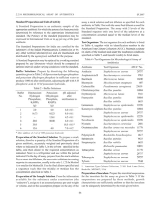
to the bromine is discharged, add 0.6 ml of formic acid, stopper
the flask and mix the contents thoroughly so as to remove any
excess of bromine from the vapour above the liquid in the
flask. After allowing to stand for 1 to 2 minutes, add 1 g of
potassium iodide and a few ml of 1 M sulphuric acid and
titrate the liberated iodine with 0.1 M sodium thiosulphate.
Perform a blank titration and make any necessary correction.
1 ml of 0.1 M sodium thiosulphate is equivalent to 0.0005172g
ofmethoxyl(CH3O).
2.3.30. Nitrogen
Use method E for substances containing 2 mg or less of
nitrogen.
MethodA
Weigh accurately the quantity of the substance under
examination specified in the individual monograph or a
quantity equivalent to about 35 mg of nitrogen into a 200-ml
long-necked flask and add 3 g of anhydrous sodium sulphate,
0.3 g of nitrogen-free mercuric oxide and 20 ml of nitrogen-
free sulphuric acid, unless otherwise specified in the
monograph.Heatthemixtureoverasmallflameuntilcolourless
and boil gently for a further 2 hours, unless otherwise directed
in the monograph, care being taken to prevent the upper part
of the flask from getting overheated. Cool, cautiously dilute
to about 75 ml with water and add a piece of granulated zinc
and a solution containing 1.5 g of sodium hydroxide per ml of
the sulphuric acid used and 2 g of sodium thiosulphate in
25 ml of water. Ensure that before distillation the mixture is
strongly alkaline by increasing, if necessary, the quantity of
sodium hydroxide. Immediately connect the flask to a
distillation apparatus, mix the contents, distil the liberated
ammonia into 50.0 ml of 0.1 M sulphuric acid and titrate the
excess of acid with 0.1 M sodium hydroxide using methyl
red-methylene blue solution as indicator. Repeat the operation
without the substance under examination. The difference
between the titrations represents the ammonia liberated by
the substance under examination.
1 ml of 0.1 M sulphuric acid is equivalent to 0.002802 g
of N.
MethodB
Weigh accurately the quantity of the substance under
examination specified in the monograph or a quantity
equivalent to about 35 mg of nitrogen into a 200-ml long-
necked flask, add 20 ml of nitrogen-free sulphuric acid, unless
otherwise specified in the monograph, and heat for 15 minutes.
Add 3 g of anhydrous sodium sulphate and 0.3 g of nitrogen-
free mercuric oxide and complete MethodA, beginning at the
words “Heat the mixture...”.
1 ml of 0.1 M sulphuric acid is equivalent to 0.002802 g
of N.
MethodC
Weigh accurately the quantity of the substance under
examination specified in the monograph or a quantity
equivalent to about 15 mg of nitrogen into a 200-ml long-
necked flask and add 1 g of a powdered mixture of 10 parts of
anhydrous sodium sulphate or potassium sulphate and 1 part
of cupric sulphate.Add 10 ml of nitrogen-free sulphuric acid,
mix, and carefully add 1 ml of hydrogen peroxide solution
(100 vol) carefully down the wall of the flask. Heat until the
solution becomes clear green in colour or almost colourless
for 30 minutes. Cool, carefully add 20 ml of water, cool again
and connect the flask to a distillation apparatus.Add 50 ml of
10 M sodium hydroxide and distil immediately by passing
steam through the flask. Collect the distillate in 25.0 ml of
0.1 M hydrochloric acid and titrate the excess of acid with
0.1 M sodium hydroxide using methyl red-methylene blue
solution as indicator. Repeat the operation using 25 mg of
anhydrous dextrose in place of the substance under
examination. The difference between the titrations represents
the ammonia liberated by the substance under examination.
1 ml of 0.1 M hydrochloric acid is equivalent to 0.001401
g of N.
2.3.30. NITROGEN
84
IP 2007
Method D (When nitrates and nitrites are present)
Weigh accurately the quantity of the substance under
examination specified in the monograph or a quantity
equivalent to about 15 mg of nitrogen into a 200-ml long-
necked flask, add 10 ml of nitrogen-free sulphuric acid in
which 0.2 g of salicylic acid has been previously dissolved
andmix.Allowthemixturetostandfor30minuteswithfrequent
shaking and add 1 g of a powdered mixture of 10 parts of
anhydrous sodium sulphate or potassium sulphate and 1 part
of cupric sulphate, mix and carefully add 1 ml of hydrogen
peroxide solution (100 vol) down the wall of the flask.
Complete Method C beginning at the words “Heat until the
solution....”.
1 ml of 0.1 M hydrochloric acid is equivalent to 0.001401
g of N.
MethodE
Apparatus:Aunit of the type generally known as semi-micro
Kjeldahl apparatus.
Method
Weigh accurately a quantity of the substance under
examination equivalent to about 2 mg of nitrogen into the
digestion flask of the apparatus.Add 1 g of a powdered mixture
of 10 parts of anhydrous sodium sulphate or potassium
sulphate and 1 part of cupric sulphate and wash down any
adhering material from the neck of the flask with water.Add 7
ml of nitrogen-free sulphuric acid and 1 ml of hydrogen
peroxide solution (100 vol) carefully down the wall of the
flask. (Do not add hydrogen peroxide during the digestion).
Heat until the solution has a clear blue colour and the sides of
the flask are free from carbonaceous matter. Cool, add carefully
20 ml of water, cool the solution and arrange for steam
distillation. Add through the funnel 30 ml of 10 M sodium
hydroxide, rinse the funnel with 10 ml of water, tightly close
the apparatus and begin the distillation with steam immediately.
Collect the distillate in 25.0 ml of 0.01 M sulphuric acid,
continue the distillation until the distillate measures about
100 ml. Titrate the distillate with 0.01 M sodium hydroxide
using methyl red-methylene blue solution as indicator. Repeat
the operation without the substance under examination. The
difference between the titrations represents the ammonia
liberated by the substance under examination.
1 ml of 0.01 M sulphuric acid is equivalent to 0.0002802 g
of N.
Method F (Determination of Protein in Blood Products)
For dried blood products prepare a solution of the preparation
as directed in the monograph.
To a volume expected to contain about 0.1 g of protein add
sufficient saline solution to produce 20 ml. To 2 ml of the
resultingsolution,ina75-mlboilingtube,add2ml ofasolution
containing 75.0 per cent v/v of nitrogen-free sulphuric acid,
4.5 per cent w/v of potassium sulphate and 0.5 percent w/v of
copper (II) sulphate, mix and loosely stopper the tube. Heat
gradually to boiling, boil vigorously for 1.5 hours and cool. If
the solution is not clear add 0.25 ml of hydrogen peroxide
solution (20 vol), continue heating until a clear solution is
produced and cool. During heating, take precautions to ensure
that the upper part of the tube is not overheated.
Transfer the solution to a distillation apparatus using three 3-
ml quantities of water, add 10 ml of 10M sodium hydroxide
and distil rapidly for 4 minutes, collecting the distillate in a
mixture of 5 ml of a saturated solution of boric acid and 5 ml of
water and keeping the tip of the condenser below the level of
the acid. Lower the collection flask so that the condenser can
drain freely and continue the distillation for a further 1 minute.
Titrate with 0.02M hydrochloric acid using methyl red mixed
solution as indicator (V1 ml).
To a further volume of the preparation under examination, or
of the solution prepared from it, expected to contain about 0.1
g of protein, add 12 ml of saline solution, 2 ml of a 7.5 per cent
w/v solution of sodium molybdate and 2 ml of a mixture of 1
volume of nitrogen-free sulphuric acid and 30 volumes of
water. Shake, allow to stand for 15 minutes, add sufficient
water to produce 20 ml, shake again and centrifuge. Using 2
ml of the resulting clear supernatant liquid repeat the procedure
described above beginning at the words ‘in a 75-ml boiling
tube…’ (V2 ml). Calculate the protein content in mg per ml of
the preparation under examination, using the expression
6.25 × 0.280 (V1–V2) and taking into account the initial dilution.
2.3.31 Nitrite Titration
The following method is suitable for the determination of most
of the pharmacopoeial sulphonamide drugs and their
preparations. It may also be used for other pharmacopoeial
drugs for which nitrite titration is recommended.
Apparatus
A suitable open vessel of about 200 ml capacity is fitted with
two similar clean platinum electrodes and a stirrer. The
electrodes may be of platinum foil 0.5 cm square and should
be placed 1.5 cm apart. They may be cleaned by immersing for
a few seconds in boiling nitric acid containing a small amount
of ferric chloride, followed by washing with water.
The polarising voltage may be obtained from a 1.5 volt dry cell
and potentiometer or other convenient device which enables
a small but definite voltage to be applied across the electrodes.
The current flowing in the system is indicated by a series
galvanometer which should have adequate sensitivity.
2.3.31. NITRITE TITRATION
85
IP 2007
Method
Weigh accurately about 0.5 g in the case of a sulphonamide or
otherwise the quantity specified in the individual monograph
and transfer to the titration vessel.Add 20 ml of hydrochloric
acid and 50 ml of water stir until dissolved, cool to about 15º.
Immerse the platinum electrodes in the solution and apply a
voltage of about 50 mV across the electrodes when polarisation
of the electrodes takes place. Place the burette tip just above
the surface of the solution and stir the solution gently,
maintaining the temperature at about 15º. The titration may be
carried out manually or by means of an automatic titrator. In
the manual titration, add 0.1 M sodium nitrite slowly and
when the titration is within 1 ml of the end point, add the
titrant in 0.1 ml portions, allowing not less than 1 minute
between additions. (The galvanometer needle deflects and
then returns to approximately its original position until the
end point is reached). At the end-point, when a slight excess
of sodium nitrite is present, the electrodes are depolarised,
current flows and a permanent deflection of the needle is
obtained.
NOTE—It will be necessary to adjust the sensitivity of the
galvanometer or the applied voltage before the titration is
begun in order to obtain an adequate deflection at the end-
point.
2.3.32 Assay of Nitrous Oxide
Apparatus
The apparatus shown in Fig 2.3.32-1 comprises a gas burette
of 100-ml capacity, connected through a two-way tap at its
upper end to two capillaries, one of which (A) is used to
introduce the gas into the apparatus, the other (B) being
connected to a vertical capillary arm to form a four-way
junction. The descending arm of the junction is connected to
a condenser (C) of about 60-ml capacity, and the right arm of
the junction is connected to a mercury manometer (M). Tap D
on the upper vertical arm of the junction opens to the air. The
lower part of the gas burette is fitted with a one-way tap
connected by a rubber tube to a mercury reservoir. The upper
2.3.32. ASSAY OF NITROUS OXIDE
(Dimensions in mm unless otherwise stated)
Fig 2.3.32-1:Apparatus forAssay of Nitrous Oxide
86
IP 2007
part of the burette is graduated from 0 to 5 ml, and the lower
part from 99.5 to 100.5 ml, both in increments of 0.1 ml.
Method
Close the three taps and immerse the condenser in liquid
nitrogen, keeping the level slightly above the upper part of
the condenser. By manipulating the two-way tap and the mobile
reservoir create a partial vacuum in the apparatus, choosing
an arbitrary pressure, Po, between 6.7 and 8 kPa (50 to 60 torr),
accurately measured. This pressure must remain constant for
10 minutes to demonstrate that the apparatus is gas-tight.
Open the two-way tap to tubeAand completely fill the burette
and tube A with mercury. Close the two-way tap. Connect a
rubber tube through a suitable pressure-relieving device to
the exit valve of the cylinder of the gas under examination and
pass a current of the gas through the rubber tube for 1 minute.
Whilst the gas is still flowing, connect the rubber tube to the
end of tube A and immediately open the two-way tap to tube
A. Allow the specified volume of the gas to enter the burette
by lowering the mercury reservoir. Disconnect the rubber tube
and expel the gas from the burette by slowly raising the mercury
reservoir above the capillary tube.Allow the specified quantity
of the gas under examination to enter the burette by lowering
the mercury reservoir and ensure that the pressure of the gas
is equal to atmospheric pressure. Close the two-way tap.
Raise the mercury reservoir slightly above tube A and lower
the level of the liquid nitrogen to the middle of the condenser.
Carefully open the tap of the burette to connect with the
condenser and allow the mercury to rise in the burette until it
reaches the tap. Close the tap. Raise the level of the liquid
nitrogen so as to totally immerse the condenser. Read the
pressure and wait until it remains steady for 2 minutes.
Place the mercury reservoir in its bottom position and open
the tap of the burette to make connection with the condenser.
Move the mercury reservoir until the manometer reading is
the same as the initial pressure Po. Close the tap of the burette
and, by means of the mercury reservoir, bring the pressure of
the gas in the burette to atmospheric pressure. The number of
ml of gas represents the non-condensable volume in the
specified volume used.
After each series of 10 determinations allow atmospheric air
to enter by opening the tap D, remove the liquid nitrogen from
the condenser and allow the condenser to warm to room
temperature.
2.3.33 Assay of Oxygen
Apparatus
The apparatus shown in Fig 2.3.33-1 comprises a gas burette
of the type described under assay of nitrous oxide (Fig 2.3.32-
(Dimensions in mm unless otherwise stated)
Fig 2.3.33-1:Apparatus forAssay of Oxygen
2.3.31. ASSAY OF OXYGEN
87
IP 2007
1) in which tube B is connected to a gas pipette comprising
two bulbs of suitable size.
Method
Charge the pipette with the reagents specified in the
monograph. With the two-way tap open to tube B, draw the
solution just to the level of the tap by moving the mercury
reservoir. Open the two-way tap to tubeA, completely fill the
burette and tube A with mercury and close the two-way tap.
Connect a rubber tube to the exit valve of the cylinder of the
gas under examination through a suitable pressure reducing
device and pass a current of the gas through the tube for 1
minute.
Whilst the gas is still flowing, connect the rubber tube to tube
A, immediately open the two-way tap to tube A, allow the
specified quantity of the gas to enter the burette by lowering
the mercury reservoir and close the two-way tap. Increase the
pressure of the gas by raising the mercury reservoir, open the
two-way tap to tube B and transfer all the gas to the pipette.
Close the tap and gently shake the pipette. After 15 minutes,
when most of the gas has been absorbed by the liquid, draw
the residual gas back into the burette and repeat the procedure
beginning at the words “Increase the pressure of the gas..”
until the volume of residual gas is constant. Measure the
volume of the residual gas in the burette.
2.3.34 Oxygen-Flask Method
NOTE — Great care must be taken in carrying out the
following method. It is advisable to wear safety glasses and
to use a suitable safety screen particularly when combustion
takes place.
Apparatus
The apparatus consists of a thick-walled, conical 500-ml iodine
flask, fitted with a ground-glass stopper to which is fused a
piece of platinum wire about 13 cm long and 1 mm in diameter
and to which is attached a piece of platinum gauze to serve as
means of holding the sample. The gauze may be about 2 cm
wide and 1.5 cm long and should comply with the dimensions
of a sieve with a nominal mesh aperture of 425 µm (No. 36
sieve). The flask must be well-cleaned and free from even
traces of organic solvents.
Method
Solid substances should be finely ground and thoroughly
mixed before the specified quantity is weighed.
For liquids place the specified quantity on about 15 mg of
ashless filter-paper flock contained in one part of a
methylcellulose capsule of a suitable size, close the capsule,
inserting one end of a narrow strip of filter-paper between the
two parts, and secure the capsule in the platinum gauze.
Ointments should be enclosed in grease-proof paper before
wrapping in filter-paper.
Weigh accurately a suitable quantity of the substance under
examination and wrap, if a solid, in a piece of halide-free filter-
paper (4 cm × 3 cm), secure the package in the platinum gauze
sample holder and insert one end of a narrow strip (1 cm × 3
cm) of filter-paper in the roll to serve as a fuse. Flush the flask
with oxygen, moisten the neck with water, place the specified
absorbing liquid in the flask, fill it with oxygen by swirling the
liquid to favour its taking up the oxygen, light the free end of
the fuse-strip and immediately insert the stopper. Hold the
stopper firmly in place when vigorous burning has begun,
invert the flask so as to provide a liquid seal but taking care to
prevent incompletely burned material falling into the liquid.
When combustion is complete, shake the flask vigorously for
about 5 minutes, place a few ml of water in the cup top,
carefully remove the stopper, and rinse the stopper, platinum
wire, platinum gauze, and sides of the flask withwater. Proceed
as directed in the following methods.
ForBromine
Burn the specified quantity of the substance under examination
in the prescribed manner using 15 ml of a mixture of 9 volumes
of 0.5 M sulphuric acid and 1 volume hydrogen peroxide
solution (100 vol) as the absorbing liquid. When the process
is complete, cool in ice for 15 minutes, add 5 ml of 2M nitric
acid and 10 ml of 0.1 M silver nitrate and titrate with 0.05 M
ammonium thiocyanate using ferric ammonium sulphate
solution as indicator and shaking vigorously as the end-point
is approached. Repeat the operation without the substance
under examination; the difference between the titrations
represents the number of ml of 0.05 M silver nitrate required.
1 ml of 0.05 M silver nitrate is equivalent to 0.003995 g of
Br.
For Chlorine
Burn the specified quantity of the substance under examination
in the prescribed manner using 20 ml of 1 M sodium hydroxide
as the absorbing liquid. When the process is complete, add
2.5 ml of nitric acid, 2.5 ml of water and 10 ml of 0.1 M silver
nitrate and titrate with 0.05 M ammonium thiocyanate using
ferric ammonium sulphate solution as indicator and shaking
vigorously as the end-point is approached. Repeat the
operation without the substance under examination; the
difference between the titrations represents the number of ml
of 0.05 M silver nitrate required. 1 ml of 0.05 M silver nitrate
is equivalent to 0.001773 g of Cl.
ForFluorine
Burn the specified quantity of the substance under examination
in the prescribed manner using 20 ml of water as the absorbing
liquid. When the process is complete, add sufficient water to
2.3.34. OXYGEN-FLASK METHOD
88
IP 2007
produce 100.0 ml.To 2.0 ml of this solution add 50 ml ofwater,
10 ml of alizarine fluorine blue solution, 3 ml of a solution
containing 12 per cent w/v of sodium acetate and 6 per cent v/
v of glacial acetic acid, 10 ml of cerous nitrate solution and
sufficient water to produce 100.0 ml. Allow to stand in the
dark for 1 hour and measure the absorbance of a 4-cm layer of
the resulting solution at about 610 nm (2.4.7), using as the
blank a solution prepared in the same manner but using 2.0 ml
of water in place of the solution and beginning at the words
“To 2.0 ml...”. Calculate the flourine content from a reference
curve prepared by treating suitable aliquots of a solution of
sodium fluoride in the manner described above, beginning at
the words “add 50 ml of water.....”.
ForIodine
Burn the specified quantity of the substance under examination
in the prescribed manner using a mixture of 10 ml of water and
2 ml of 1 M sodium hydroxide as the absorbing liquid. When
the process is complete, add an excess (5 to 10 ml) of acetic
bromine solution and allow to stand for 2 minutes. Remove
the excess of bromine by the addition of formic acid (5 to 10
ml), rinse the sides of the flask with water and sweep out any
bromine vapour above the liquid with a current of air.Add 1 g
of potassium iodide and titrate with 0.02 M sodium
thiosulphate using starch solution, added towards the end
of the titration, as indicator.
1 ml of 0.02 M sodium thiosulphate is equivalent to 0.000423
g of I.
ForSulphur
Method I (in the absence of halogens and phosphorus) —
Burn the specified quantity of the substance under examination
in the prescribed manner using 10 ml of water and 0.1 ml
hydrogen peroxide solution (100 vol) as the absorbing liquid.
When the process is complete, cool the solution in ice for
about 15 minutes. Gently boil for 2 minutes, cool and add 50 ml
of ethanolic acetic-ammonia buffer pH 3.7. Titrate with 0.05
M barium perchlorate using 0.3 ml of alizarin red S solution
as indicator, until the solution becomes orange-pink in colour.
1 ml of 0.05 M barium perchlorate is equivalent to 0.001603 g
of S.
Method II (in the presence of halogens or phosphorus) —
Burn the specified quantity of the substance under examination
in the prescribed manner using 15 ml of water and 1 ml of
hydrogen peroxide solution (20 vol) as the absorbing liquid.
When the process is complete, boil the solution for 10 minutes,
cool and add 60 ml of ethanol (95 per cent). Titrate the solution
with 0.01 M barium perchlorate, using a 0.1 ml of 0.2 per cent
w/v solution of thoron and 0.1 ml of a 0.0125 per cent w/v
solution of methylene blue as indicator, until the yellow colour
changes to pink.
1 ml of 0.01 M barium perchlorate is equivalent to 0.0003206
g of S.
If the temperature at which the determinations prescribed under
Method I and II are performed differs from that at which the
barium perchlorate solution was standardised, the titrant
volumes are corrected by applying the expression
Vc = V [1 + 0.0008(t1 - t2)]
where, Vc = the corrected volume of titrant,
V = the volume of titrant used,
t1 = the temperature of the titrant during
standardisation,
t2 = the temperature of the titrant during the
determination.
2.3.35. Peroxide Value
The peroxide value is the number of milliequivalents of active
oxygen that expresses the amount of peroxide contained in
1000 g of the substance.
Method
Unless otherwise specified in the individual monograph, weigh
accurately about 5 g of the substance under examination,
transfer to a 250-ml glass-stoppered conical flask, add 30 ml of
a mixture of 3 volumes of glacial acetic acid and 2 volumes of
chloroform, swirl until dissolved and add 0.5 ml of saturated
potassium iodide solution.Allow to stand for exactly 1 minute,
withoccasionalshaking,add30mlofwaterandtitrategradually,
with continuous and vigorous shaking, with 0.01 M sodium
thiosulphate until the yellow colour almost disappears. Add
0.5 ml of starch solution and continue the titration, shaking
vigorously until the blue colour just disappears (a ml). Perform
ablankdeterminationomittingthesubstanceunderexamination
(b ml). The volume of 0.01 M sodium thiosulphate in the
blank determination must not exceed 0.1 ml.
Calculate the peroxide value from the expression
Peroxide value = 10 (a - b)/w
Where, w = weight, in g, of the substance.
3.3.36. Phenol in Vaccines andAntisera
Homogenise the preparation under examination. Dilute an
appropriate volume with water to give a solution containing
about 15 µg of phenol per ml.To 5.0 ml of the resulting solution
add 5 ml each of buffer solution pH 9.0, 4-aminophenazone
solution and potassium ferricyanide solution. Allow to stand
for 10 minutes and measure the absorbance of the resulting
solution at about 546 nm (2.4.7). Calculate the phenol content
from the absorbance obtained, using a calibration curve
2.3.35. PEROXIDE VALUE
89
IP 2007
prepared by repeating the operation using 5 ml of each of a
series of solutions containing 5 µg, 10 µg, 15 µg, 20 µg and 30
µg of phenol per ml respectively.
2.3.37. Saponification Value
The saponification value is the number of milligrams of
potassium hydroxide necessary to neutralise the free acids
and to saponify the esters present in 1 g of the substance.
Method
Unless otherwise specified in the individual monograph,
introduce about 2 g of the substance under examination,
accurately weighed, into a 200-ml flask of borosilicate glass
fitted with a reflux condenser.Add 25.0 ml of 0.5 M ethanolic
potassium hydroxide and a little pumice powder and boil under
reflux on a water-bath for 30 minutes. Add 1 ml of
phenolphthalein solution and titrate immediately with 0.5 M
hydrochloric acid (a ml). Perform a blank determination
omitting the substance under examination (b ml). Calculate
the saponification value from the expression
Saponification value = 28.05 (b - a)/w
where, w = weight, in g, of the substance.
NOTE — If the oil has been saturated with carbon dioxide
for the purpose of preservation, gently reflux the solution of
the oil in ethanol (95 per cent) and ether for 10 minutes before
titration. The oil may be freed from the carbon dioxide by
exposing it in a shallow dish in a vacuum desiccator for 24
hours before weighing the sample.
2.3.38. Assay of Steroids
Test solution. Prepare as directed in the individual monograph.
Standard solution. Weigh accurately a suitable quantity of
the reference substance specified in the individual
monograph, previously dried under the conditions specified
in the monograph, and dissolve in a suitable volume of
aldehyde-free ethanol. Dilute quantitatively and stepwise with
aldehyde-free ethanol to obtain a solution containing about
10 µg of the steroid per ml.
Method
Into a glass-stoppered, 50-ml conical flask add 20.0 ml of the
test solution. Into two similar flasks add 20.0 ml of the standard
solution and 20.0 ml of aldehyde-free ethanol (blank),
respectively. To each flask add 2.0 ml of blue tetrazolium
solution and mix; to each flask add 2.0 ml of a mixture of
tetramethylammonium hydroxide solution (10 per cent) and
90 volumes of aldehyde-free ethanol, mix and allow to stand
in the dark at a temperature between 25º and 35º.At the end of
exactly 90 minutes add to each flask 1.0 ml of glacial acetic
acid and mix. Measure the absorbances of the solutions
obtained from the test solution and the standard solution at
about 525 nm against the blank (2.4.7).
Calculate the quantity, in mg, of the steroid in the 20-ml aliquot
of the test solution from the expression
At /As x Cs where, At is the absorbance of the test solution,As
is the absorbance of the standard solution and Cs is the
quantity, in mg, of the reference substance in the 20-ml aliquot
of the standard solution.
Calculate the quantity of the steroid in the substance under
examination on the basis of the aliquot of the test solution
taken for the assay and from the declared content of the steroid
in the appropriate reference substance.
2.3.39. Unsaponifiable Matter
The unsaponifiable matter consists of substances present in
oils and fats which are not saponifiable by alkali hydroxides
and are determined by extraction with an organic solvent of a
solution of the saponified substance under examination.
Method
Unless otherwise specified in the individual monograph,
introduce about 5 g of the substance under examination,
accurately weighed, into a 250-ml flask fitted with a reflux
condenser.Add a solution of 2 g of potassium hydroxide in 40
ml of ethanol (95 per cent) and heat on a water bath for 1
hour, shaking frequently. Transfer the contents of the flask to
a separating funnel with the aid of 100 ml of hot water and,
while the liquid is still warm, shake very carefully with three
quantities, each of 100 ml, of peroxide-free ether. Combine the
ether extracts in a second separating funnel containing 40 ml
of water, swirl gently for a few minutes, allow to separate and
reject the lower layer. Wash the extract with two quantities
each of 40 ml, of water and with three quantities, each of 40 ml,
of a 3 per cent w/v solution of potassium hydroxide, each
treatment being followed by a washing with 40 ml of water.
Finally, wash the ether layer with successive quantities, each
of 40 ml, of water until the aqueous layer is not alkaline to
phenolphthalein solution. Transfer the ether layer to a
weighed flask, washing out the separating funnel with
peroxide-free ether. Distill off the ether and add to the residue
6 ml of acetone. Remove the solvent completely from the flask
with the aid of a gentle current of air. Dry at 100º to 105º for 30
minutes. Cool in a desiccator and weigh the residue. Calculate
the unsaponifiable matter as per cent w/w.
Dissolve the residue in 20 ml of ethanol (95 per cent),
previously neutralised to phenolphthalein solution and titrate
with 0.1 M ethanolic potassium hydroxide. If the volume of
0.1 M ethanolic potassium hydroxide exceeds 0.2 ml, the
2.3.39. UNSAPONIFIABLE MATTER