Blood Supply of GIT
Dr. Komal Parmar
Total Number of Slides: 45
Aims and Objectives
• Understanding the three dimensional topography of vascular
system inside the abdominopelvic cavity.
• Detailed description of the blood supply of gastrointestinal
tract.
• Clinical and pathological correlation.
2
Blood Supply of Oropharynx
3
Blood Supply of Oesophagus
4
Entry into the Abdominal Cavity
• T8: Caval Opening
• IVC
• Right Phrenic Nerve
• T10: Esophageal Hiatus
• Esophagus
• Vagal Trunks
• Esophageal Vessels
• T12: Aortic Hiatus
• Aorta
• Thoracic Duct
• Azygous Vein 5
Thoracoabdominal
Interface
6
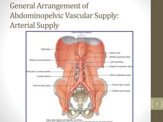
General Arrangement of
Abdominopelvic Vascular Supply:
Arterial Supply
7
Venous System
8
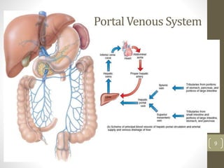
9
Portal Venous System
Arterial
Supply
10
Abdominal Aorta
• Begins at Aortic hiatus of
Diaphragm: T12
• Bifurcates into two common iliac
arteries at L4/5 intervertebral disc.
• Mean diameter=
• 19-21mm in males
• 16-18mm in females
• Relations:
• Anterior= Coeliac trunk, Autonomic nerve
plexuses, lymphatics, pancreas, 3rd part of
duodenum, root of mesentry
• Posterior: Vertebrae, I/V disc and
ligaments, Lumbar Arteries
• Right: Thoracic Duct, Azygous Vein, Right
Crus of Diaphragm, IVC, Right Coeliac
Ganglion.
• Left: Left Crus of Diaphragm, Left Coeliac
Ganglion, 4th part of Duodenum, Left
Sympathetic Trunk, Inferior mesenteric
Vein. 11
12
Branches of Abdominal Aorta
• Anterior group
(unpaired)
• Coeliac Trunk
• Superior Mesenteric A
• Inferior Mesenteric A
• Lateral Group
• Middle Suprarenal A
• Renal A (R>L)
• Gonadal A
• Dorsal Group
• Inferior Phrenic A
• Lumbar A (4) (*** Artery of
Adamkewicz)
• Median Sacral A (1) 13
Coeliac Trunk
• T12
• 1-3 cm long.
• Trifucration and ramification
• Related to respective crus of diaphragm and coeliac ganglion on
each side.
14
15
Splenic Artery
• Tortous. 8-32cm long.
• Largest branch of the coeliac trunk.
• Runs along the upper border of
pancreas.
• Branches
• Pancreatic
• Short Gastric
• Left Gastroepiploic
16
Common Hepatic Artery
17
18
Superior Mesenteric Artery
• Originates 1 cm below
the Coeliac trunk
• Lower border of L1,
Transpyloric plane.
• Posterior relations:
uncinate process of
Pancreas, 3rd part of
Duodenum, left renal vein,
IVC, right ureter, right psoas.
• Anterior Relations:
Splenic vein and Body of
Pancreas.
• ***Marginal Artery
19
20
Inferior Mesenteric Artery
• Arises behind the inferior border of 3rd part of Duodenum.
• 3-4 cm above the aortic bifurcation
21
22
Rectal Arteries
23
24
25
26
Veins
Mesenteric Veins
Inferior Mesenteric
• Begins as Superior
Rectal Vein.
• Receives
• Sigmoidal Veins and
• Left Colic Veins
• Drains into Splenic Vein
eventually.
Superior Mesenteric
• Tributaries
• Right Gastroepiploic V
• Inferior
Pancreaticoduodenal V
• Middle colic V
• Right Colic V
• Ileocolic V
• Ileal V
• Jejunal V
• Drains into: Portal Vein
27
28
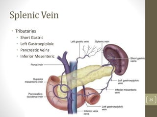
Splenic Vein
• Tributaries
• Short Gastric
• Left Gastroepiploic
• Pancreatic Veins
• Inferior Mesenteric
29
Portal Venous Circulation
30
Portal Vein
• Formed behind the neck of Pancreas at the level of L1/2
intervertebral disc.
• Superior mesenteric Vein + Splenic vein
• 8cm
• Ascends behind the 1st part of Duodenum, CBD and
gastroduodenal artery.
• Enters the right border of Lesser Omentum and reaches the
right end of Porta hepatis behind CBD and Hepatic Artery.
• Divides into R and L branches which accompany the
corresponding Hepatic Artery.
• Tributaries: Left gastric and Posterior Superior
Pancreaticoduodenal. Cystic veins, Para Umbilical veins. 31
32
33
34
Hepatic
Lobule
Hepatic Veins
• The hepatic veins are divided
into upper and lower groups
• The upper three drain the
central veins from the right,
middle, and left regions of the
liver and are larger than the
lower group of veins.
• The lower group of from six to
twenty smaller hepatic veins
come from the right lobe and
the caudate lobe, are in contact
with the hepatic tissue, and are
valveless. All the veins empty
into the inferior vena cava at
the back of the liver. 35
Hepatic veins
draining into IVC
Inferior Vena Cava
36
IVC
• Formed by the junction of the common iliac veins anterior to
the 5th lumbar vertebral body.
• Tributaries
• Lumbar veins
• Right Gonadal vein
• Renal veins (L>R)
• Right Suprarenal vein
• Inferior phrenic veins
• Hepatic Veins
37
38
• Portosystemic Shunt:
Occur at the sites of Porto-
Caval Anastamoses
39
Clinical Correlates
Aortic Aneurysm
40
Gastrointertinal Bleeding
41
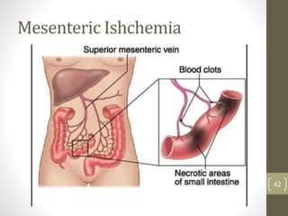
Mesenteric Ishchemia
42
Intestinal
Resection and
Anastomoses
43
Conclusions
• Arterial system and Venous system do not follow an exactly
parallel system.
• Importance of Portal Venous system and Liver as the focal site
of metabolism.
• Correlation of anatomical intricacies with the Clinical
requirements.
• Tip for consolidating the information: Look at the figures and
approach it conceptually.
44
45