Dr Sai L Daayana
MBBS, MRCOG, MD, Sub-specialty training in
Gynaecology Oncology (UK)
Consultant in Surgical Gynaecology Oncology
Apollo Cancer Institutes, Hyderabad
VULVA - BENIGN, PRE-MALIGNANT &
MALIGNANT CONDITIONS
WHAT IS VULVAL INTRAEPITHELIAL
NEOPLASIA?
Vulval (or vulvar) intraepithelial neoplasia is a pre-cancerous skin
lesion of any part of the vulva. Vulval intraepithelial neoplasia
(VIN) is now also called vulval squamous intraepithelial lesion
(SIL).
VIN is not invasive cancer but vulval squamous cell cancer (SCC)
occurs in about 15% of women if VIN is left untreated.
Epidemiology of VIN
Uncommon premalignant condition of the vulva
Increasing in incidence
Associated with HPV in young women
May co-exist with CIN, immunosuppression is risk factor
Clinical Features of VIN
Itch, soreness, burning, asymptomatic
Leukoplakic, erythematous, ulcerated or pigmented lesions
on the vulva
Pathology of VIN
Maturation disorder, cellular atypia, abnormal mitotic figures,
increased nuclear:cytoplasmic ratio, graded VIN 1 to VIN 3
VIN – high grade
Vulval Intraepithelial Neoplasia
§ Is usually asymptomatic True/False
§ Frequently progresses to invasive carcinoma if left untreated
True/False
§ Is characterised by blunting of rete ridges in the epidermis
True/False
§ Presents with parchment-like appearance of the vulva in a figure of
eight distribution, with loss of vulval architecture and atrophy
True/False
§ Is commonly seen in women with a history of abnormal cervical
cytology True/False
VULVAL INTRAEPITHELIAL
NEOPLASIA
TREATMENT OF VULVAL INTRAEPITHELIAL
NEOPLASIA
§  Low-grade – asymptomatic – regular vulvoscopy every 6
months
§  Low-grade – symptomatic – treatment offered
§  High-grade – treatment offered to reduce the risk of developing
invasive cervical cancer
TREATMENT FOR VULVAL
INTRAEPITHELAIL NEOPLASIA
Unilocular lesion – Surgical treatment – wide local excision with a
margin of normal tissue; or skinning vulvectomy
LASER ablation (under GA) – if no malignancy suspected,
multifocal lesions
MEDICAL MANAGEMENT OF VIN
Large areas of VIN; multiple previous excisions can be managed medically with:
1. Imiquimod cream, applied 3 times weekly for 12 to 20 weeks. This results in red,
inflamed and eroded tissue often accompanied by considerable discomfort.
2. 5-fluorouracil cream, applied twice daily for several weeks. This causes quite severe
inflammation (several weeks) and will not be tolerated by all women. It is less effective
than imiquimod cream.
3. Photodynamic therapy (PDT) requires specialised equipment and can also be very
painful.
4. Cidovir has been described to be useful in some patients.
IMIQUIMOD CREAM
¡ Immunomodulator
¡ Acts on Toll-like
receptors
¡ Enhances local immune
responses
CIDOFOVIR
MEDICAL MANAGEMENT OF VIN
§  Anti-viral drug
§  Anti-tumour agent
5-FLUORO URACIL CREAM
¡ Anti-metabolite
¡ Given systemically and
topically in cancer
treatment
PHOTO DYNAMIC THERAPY
MEDICAL MANAGEMENT OF VIN
¡ Uses a drug, called a
photosensitizer or
photosensitizing agent,
and a particular type of
light. They produce a
free radical oxygen
molecules that kills
nearby cells.
RECURRENCE OF VIN
Recurrence rates of VIN after treatment range from 9% to 50%
Risk factor for recurrence:
•  positive excision margins
•  Multi-focal disease
•  Smoking
•  Immunosuppression
FOLLOW-UP FOR VIN
Self-examination of the vulva and regular vulvoscopy follow-up is
prudent
Women with a complete response to therapy and no new lesions
at follow-up visits scheduled 6 months and 12 months after initial
treatment should be monitored by visual inspection of the vulva
annually thereafter.
Lichen Sclerosus
§ Epithelial thinning, inflammation, histological changes in
the dermis (loss of rete ridges)
¡ Clinical features
§ Intractable itch, soreness, dyspareunia, asymptomatic
§ Parchment-like appearance, figure of eight distribution,
loss of vulval architecture, vulval atrophy
§ Adults, occasionally children
§ 9% incidence of progression to SCC of vulva
OTHER DERMATOLOGICAL
CONDITIONS OF THE VULVA
Lichen Sclerosus
Lichen Sclerosus
Lichen Planus
•  Chronic inflammatory dermatitis of unknown
aetiology
•  Characterised by flat topped violaceous shiny
papules
•  Clinical features range from white reticulated
papules to an erosive, desquamating process
DERMATOLOGICAL
CONDITIONS (2)
Lichen Planus
DERMATOLOGICAL CONDITIONS (3)
Squamous Cell Hyperplasia
•  Diagnosis of exclusion
•  Chronic reaction to fungal vulvitis, allergies or unknown stimuli
•  Thickened skin with white keratotic patches, excoriations &
fissures
¡ Psoriasis
¡ Hidradenitis Suppurativa
§ Chronic suppurative disorder of apocrine glands
§ Deep painful subcutaneous nodules that ulcerate
and drain leading to open sinuses and extensive
scarring
DERMATOLOGICAL
CONDITIONS (4)
Psoriasis
Hidradenitis Suppurativa
¡ Can be solitary or multiple, painful or non painful
¡ Seen more frequently in GUM clinic
¡ Solitary, non painful ulcers include: syphilis,
lymphogranuloma venerum, cancer
¡ Multiple painful ulcers include: herpes, Behcet’s disease,
Crohn’s disease
¡ Investigations: serology, culture, biopsy
¡ Treatment depends on cause
ULCERATIVE DERMATOSES
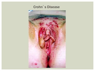
Crohn’s Disease
¡  Rare condition that typically affects postmenopausal Caucasian women
¡  Presents as crusty, erythematous area on vulva
¡  Associated with pruritis vulvae
¡  Often misdiagnosed as contact dermatitis or eczema
¡  Biopsy is necessary for diagnosis
¡  90% of cases are primary intraepithelial malignancies & treatment is
aimed at symptom control
¡  10-15% are associated with an underlying adenocarcinoma of the vulva
¡  30% have malignancies at non-vulval sites, including breast, colon, &
bladder
¡  Need to exclude invasive disease in vulva and exclude underlying
internal carcinoma from another site (e.g. by colonoscopy, CXR,
mammogram, hysteroscopy etc)
EXTRA MAMMARY PAGETS DISEASE
Extramammary Paget’s
Disease of the Vulva
§ Uncommon cancer
§ Most patients are elderly (60-75 yrs)
§ Squamous carcinoma most common
§ Melanoma second most common (4-9% of cases, post menopausal
Caucasian women)
§ Basal Cell Carcinoma has excellent prognosis
§ Adenocarcinoma of Bartholin’s gland or in conjunction with
Paget’s disease (20%) is rare
VULVAL CANCER (1)
INCIDENCE OF VULVAL CANCER
Incidence of malignant diseases the vulva:
3 - 4% of all gynecologic malignancies.
- The incidence increases with age.
- Recently there was a rise in the incidence, due to
•  Longevity
•  Increased HPV infections.
•  Increased smoking habits
RISK FACTORS FOR CARCINOMA OF
THE VULVA
1- Human papillomavirus infection.
–  Genital condylomas: these are detected in 5 % of vulvar
cancer
–  Vulvar intraepithelial neoplasia (VIN) and also CIN
2- Medical history of:
– Vulvar dystrophy
– Chronic vulvar pruritus
3- Patients with a history of squamous cell carcinoma of the
cervix or vagina
4- Chronic immunosuppression
5- Smoking
¡ Diagnosis
§ Vulval itch & soreness
§ Painful bleeding mass
§ Asymptomatic lump
¡ Sites involved
§ Labium majus (50%)
§ Labium minus (20%)
§ Clitoris & Bartholin’s Gland (uncommon)
VULVAL CANCER (3)
Squamous Cell Carcinoma of Vulva
Basal Cell Carcinoma of vulva
Malignant Melanoma of vulva
VULVAL CANCER (4)
§  Disease Spread
Via the lymphatics to the superficial inguinal lymph nodes;
metastasizes to pelvic lymph nodes late
Direct spread to lower vagina, urethra & anus
FIGO STAGING OF VULVA
CARCINOMA
Stage 1 2 cm lesion
size Or less
Confined to the vulva or perineum nodes
histo-Logically negative.
Stage 2 > 2cm lesion
size
Confined to the vulva or perineum nodes
histo-Logically negative.
Stage 3   Tumor of any size spread to lower urethra
vagina anus +/- Unilateral metastasis
Stage 4 A Involvement of :
Upper urethra
Bladder mucosa
Rectal mucosa
Pelvic bone
Bilateral L.N.metastasis
  B Distant metastases and / or pelvic nodes
VIN	 Vulval	cancer
VIN
Vulval	carcinoma
The overall 5 years survival rate for vulval cancer
is 70% for all operable cases,
This depends on:
1.  L.N Involvement:
This is the most prognostic factor
¡  Metastatic involvement of groin nodes
decreases the 5 years survival rate to below
50% as opposed to the 90% when L.N are not
involved.
¡  Once pelvic nodes are involved the 5 years
survival rate is 15%.
PROGNOSIS OF VULVAL CANCER
¡  		Detec2on	and	management	of	VIN.	
¡  		Proper	management	of	all	cases	with	pruiritus	vulvae.		
¡  		All	vulval	lesions	should	be	diagnosed	accurately	especially	
those	arising	a?er	menopause.		
¡  		All	pigmented	vulvar	lesions	should	be	removed	for	biopsy.		
PROPHYLAXIS…A	HIGH	INDEX	OF	
SUSPICION
Treatment OptionStage
Partial Vulvectomy excision of the tumor, with a 1 cm safe margins. No need
for node removal.
Ia
Ib
Modified radical vulvectomy with either of the following:
1) Ipsilateral groin lymph node dissection: in cases of lateralized
lesion
2) Bilateral groin node dissection: in cases of centralized lesions
Modified radical vulvectomy with bilateral groin node dissection.
II
- Combined approach:
1- Preoperative external beam radiation therapy.
2- Chemotherapy (e.g. 5-fluorouracil, cisplatin).
3- Radical excision with bilateral inguinal & femoral node dissection.
4- Preoperative RT, then surgical excision of the tumor.
- Pelvic exenteration.
III
Individualized
IV
TREATMENT OF VULVAL CANCER
VULVECTOMY:	
Radical vulvectomy can be complete or partial.
When part of the vulva, including the deep tissue, is removed, the operation is
called a partial vulvectomy.
In a complete radical vulvectomy, the entire vulva and deep tissues, including the
clitoris, are removed.
An operation to remove the lymph nodes near the vulva is called
a en block dissection. It is important to remove these lymph
nodes if they contain cancer.
Right hemi-vulvectomy
Anterior vulvectomy with bilateral groin node dissection
¡  		Detec2on	and	management	of	VIN.	
¡  		Proper	management	of	all	cases	with	pruiritus	vulvae.		
¡  		All	vulval	lesions	should	be	diagnosed	accurately	especially	
those	arising	a?er	menopause.		
¡  		All	pigmented	vulvar	lesions	should	be	removed	for	biopsy.		
PROPHYLAXIS…A	HIGH	INDEX	OF	
SUSPICION
What is this lesion?
Vulval Intraepithelial Neoplasia
What is this lesion?
Lichen Sclerosus
The most effective treatment for pruritus vulva
associated with lichen sclerosus is:
1. Acqueous cream
2. Topical clotrimazole
3. Anti-histamines
4. Imiquimod
5. Topical steroid creams
The most effective treatment for pruritus vulva
associated with lichen sclerosus is:
1. Acqueous cream
2. Topical clotrimazole
3. Anti-histamines
4. Imiquimod
5. Topical steroid creams
What is this lesion on the vulva?
Squamous Cell Carcinoma of Vulva
What is this lesion on the vulva?
Malignant Melanoma of vulva
¡  Vulval Intraepithelial Neoplasia
§ Is usually asymptomatic True/False
§ Frequently progresses to invasive carcinoma if left untreated
True/False
§ Is characterised by blunting of rete ridges in the epidermis
True/False
§ Presents with parchment-like appearance of the vulva in a figure of
eight distribution, with loss of vulval architecture and atrophy
True/False
§ Is commonly seen in women with a history of abnormal cervical
cytology True/False
¡  Vulval Intraepithelial Neoplasia
§ Is usually asymptomatic True/False
§ Frequently progresses to invasive carcinoma if left untreated
True/False
§ Is characterised by blunting of rete ridges in the epidermis
True/False
§ Presents with parchment-like appearance of the vulva in a figure of
eight distribution, with loss of vulval architecture and atrophy
True/False
§ Is commonly seen in women with a history of abnormal cervical
cytology True/False