Dr. Mamoon Ameen
Definitions
Surgical opening of the trachea.
Creation of a stoma at the skin surface which
leads into the trachea.
• The Tracheostomy is one of the oldest
surgical procedure.
• It can be traced back to Egyptian tablets
from 3600 B.C.
• 1546 : first well-documented tracheostomy
by Antonius Musa Brasavola,
• 1921: Chevaliar Jackson – standardized
the technique of the tracheostomy .
• Modern percutaneous tracheostomy (PCT)
developed by Toye and Weinstein in
1969.
• Trachea lies in midline of the
neck extending from cricoid
cartilage (C6) superiorly to the
tracheal bifurcation at the
level of sternal angle (T5).
• Comprises 16-20 C shaped
cartilage rings.
• Becomes intra-thoracic at 6th
cartilaginous ring.
• Length 10-12cm.
• Diameter 15-20mm.
Anterior relations
 Esophagus lies
Posterior
 Note Trachealis
muscle
• Elective /routine
• Emergency
• Permanent
• Temporary
• High
• Mid
• Low
• The trachea is
permanently
disconnected from the
pharynx and the
proximal end of the
trachea is sutured to the
skin.
• Permanent tracheostomy
is an elective procedure
carried out as part of an
operation
• Involving removal of the
larynx, such as a
laryngectomy or
laryngopharyngectomy
• A temporary tracheostomy
may be in use permanently;
however, it differs from a
permanent tracheostomy in
that there is still a
communication between the
pharynx and the lower
airway via the larynx. In a
permanent tracheostomy the
only access to the lower
airway is via the
tracheostome.
1. Upper Airway Obstruction.
2. Pulmonary Ventilation.
3. Pulmonary Toilet.
4. Elective Procedure
• Tumors (of oropharynx, larynx, upper
trachea)
• Infections (epiglottitis, severe
tracheobronchitis)
• Bilateral Vocal Cord Paralysis
• Trauma (laryngeal, maxillofacial fractures)
• Foreign body obstruction
• Subglottic or tracheal stenosis
• Tracheostomy should be performed in
a patient still requiring ventilation
through an endotracheal tube for
more than a one week.
• congestive cardiac failure, infection,
pulmonary edema and bulbar palsy
• Those who cannot cough and clear
their chest
• For major head and neck operations
that effect the patency of airway
• In patients with uncertain general
conditions particularly cardiovascular
or pulmonary defficency pt.
• Better too often than too late
• HISTORY
• Physical assessment
• Anesthesiological assessment
• CBC
• caugulation profile
• informed consent
1) Cricothyroidotomy
2) open tracheostomy
3) Percutaneous procedure
• Emergency procedure
• When endotracheal intubation is impossible
• Contraindicated
o In children less then 11 years
o Truama to larynx or cricoid cartillage
• Subglotic oedema & stenosis are very likely
• Keep only for 3-5 days
supine position, neck extended, L.A, stabilze larynx
Thyroid cartilage is gripped between thumb and
middle finger
move your finger down to palpate cricoid cartilage
space between thyroid and cricoid cartilge is cricothyroid
membrane
1cm vertical incision thru skin and
sub cut. tissue
use curved hemostat for blunt dissection thru planes
use horizontal incision on cricothyroid membrane
insert trousseau dilator and dilate membrane
vertically
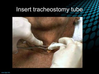
Insert tracheostomy tube
inflate cuff with 10cc syringe
attach bag valve unit and ventilate pt.
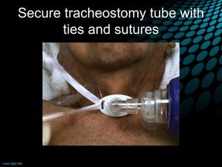
Secure tracheostomy tube with
ties and sutures
1.Airway control
2.Patient position-
supine ,neck extended
,pillow under the shoulder
3. Anesthesia
• Not necessery if pt is
unconscious or n emergency
situations
• If conscious ,1-2% lignocain
+epinephrine is infiltrated in the
line of incision and area of
dissection
• Sometime general anesthesia
with intubation is used
Identify the landmarks
a tranverse Incision 1 cm below the cricoid
or halfway between the cricoid and the
sternal notch.
Retractors are placed, the skin is retracted, and the strap
muscles are visualized in the midline. The muscles are
divided along the raphe, then retracted laterally
•The thyroid isthmus lies in the field of
the dissection.
• Typically, the
isthmus is 5 to
10 mm in its
vertical
dimension.
• Retract it up.
Identify trachea.
Anesthetist should remove any tapes used to secure the
endotracheal tube and prepare to withdraw the tube slowly under
direct vision by the surgeon.
Then place the tracheal incision in the second or third tracheal
interspace.
Tube is inserted and secured
Bjork flap
Pediatric tracheostomy
• Better done under general anesthesia
• Neck shoudnt be extended too much
• Always divide the thyroid isthmus
• Vertical incision in trachea b/w 2nd and 3rd
ring.
• No excision of ant. Wall of trachea
• Margins of tracheal incision sutured to skin
Percutaneous Dilatational
Tracheostomy
• ICU Bed Side Tracheostomy
• Use of guide wire and Dilators
• Under the vision of Bronchoscope through
endotracheal tube
• Less time ,Less Expensive
• Not suitable for thick neck and children
and emergency
Percutaneous Dilatational
Tracheostomy
 Several variants of the percutaneous
tracheostomy technique have been developed.
Using a wire guided sharp forceps(Griggs
technique)
using a single tapered dilator (BlueRhino)
passing the dilator from inside the trachea to the
outside (Fantoni’s technique);
 using a screw like device to open the trachea wall
(PercTwist).
Patient is placed like that in open tracheostomy.
1st ,2nd ,3rd tracheal ring identified .
local anesthesia is given subcutaneously .
• 1.5 cm vertical incision is made and blunt dissection
is performed to expose the pretracheal fascia.
The trachea is palpated and the intended site is
punctured with a 14G intravenous cannula in a
postero-caudal direction.
The entry of the IV cannula in trachea is confirmed
by aspiration of air into a saline filled syringe.
A guide wire is inserted through the cannula, and
the cannula is withdrawn,
→The tracheal opening is dilated over the guide wire until
a stoma of sufficient size to accommodate the
tracheostomy tube is created.
A tracheostomy tube is placed over the guide
wire and dilator through the passage created.
X-Ray soft tissue neck
Analgesics
Antibiotics
IV fluid until able to tolerate orally
• Age: infants and adults over 75
• Obesity
• Smoking
• Poor nutrition
• Recent illness, especially an upper-
respiratory infection
• Alcoholism
• Chronic illness
• Diabetes
–Haemorrhage
–Air embolism
–Apnea
–Local damage (thyroid cartilage ,cricoid
cartillage, recurrent laryngeal nerve)
–Cardiac arrest
–Pneumothorax/pneumomediastinum
–Dislodgement/displacement of the tube
–Subcutaneous emphysema
–Pneumothorax/pneumomediastinum
–Scabs and crusts
–Infection
–Tracheal necrosis
–Trhacheo-esophageal fistula
–dysphagia
–Tracheal stenosis
–Difficulty with decannulation
–Tracheocutaneous fistula /scar
• Plastic and metal
• Cuffed and uncuffed
• Fenestrated and unfenestrated
• Single and double lumen
 Metal tubes are constructed of silver or stainless
steels.
 Metal tubes are not used commonly because they
are
→ expenseive,
→ rigid construction
→ uncuffed
→lack connector to
Ventilator
• Can be made with cuff
• It has connector to
anesthetic machine and
ventilator
• Cause less mechanical
damage to trachea
• To protect airway
uncuffed cuffed
• Allow patient to
ventilate past tube
via upper airway
• Allow speech
• Double lumen allows easy cleaning
 Single lumen has a greater internal diameter
•
• Regular gentle suctioning
• Not aggressive and not too much deep
•
• Meticulous wound and stoma care
• To prevent irritation and secondary inflammation due to
discharge
•
• Once or more daily removed and clean.
Artificial nose” to prevent crusting of secretions
To prevent decubitus of trachea
Not to cover with blanket!!
• When to inflate the cuff
• • Immediately post-operatively - to prevent aspiration of
blood or serous fluid from the wound
• • To seal the trachea during mechanical ventilation
• • To prevent aspiration of leakage from tracheo-
oesophageal fistula
• • To prevent aspiration due to laryngeal incompetence
• •Deflate:
• first suction the oropharynx.
• Cuff should be deflated atleast 5mins every hr.
Indications: soiled,, blocked, cuff rupture
Changed to smaller size or
another type
• Avoid within 1st week.
• First tube changed by the surgeon.
• Difficult cases (obese, short and thick neck), be
prepared for endotracheal intubation.
• Education and training of the attendant
• Should have suction catheter and suction
machine
• Educate them When to come to hospital
• Should be left in place no longer than necessary
• As soon as the patient's condition permits,
reduced the size of tube to avoid physiologic
dependence on a large tube,
• Check for adequacy of the airway, ability to
swallow and handle secretions for 24 hrs and
then plug the tube.
• If Occlusion tolerated for 24 hrs, the tube is
removed & the tracheocutaneous fistula is taped
shut.
• Bronchoscopy before decannulation in the
pediatric patient,
• Immediately after decannulation, the patient
must be closely observed, and means for
reestablishing the airway must be at hand.
• Healing of the wound take place in few days or
week.
• Rarely secondary closure of the wound is
required.
Vertical stab incision made through the
cricothyroid membrane under local anesthesia
allows the insertion of a 4 mm cannula to
provide ready access and delivery of oxygen
Described by Mathews and Hopkinson in 1984
Indications
To remove chest secretions (thoracotomy)
Respiratory failure
M
Tracheostomy