PLACENTA ACCRETA
SYNDROME Risk Factors and
Management
PRESENTED BY:
DR. KIRAN PANDEY
Prof. & HOD
Dept. of Obs. & Gyne.
GSVM MEDICAL COLLEGE KANPUR
DR. PAVIKA LAL
Assistant Professor
Dept. of Obs. & Gyne.
GSVM MEDICAL COLLEGE KANPUR
DR. KIRAN PANDEY
Head of department, dept of OBG,
GSVM Medical College,Kanpur.
Secretary upsc AGOI 2017-2019
President 2016-2018,Kanpur obs&gynaesociety
Organizing secretary: WWWCON-2018
Organizing chairperson: Adolescent Workshop,
Emergency Obstetrics Workshop- oct 2018
Organizing chairperson: National Adolescent
Conference Youth Summit and CME 2017
Organizing Secretary,National conf Obs&Gynae 2015
Organizing chairperson,Urogynecology,NDVH,Pelvic
floor
PLACENTA PLACENTA ACCRETA
ACCRETA SYNDROME
of an abnormal placental
implantation and firm adherence which are
classified according to the depth of invasion in
to the uterus.
PLACENTA
ACCRETA
PLACENTA ACCRETA
SYNDROME
ADHERENT PLACENTA syndrome
Accreta Increta Percreta
TOTAL
PLACENTA
ACCRETA
(INVOLES ALL
LOBES)
FOCAL
PLACENTA
ACCRETA
(INVOLVES
SINGLE LOBE)
PARTIAL
PLACENTA
ACCRETA
(INVOLES
ONLY TWO
LOBES)
Partial or total absence of the decidua basalis and
defective development of the fibrinoid or
Nitabuch layer results in abnormally firm adherence
defect of the biological functions of the
trophoblast, leading to excessive invasion of the
myometrium
shift in placental blood supply from a spiral artery
as found in normal placentation to a supply from a
larger, deeper radial or arcuate artery
High velocity blood flow in uteroplacental
circulation in placental lacunae
Hypervascularisation pattern under
placental blood flow in color doppler
sharp rise over the years, which may be attributed to rising
number of cesarean deliveries
.
Risk
factors
Placenta previa
(5-10% risk)
Previous LSCS
Advanced
maternal age
Previuos history of
uterine surgeries -
hysterotomy,
myomectomy.
Endometrial
thermal ablation
Vigorous multiple
curretage
Multiparity
Uterine artery
embolization
Uterine pathologies
(adenomyosis,
bicornuate uterus,
submucous
fibroid,Asherman
syndrome)
Elevated first trimester PAPP-A
Elevated second trimester
MSAFP and free beta hCG
Downreguation of
specefic micro
RNA
Decreased TNF- related
apoptosis including ligand
(TRAIL) receptors level
Myometrial fibers
in thebasal
plate(BPMYO)
Maternal cell free beta hCG
FEWRECENTLY
ASSOCIATED
BIOCHEMICAL
RISKFACTORS
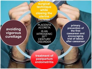
PLACENTA
ACCRETA
IS AN
IATROGENIC
20TH
CENTURY
DISORDER
Correct
surgical
technique
while
closing the
uterine
incision
primary
prevention of
the first
caesarean and
encouraging
trial of labour
after caesarean
treatment of
postpartum
endometritis
avoiding
vigorous
curettage
 Transvaginal scans are more sensitive in
confirming placental location and diagnosing
placenta previa at 20th week of gestation than
transabdominal scans.
has been the cornerstone in
diagnosing placenta accreta due to its wide
availability and high accuracy-
GREY SCALE
LOW LYING GESTATIONAL SAC
Findings suggestive of placenta accreta are-
 Hypervascularity of serosa–bladder interface.
 Diffuse or focal lacuna flow vascular lakes with turbulent
flow (Peak systolic velocity over 15 cm/s).
 Markedly dilated vessels over peripheral
subplacentalzone.
OBLITERATION OF CLEAR
SPACE BETWEEN PLACENTA &
UTERINE WALL
MOTH -EATEN /SWISS
CHEESE APPEARANCE
DUE TO LACUNAE
HYPERVASCULARITY
3-D power doppler has been shown to be superior
In diagnosing placenta percreta from accreta.
Findings on 3-D power doppler are:
Numerous coherent vessels involving the whole
uterine serosa–bladder junction (basal view)
Inseparable cotyledon and intervillous circulations
Transverse
transvaginl
USG
multiple
tortuous
hypoechoeic
structures
within the
placenta
Transverse
transabdo
m- inal
2D-Doppler
USG
Confirm the
hypoechoeic
pattern
3D-Doppler
ultrasound
For diagnosis of
bladder invasion
in placenta
percreta, seen
as positive
vasculari-zation
When to recommend MRI for diagnosis of
Placenta Accreta -
 equivocal USG findings of abnormal
placentation
 evaluation of posterior placenta in patients with
risk factors
 obese patients
 complementary role in specifically delineating
the
extent of an USG-diagnosed placenta percreta
Imaging features of normal placentation
on MRI
 Homogeneous T2-intermediate signal intensity of placenta
 Subtle, thin, regularly spaced placental septi.
 Normal subplacental vascularity.
 Triple-layered sandwich appearance of myometrium.
 Pear-shape of normal gravid uterus with smooth contour.
 Dark intra-placental bands on
T2-weighted images.
 Abnormal disorganized placental
vascularity.
 Focal interruptions of the
myometrial wall or extension
through the myometrium with
occasional invasion of adjacent
structures.
Heterogencity with in placenta
.disruption of pear shaped pattern
(lower segment wider than fundus)
 G3p1+1,24years female,GA 37 weeks 3 days presented with
leaking PV.
 History of preterm LSCS 3 years back and spontaneous
abortion f/b D&C 1 years back.
 No ANC visit single 3rd trimester USG showing SLIUF
appropriate for GA,placenta posterior and low lying
 The case was opened by obstetrician.
 Delivery of the fetus f/b repeated attempts to remove placenta
but within few minutes it was realised that it is adherent
placenta as the patient started bleeding
profusely(HORRIFYING !!!)
Hysterectomy was done but patient went into
irreversible shock and could not be revived back
Forcible and unneccessary attempt to claw off placenta
should not be done(may provoke further torrential
hemorrhage).
Multidisciplinary approach
senior obstetrician,
pelvic surgeon,
uro-surgeon (if needed)
neonatologist,
anesthetist,
TRIPLE P PROCEDURE
 A novel uterine sparing procedure for PAS
 Steps
1. Peri-operative placental ultra sound localization
of the superior edge of the placenta
2. Pelvic devasularization – pre operative
placement of intra arterial balloon catheters
3. No attempt to remove the entire placenta
*FIGO consensus guidelines on placenta accreta spectrum disorders
 25 years, unbooked, G4P1+2 with h/o FTLSCS 4 year
back f/b 2 spontaneous abortions(D&C done) came in
active stage of labor at 37wks + 2 days at CHC Kanpur
Dehat.
 Em LSCS was done and intraoperatively it was
found to be placenta percreta with bladder
involvement. Pt bled profusely.
 Subtotal hysterectomy was done along with ligation
of blood vessels going into the bladder but all in vain
as the patient’s vitals deteriorated, pt developed DIC &
anuria with ARF in postoperative period(d/t massive
blood loss)
CLINICAL SCENARIO 2
HOW COULD
SHE BE
BETTER
MANAGED??
Previous LSCS – Regular antenatal check ups
 USG to diagnose placenta accreta.
Elective cesarean is the choice
ICU CARE- For proper management of complications (
DIC,ARF,Transfusion reactions , ARDS, Electrolyte imbalance).
RCOG 2011
Elective ceaserean section in asymptomatic
women is not recommended before 38 weeks
of gestation for placenta previa or before 36-
37 weeks of gestation for suspected placenta
accreta.
ACOG 2014
Elective delivery by ceaserean section at 34-
35 weeks of gestation for suspected placenta
accreta
What makes hysterectomy different and difficult
?
Genitourinary tract inuries
.
Prevention
 Avoid cutting placenta => trans
fundal approach
 PREVENTIVE SURGICAL OR
RADIOLOGICAL
DEVASULARIZATION
Placement of balloon occlusion
catheter.
Placement of pelvic pressure
packing like laprotomy devices or
balloon tamponade.
Causes
 unplanned Hysterectomy
 Emergency hysterectomy in
an undiagnosed placenta
accreta
Inadvertent attempt at
removal of placenta
 Delivery of baby by cutting
through the placenta.
MASSIVE
BLOOD
LOSS
(3-5 ltrs)
Inspection
 Presence of any vascular channels
noted(should not be disturbed.
Uterine incision
 CLASSICAL/TRANSFUNDAL
cesarean section to avoid cutting
through the placenta f/b delivery of
fetus f/b closure of uterine incision by
WHIP stitch
B/L anterior division of internal iliac artery ligation done f/b uterine
artery ligation.
cesarean hysterectomyCESAREAN HYSTERECTOMY
IS CONSIDERED AS GOLD
STANDARD TREATMENT FOR
INVASIVE ACCRETA ALTHOUGH
HIGH RATES OF SEVERE MATERNAL
MORBIDITY(40-50%)
AND MORTALITY(7%)
Postoperative
management
.
 G6P1+4(none alive) GA 36 weeks with H/O
previous 1 LSCS and 3 D&C was refferred to our
hospital with the USG(3D Doppler)findings of
complete central placenta with focal placent
increta
 Elective LSCS was done at 37 weeks
 B/L uterine artery were ligated and gentle
removal of placenta as much as possible with
leaving the adherent part of placenta “insitu
approach” closure of uterine incision.
CLINICAL SCENARIO 3
Saving the uterus was our priority
 Postoperatively 2 units of blood was transfused along with
methotrexate adjuvant treatment* given.
 Patients managed successfully
 Key points during conservative management-
 Gentle attempt to remove the “non accreta” portion of placenta
, thus the volume of villous tissue left insitu
 Preventive radiological and surgical devascularization
(uterine/hypogastric artery)
prevents secondary hemorrhage
placental reabsorption
 Methotrexate hastens placental resolution although the risk of
neutropenia
sepsis
secondary hemorrhage=> hysterectomy
Conservative management should
always be
attempted in patients who agree to long
term
monitoring,strongly desired to
preserved their
fertility with adequate expertise
Insufficient evidence for the use of MRI and/or serum beta HCG for the
monitoring of conservative management of PAS.
* Use of methotrexate is not routinely recommended and therefore
should only be given judiciously.
FIGO consensus guidelines on placenta accreta spectrum disorder
Int J Gynecol Obstet 2018;140:291-298
One step conservative surgical
approach for PAS
 Vascular diconnection of newly formed feeder
vessels and seperation of invaded Ut. Tissue
from invaded bladder tissue
 Upper segment hysterotomy and delivery of
fetus
 Resection of all invaded myometrium with
placenta in one piece after local vascular control
 Surgical procedure for hemostasis
 Myometrial reconstruction in 2 planes
 Bladder repair if necessary.
*FIGO consensus guidelines on placenta accreta spectrum disorders
 Though UAE is latest interventional technique in cases of
accreta,but its not available in many of our set-ups, therefore
uterine artery ligation may be life saving.
 The role of interventional radiological procedure though looks
promising, further research and prospective larger studies
are required.
Pearls of wisdom
My personal experience….
 In cases requiring hysterectomy I personally use double set
of clamps to rapidly secure all bleeding points and remove
the uterus as soon as possible and ligate the pedicles later.
reduces blood loss effectively
 I have found that uterine artery ligation prior to attempt of
removal of placenta in cases of placenta accreta may be life
saving.
Feasible alternative to uterine artery embolization in
low
resource settings .
THE DEADLY D’S of Accreta
 DELAYED:
 Delayed referral
 Delayed caesarean: i.e. emergency (not elective)
 Delayed decision for hysterectomy
 DEFICIENCY:
 Deficiency of time availability: extensive adhesions,
no time for pre-op devascularization
 Deficiency of blood and blood products
 Deficiency of ICU beds
 Deficiency of multi-disciplinary senior team.
PREOPERATIVE DIAGNOSIS
ANEMIA CORRECTION
PATIENT COUNSELLING
REGULAR FOLLW UP
UTERINE ARTERY LIGATION
HYSTERECTOMY WHEN NEEDE
CAN SAVES LIFE!!!!