PAIN MANAGEMENT
NUR AINA BINTI AB KADIR
CONTENTS
1) Introduction to pain
2) Anatomy and Physiology of Pain
Pathway
3) Physiology of Nociception
4) Groups of drugs used in anesthesia
INTRODUCTION
 Def: pain is an unpleasant sensory and
emotional experience associated with actual or
potential tissue damage, or described in terms
of
such damage.”
 The term nociception is noci (Latin for harm or
injury)  neural responses to traumatic or
noxious stimuli.
INTRODUCTION
 Acute pain, primarily due to nociception
 Chronic pain nociception(psychological &
behavioral factors)
 Pain may also be classified
 Pathophysiology : nociceptive or neuropathic
pain
 Etiology : arthritis or cancer pain
 Affected area : headache or low back pain
ANATOMY AND PHYSIOLOGY OF
PAIN
FIRST ORDER NEURONS
 Pain fibers originating from the head are
carried by the trigeminal (V), facial
(VII),glossopharyngeal (IX), and vagal (X)
nerves and the cell bodies are located in
different ganglias respectively.
 The proximal axonal processes of the first
order neurons reach the brainstem nuclei
via their respective cranial nerves, where
they synapse with second-order neurons
in brainstem nuclei.
SECOND ORDER NEURONS
 As afferent fibersspinal
cordsegregate(size)
 large, myelinated fibers: medial
 small, unmyelinated fibers: lateral.
 In many instances they communicate with
second-order neurons through
interneurons.
SECOND ORDER NEURONS
 The gray matter was divided by Rexed into 10
laminae. The first 6 which make up the dorsal
horn receive all afferent neural activity and is the
principal site of modulation of pain.
 2nd order neurons are either:
 nociceptive-specific (only noxious stimuli)
lamina 1
 wide dynamic range (WDR) neurons (also
receive nonnoxious afferent input from Aβ, Aδ
and C fibers.) lamina V-abundant
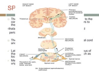
SPINOTHALAMIC TRACT
 The axons of most 2nd order neurons cross the midline to the
contralateral side of the spinal cord and send their fibers to
the :
-thalamus, reticular formation, nucleus raphe magnus and
periaqueductal gray.
 The tract lies anterolaterally in the white matter of spinal cord
and can be divided into a lateral and medial tract.
 Lateral – projects mainly to ventral posterolateral nucleus of
thalamus and carries discriminative aspects of pain such as
location, intensity and duration.
 Medial – projects to medial thalamus- autonomic and
unpleasant emotional perceptions of pain.
ALTERNATE PAIN PATHWAY
 Spinoreticular tract – mediate arousal and autonomic
responses to pain
 Spinomesencephalic tract – activating antinociceptive
descending pathways
 Spinohypothalamic and spinotelencephalic tracts –
activate hypothalamus and evoke emotional behaviour.
 Spinocervical tract -ascends uncrossed to the lateral
cervical nucleus, which relays the fibers to the
contralateral thalamus
 Some fibers in the dorsal columns (which mainly carry
light touch and proprioception) are responsive to pain.
THIRD ORDER NEURONS
 These neurons are located in the thalamus and send fibers to
somatosensory areas I and II in the postcentral gyrus of the
parietal cortex and the superior wall of the sylvian fissure,
respectively.
 Perception and discrete localization of pain take place in
these cortical areas.
 Neurons from lateral thalamic nuclei – project to primary
somatosensory cortex.
 Neurons from intralaminar and medial nuclei – project to
anterior cingulate gyrus (mediate suffering and emotional
components of pain).
PHYSIOLOGY OF
NOCICEPTIONS
 NOCICEPTOR
 Characterized by a high threshold for activation and encode the
intensity of stimulation by increasing their discharge rates in a
graded fashion.
 Noxious sensations
a)fast, sharp and well-localized sensation (‘first pain’) by Aδ tested by
pin-prick.
b)slower onset, duller and often poorly localized sensation (‘second
pain’) conducted by C fibers.
 Most nociceptors are free nerve endings that sense heat,
mechanical and chemical tissue damage.
-mechanonociceptors which respond to pinch and pinprick
-silent nociceptors; respond only in the presence of inflammation
-polymodal mechanoheat nociceptors ( most prevalent)
NOCICEPTORS
 CUTANEOUS: present in the both somatic and
visceral
 Somatic nociceptors include skin (cutaneous) and
deep tissues (muscle,tendons, fascia, and bone)
 Visceral nociceptor(internal organs)
 The cornea and tooth pulp: Aδ and C fibers
 DEEP SOMATICS: less sensitive to noxious
stimuli than cutaneous nociceptors
 easily sensitized by inflammation,dull and poorly
localized,muscles and joint capsule
 VISCERAL:Visceral organs are generally
insensitive tissues that mostly contain silent
CHEMICAL MEDIATORS OF
PAIN
 Several neuropeptides and excitatory amino
acids function as neurotransmitters for afferent
neurons subserving pain
MODULATION
OF
PAIN
PERIPHERAL CENTRAL
PERIPHERAL MODULATION OF
PAIN
 Nociceptors and their neurons display sensitization
following repeated stimulation.
 Primary Hyperalgesia
 Sensitization of nociceptors results in a decrease in
threshold, an increase in the frequency response to
the same stimulus intensity, a decrease in response
latency and spontaneous firing even after cessation of
the stimulus.
 Secondary Hyperalgesia
 Also called neurogenic inflammation is manifested by
the ‘triple response (of Lewis)’ of a red flush around
the site of injury (flare), local tissue edema and
sensitization to noxious stimuli.
CENTRAL MODULATION OF
PAIN
A)Facilitation
 Sensitization of second-order neurons
 Receptor field expansion
 Hyperexcitability of flexion reflexes.
 Neurochemical mediators : Substance P,
CGRP, vasoactive intestinal peptide,
cholecystokinin, angiotension, galanin, L-
glutamate and L-aspartate.
CENTRAL MODULATION OF
PAIN
B) Inhibition
1)Segmental inhibition
 Activation of large afferent fibers subserving
sensation inhibits WDR neuron and spinothalamic
tract activity.
 Activation of noxious stimuli in noncontiguous
parts of body inhibits WDR neurons at other
levels.
 Glycine and γ-aminobutyric acid (GABA) –
inhibitory neurotransmitters.
 Adenosine A1 receptor also has antinociceptive
2)supraspinal inhibition
 Inhibitory adrenergic pathways originate primarily from the
PGA and the reticular formation.
 Norepinephrine mediates this action via activation of
presynaptic or postsynaptic alpha-2 receptors.
 Serotonergic fibers from NRM relay the inhibiton to dorsal
horn neurons.
 The endogenous opiate system (primarily the NRM and
reticular formation) act presynaptically to hyperpolarize
primary afferent neurons and inhibit release of substance P;
some postsynaptic inhibiton.
 Exogenous opioids act postsynaptically on 2nd order neurons
or interneurons in substantia gelatinosa.
DRUGS USED IN PAIN
MANAGEMENT
 Acetaminophen
 It inhibits
prostaglandin
synthesis but lacks
significant
antiinflammatory
activity.
 Hepatotoxic at high
doses.
 Max dose daily in
adult – 3000mg/d.
 NSAIDs
 Inhibit prostaglandin
synthesis (COX).
 Analgesic, antipyretic
and antiinflammatory.
 Egs : Diclofenac,
ibuprofen, celecoxib,
ketorolac
DRUGS USED IN PAIN
MANAGEMENT
 Antidepressants
 Most useful for patients
with neuropathic pain.
 Blockade of presynaptic
reuptake of serotonin,
norepinephrine, or both.
Potentiate the action of
opiods.
 Egs: amitriptyline,
bupropion, citalopram,
clomipramine,
paroxetine.
 Serotonin and
Norepinephrine
Reuptake Inhibitors
(SNRIs)
 Duloxetine
 Treatment of
neuropathic pain,
depression and
fibromyalgia.
DRUGS USED IN PAIN
MANAGEMENT
 Neuroleptics
 For patients with
neuropathic pain, most
helpful in pts with
marked agitation or
psychotic symptoms.
 Egs: fluphenazine,
haloperidol,
chlorpromazine,
perphenazine.
 Blockade of
dopaminergic receptors
in mesolimbic sites.
 Antispasmodics and
muscle relaxants
 Helpful for pts with
musculoskeletal sprain
and pain associated
with spasm or
contractures.
 Egs: Tizanidine,
cyclobenzaprine,
baclofen
DRUGS USED IN PAIN
MANAGEMENT
 Corticosteroids
 Anti-inflammatory and
possibly analgesic
actions.
 Given topically, orally
or parenterally.
 Egs: Hydrocortisone,
prednisone,
dexamethasone,
betamethasone.
 Local Anesthetics
 Systemic infusion
produces sedation
and central analgesia
and is occasionally
used in treatment of
pts with neuropathic
pain.
 Egs: Lidocaine,
procaine and
chloroprocaine.
DRUGS USED IN PAIN
MANAGEMENT
 α2- Adrenergic agonists
 Activation of descending
inhibitory pathways in the
dorsal horn.
 Eg: clonidine
 Opioids
 Interacting with opiod
receptors – mu, delta and
kappa.
 Located at spinal,
supraspinal and peripheral
areas.
 Egs: codeine, oxycodeine,
hydrocodone, tramadol,
morphine, methadone,
fentanyl.
 Parenteral, Intravenous
and spinal opioid
administration.
DRUGS USED IN PAIN
MANAGEMENT
 Botulinum toxin (Botox)
 Proposed mechanisms of
analgesia include
improved local blood flow,
relief of muscle spasms
and release of muscular
compression of nerve
fibers.
 Anticonvulsants
 They are useful for
patients with neuropathic
pain, especially trigeminal
neuralgia and diabetic
neuropathy.
 These agents block
voltage-gated calcium or
sodium channels and can
suppress the spontaneous
neural discharges.
 Egs: phenytoin,
carbamazepine, valproic
acid, clonazepam,
gabapentin, pregabalin.
DRUGS USED IN PAIN
MANAGEMENT
 Inhalational
agents
 They directly bind
to cellular proteins
altering their
enzymes.
 Egs: halothane,
isoflurane, nitrous
oxide
Morgan and Mikhail’s textbook of
Anesthesiology
REFERENCES