Mohamed Said Ibrahim
mm.said2004@ymail.com
‫مع‬ ‫المخروطية‬ ‫القرنية‬ ‫يوم‬ ‫فاعليات‬
‫إبراء‬ ‫ومستشفي‬ ‫العماني‬ ‫البصريات‬ ‫نادي‬
-
‫مان‬ُ‫ع‬
17-11-2021
Presenter
Mohamed said Ibrahim
Careers:
• Key Account sales Manger and training, Nikon Lenswear middle east
• Sr.Optometrist, Hadi Hospital
• International Member of the American Optometric Association
• Certified International Professional Trainer (CIPT)
• Ambassador of the Arab British Academy for Higher Education
• Advanced Member of the UK Professional Board
• Trainer of BARAKA AND C&CO. Optics (earlier)
• Demonstrated and lecturer of the High optics technology institute (earlier)
Educational:
 PhD in visual behavioural rehabilitation for vision defects
 Master Degree of projects management in optics and vision sciences by the
faculty of projects management at Maryland University, USA
 BS.C in optics technology and vision science
 achieved degree of many short diplomas in health, patient care, customer
service, business administration.
Experience:
 More than 15 years of experience for optical shops, optical products, and
optometry clinic
Mohamed Said Ibrahim
mm.Said2004@ymail.Com
Mohamed Said Ibrahim
mm.Said2004@ymail.Com
Keratoconus
 Cornea
 Corneal Topography
 Corneal Disorders
 Keratoconus : Definition, Symptoms
 Keratoconus : Pathophysiology
 Keratoconus : Methods Of Measurement
 Keratoconus : Signs
 Keratoconus : Classification
 Keratoconus : Management
 Keratoconus : Eye Refraction
Mohamed Said Ibrahim
mm.said2004@ymail.Com
Topics
Mohamed Said Ibrahim
mm.Said2004@ymail.Com
CORNEA
 It is a transparent Tissue, the most sensitive part of the body.
 it is the most powerful refractive where its power is two-thirds of
the power of the eye, equivalent to about 40.0 to 45.0 dioptres, the
refractive index is (n = 1.376)
 A protective barrier from infection and trauma.
 The corneal epithelium is one of the fastest healing tissues in the body.
 The stroma makes up 90% of the corneal structure.
 The Endothelium acts as a water pump to maintain proper tissue
hydration.
 Both corneal segments and layers are designed towards one end
which is the clarity and transparency of the cornea.
Mohamed Said Ibrahim
mm.Said2004@ymail.Com
CORNEA
 The front surface is elliptical (horizontal at 180 degrees equals
approximately 11.7 mm, vertically at 90 degrees equals
approximately 10.6 mm).
 The back surface is circular in shape (11.7 mm).
 The curvature of the surface or curvature is expressed by the
word keratometry, which shows the strength of each direction
on the surface of the cornea (the average anterior diameter of
the curvature is 7.2-8.4 millimetres). .
 The thickness of the center (about 535 microns, which is
equivalent to about half 0.5 millimetres), which is thinner than
the peripheral circumference (about 660 microns = 0.66 mm).
Mohamed Said Ibrahim
mm.Said2004@ymail.Com
CORNEAL TOPOGRAPHY
 Cool colours (Violet, blue, azure) Flatter surfaces
 Warm colours (orange, red) Steeper surfaces
 Normal (green, yellow) Normal surfaces
COLOUR MAP
Mohamed Said Ibrahim
mm.Said2004@ymail.Com
CORNEA
 The Pentacam uses Scheimpflug imaging and, like the Orbscan,
measures both elevation and pachymetry using the same data set.
 The Pentacam has been shown to have excellent agreement with
the ultrasound pachymeter with regard to central corneal
pachymetry in both pre and post LASIK eyes
 Normal corneal thickness of 535 to 555 micron
Pachymetry
Mohamed Said Ibrahim
mm.Said2004@ymail.Com
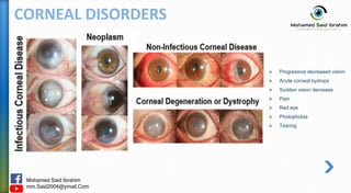
CORNEAL DISORDERS
 Microbial keratitis
 Acanthamoeba keratitis
 Fungal keratitis
 Herpes simplex keratitis
 Herpes zoster ophthalmicus
 Thygeson’s superficial punctate
keratopathy
 Recurrent corneal erosion syndrome
(RCES)
 Persistent epithelial defects
 Limbal epithelial stem cell deficiency
 Corneal degenerative
 Corneal dystrophies
 Keratoconus
 Other corneal ectasias
 Peripheral ulcerative keratitis (PUK)
 Other peripheral corneal diseases
• Injuries
• Allergies
• Keratitis
• Dry eye
• Corneal dystrophies
• Corneal
degenerative
Mohamed Said Ibrahim
mm.Said2004@ymail.Com
CORNEAL DISORDERS
 Progressive decreased vision
 Acute corneal hydrops
 Sudden vision decrease
 Pain
 Red eye
 Photophobia
 Tearing
Mohamed Said Ibrahim
mm.Said2004@ymail.Com
CORNEAL DISORDERS
 keratoconus
 Pellucid marginal degeneration
 Keratoglobus
 Post refractive surgery ectasia
CORNEAL ECTASIAS
Mohamed Said Ibrahim
mm.Said2004@ymail.Com
KERATOCONUS (Kc)
 The Cornea Assumes A Conical Shape Because Of Thinning
And Protrusion.
 A Progressive, Non-inflammatory, Bilateral (But Usually
Asymmetrical) Ecstatic Corneal Disease, Induces Irregular
Astigmatism, Myopia Leading To Mild To Moderate Impairment
In The Quality Of Vision.
 Onset is age 16 years, Shows no gender predilection.
Definition
 Ghosting/ monocular diplopia
 Glare at night , Haloes around lights
 Blurred/ distorted vision
 Frequently changing spectacle Rx and axis of astigmatism
 Poor repeatability of subjective refraction
Symptoms
Mohamed Said Ibrahim
mm.Said2004@ymail.Com
KERATOCONUS
PATHOPHYSIOLOGY
Mohamed Said Ibrahim
mm.Said2004@ymail.Com
KERATOCONUS METHODS OF MEASUREMENT
Keratometer
(Ophthalmometer) Keratoscope Placido disc Computer assisted
topographic analysis
Slit Lamp
Mohamed Said Ibrahim
mm.Said2004@ymail.Com
KERATOCONUS
BCVA less than 20/20
with normal retina
Signs
Scissors reflex
irregular retinoscopy reflex
Egg-shaped mires
on keratometry
Mohamed Said Ibrahim
mm.Said2004@ymail.Com
KERATOCONUS Signs
Vogt striae
vertical tension lines
in posterior cornea
Fleischer ring
iron ring around base of cone
Munson sign
bulging of lower lid
on downgaze
Rizutti sign
conical nasal reflection
with temporal illumination
Mohamed Said Ibrahim
mm.Said2004@ymail.Com
KERATOCONUS Signs
(I:S) value >1.4
K > 47.2
Inferior corneal steepening:
inferior-superior dioptric asymmetry
Mohamed Said Ibrahim
mm.Said2004@ymail.Com
KERATOCONUS
 Based on type of cones :
1. Round or nipple :
Most common, less than 5 mm in diameter , Cone-lies in
centre towards inferior nasal quadrant.
2. Oval cone :
Diameter > 5 mm; often displaced inferiorly.
3. Globus cone :
diameter more than 6 mm diameter, overall steepening , 75%
of cornea affected.
CLASSIFICATION
Mohamed Said Ibrahim
mm.Said2004@ymail.Com
KERATOCONUS CLASSIFICATION
 Based on severity of curvature :
1. Mild < 45 D in both meridians
2. Moderate 45-52 D in both meridians
3. Advanced > 52 D in both meridians
4. Severe > 62 D in both meridians
Mohamed Said Ibrahim
mm.Said2004@ymail.Com
KERATOCONUS Management
- The Routine Follow Up Is The Mandatory For All Keratoconus Patients
- All Optical Solutions (Eyeglasses & C.L) Are Present Pre And Post Surgeries
 Based on Keratoconus Severity :
MILD MODERATE ADVANCED
 Spectacles
 contact lenses:
Soft or hard or Hybrid
 Collagen Cross-linking (CXL)
 Spectacles
 contact lenses:
Soft or hard or Hybrid
 Collagen Cross-linking (CXL)
 Intracorneal ring segments (ICRS)
 Penetrating keratoplasty (PK)
 Deep Anterior Lamellar (DALK)
 Descemet Stripping Automated
Endothelial (DSAEK)
 Descemet Membrane
Endothelial (DMEK)
Mohamed Said Ibrahim
mm.Said2004@ymail.Com
KERATOCONUS Management
 CONTACT LENSES
 Soft contact lenses : single soft C.L
 Rigid gas permeable lenses : single hard C.L
 Combined lens system : Soft + Hard
- Piggy back system : Rigid lens fitted over a hydrogel
lens
- Hybrid lens system : fuse a soft rim onto a hard
central portion
 Fully Keratonic Family Lenses :
- Rose-k lenses
- Scleral and mini scleral lenses
PIGGY BACK SYSTEM
Soft C.L
Scleral/mini scleral
Hard C.L
Mohamed Said Ibrahim
mm.Said2004@ymail.Com
KERATOCONUS Management
 CONTACT LENSES FITTING PHILOSOPHY
FLAT FITTING
- early keratoconus
- The Entire Weight Of The Lens On The
Cone.
- Apical Touch, Good visual acuity.
- acceleration of apical changes.
- corneal abrasions
APICAL CLEARANCE
- Resting on the paracentral cornea.
- clears the central cornea.
- minimize trauma to the central cornea.
- poor tear film, bubbles under the lens,
poor visual acuity.
- the weight of the contact lens between the
cone and the peripheral cornea.
- a thin band of touching near the edge of the
lens
THREE-POINT TOUCH
Mohamed Said Ibrahim
mm.Said2004@ymail.Com
KERATOCONUS Management
 COLLAGEN CROSS-LINKING (CXL)
Mohamed Said Ibrahim
mm.Said2004@ymail.Com
KERATOCONUS Management
 INTRACORNEAL RING SEGMENTS (ICRS)
Mohamed Said Ibrahim
mm.Said2004@ymail.Com
KERATOCONUS Management
 PENETRATING KERATOPLASTY
Penetrating keratoplasty Deep Anterior Lamellar
Descemet Stripping Automated Endothelial Descemet Membrane Endothelial
Mohamed Said Ibrahim
mm.Said2004@ymail.Com
KERATOCONUS Eye Refraction
1. Visual Acuity (VA) :
- Distance unaided VA, monocular VA, binocular VA.
- Distance aided VA, monocular VA, binocular VA.
- Pinhole Visual Acuity (PH VA), aided monocular VA is worse.
- Near VA, aided VA, binocular VA.
2. Best Vision Sphere Refraction :
- Most common is Minus to correct Myopia but trial of Plus is also require regarding Irregular
Astigmatism.
- Best Visual Acuity with more plus or less minus.
3. Sphero-Cylindrical Refraction :
- Jack son cross cylinder (Test 4 main Axis – 90/180 and 35/145)
- JCC cylinder power ( add -0.50cyl will add +0.25sph).
- Refine the axis again by JCC.
4. +1 Test and Binocular Balance :
- Refine SPH Monocular and binocular for BCVA.
5. Near Refraction :
- Near VA, new Rx VA, binocular VA.
- Adjust Powers Sph & Cyl and Axis for Near BCVA.
Visual Acuity
Best Vision
Sphere
Sphero-
Cylindrical
+1Test/Binocular
Near Refraction
 Based on Keratoconus Severity – Subjective Refraction (Trial and Error) :
Mohamed Said Ibrahim
mm.Said2004@ymail.Com
REFERENCES
 Management of Optical measurements in ophthalmic health care, Mohamed Said Ibrahim, 2020
 Step by Step Reading Pentacam Topography, second edition, Dr. Mazen M Sinjab, 2015
 Oxford Handbook of Ophthalmology, Fourth edition International edition,2018
 Refractive Error Training Package, Student Manual, Brien Holden Vision Institute, Edition 2012
 American Academy of ophthalmology, Keratoconus – Europe, 2015
 National Eye Institute, www.nei.nih.gov
 medical-dictionary.thefreedictionary.com
 Keratoconus pptx, Slideshare.com
 Images and diagrams, google images search.
Mohamed Said Ibrahim
mm.Said2004@ymail.Com