Method of Haemoglobin
Estimation
Haemoglobin
estimation method

Automated

Manual

Cyanmethaemoglobin
method

Acid Haematin
method

Coulter counter
Procedures
a) Sample:
Both capillary and venous blood may be used for this test.
Procedures
b. Method:
1.

Cyanmethaemoglobin method:

o Accurate.
o Commonly used.
o Recommended by ICSH (international committee for
standardization in haematology).
a)

Principle of cyanmethaemoglobin method:

Blood + diluent (Drabkin’s solution)
“ potassium ferricyanide + potassium cynaide”
Converts: Haemoglobin (Hb) and Methaemoglobin (Hi)

Cyanmethaemoglobin (HiCN)
Measure the absorbance of the solution by using a calorimeter at
a wavelength = 540nm. Then compare it with the standard
solution of HiCN.

Spectrophotometer
Reagent and equipment for Cyanmethaemoglobin method:

b)

•
•
•
•
•

Diluent (Drabkin’s solution)
5 ml pipette.
Cuvettes.
Test tube.
20 micro liter pipettes.
c)

Procedure of Cyanmethaemoglobin method:

20ul blood + 4ml diluent

HiCN.

mix, 5-10min

sample

Measured by spectrophotometer at 540nm
standard

Use the calculator:
Hb (g/dl)= Absorbance of test
X Conc of standard
Absorbance of standard
2. Acid haematin method:
a)

Principle:

Blood + 0.1 N HCL

Acid Haematin.

(then match the color of solution with reference solution
colorimeter or colored strip)
i.e. “SAHLI’S haemoglobinometer”

However this method is inaccurate.
b) Reagent and equipments for Acid Haematin method:
o
o
o
o

Sahli’s haemoglobinometer.
Sahli’s pipette or Micropipette.
0.1 N HCL.
Dropping pipette.
• The standard set of Haemometer consists of:
A black counting chamber, round Hb Tube, 20ul Pipette, Rubber
tube with mouth piece, Cleaning brush, Glass dropper with rubber
teat, Glass rod, Amber bottle.
c)

Procedure of Acid Haematin method :

100ul HCL + 20ul blood mix in a graduated Tube (keep for 5min)
Acid Haematin

How can we read Hb value?
Compare the color of solution in the graduated tube with that of reference
strip on either side of haemoglobinometer.
Graduated tube has two scales: % and g/100 ml of whole blood.
• If the color of graduated tube is Darker add either 0.1N Hcl or D.W 7
-drop by drop- with pipette; mix with glass rod until the color matches with
reference strip.
• The reading in graduated tube refers to Hb level in g/dl

(some tubes give reading in %; to convert into g/dl X 0.146
So, e.g. 10% X 0.146 = (14.6 g/dl).
Precautio
n
 Before the sample is read the solution should be clear.
 If high WBC in specimen centrifuge the specimen then use the
supernatant.
 In case of Hb S or C dilute the mixture in 1:1 ratio with DW then
read in colorimeter.
 In case of abnormal globins add 0.1g of potassium carbonate to
the solution.
Q. What is the unite of measurement for Hb?
Whole blood Hb concentration is in g/dl.
Reference Values

Adult male:
13 - 18 g/dl

10y old child
11 - 15 g/dl

Adult female:
12 - 16 g/dl

6 m old child
11 - 14 g/dl

New born
14 - 22 g/dl
Interpretation:
Reference values for Hb are variable.

Clinical significance:
Hb value

Anemia
+
RBC count and indices (upcoming labs)
Stage formation of RBCs
1.

Proerythroblast.

2.

Basophilic normoblast (early stage).

3.

Polychromatic normoblast (intermatiedate stage ).

4.

Orthochromatic erythroblast or Nucleated RBC (late stage ).

5.

Reticulocyte.

6.

Normal erythrocytes
Stage formation of RBC:
1. Proerythroblast
The earliest precursors of
erythropoiesis and do not contain
hemoglobin.
Nucleus: The nucleus has a dense,
finely honeycombed chromatin
structure with pale blue nucleoli,
which disappear as the cell matures.
Cytoplasm : darkly basophilic.
Seen in BM
2.

Basophilic Normoblast (early stage)

These cells tend to be smaller than
proerythroblasts.

Like proerythroblast in general
character.
 The nuclear-cytoplasmic ratio is
shifted in favor of the cytoplasm.
Seen in BM.
3.

Polychromatic normoblast
(intermatiedate stage )
Nucleus appears coarse and
smudgy, and there is partial
clumping of the nuclear
chromatin.
 Cytoplasm loses more
of its basophilic with a greater
abundance of hemoglobin .
Seen in bone marrow
4.

Orthochromatic erythroblast or Nucleated RBC
(late stage )
Red blood cell with nucleus
The nuclear- cytoplasmic ratio
is shifted in favor of the
cytoplasm, which acquires an
increasingly red tinge ultimate
Seen in bone marrow and blood
Seen in sickle cell disease
,AIHA ,and beta-thalassaemia
5.

Reticulocyte

Immature RBCs that contain
cytoplasmic RNA and organelles
such as mitochondria and
ribosomes in various stages of
maturity.
The more filamentous reticula are
characteristic of younger cells
(brilliant cresyl blue stain)
Seen in bone marrow and blood
Seen in haemolytic anaemia
6.

Normal erythrocytes (Normochromic)

•Cells are uniform size & shape
•Normal hemoglobin conc.
•small, central pallor which is
Less than one-third of the total cell
volume.
Sever Hemorrhage

Systemic diseases e.g. leukemia,
lymphoma, uremia, cirrhosis,
hyperthyroidism, carcinomatosis and
systemic lupus erythematosis.
Haemolysis due to transfusion of
incompatible blood, reactions to
chemicals and drugs, bacteraemia, and
artificial heart valves

Anaemia
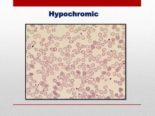
Hypochromic erythrocytes

IDA, Thalassemia &
Sideroblastic anemia
Hypochromic
Polycythaemia

High altitudes

Congestive cardiac failure
(CCF)
Haemoconcentration states of blood
e.g. sever burns
Chronic obstructive pulmonary disease
(COPD)
Polychromasia
• Mature RBCs with increased staining with basic stain and Hb staining.
Occurs in red cells have high RNA content with Hb synthesis is not yet
complete.
Polychromasia and Normoblasts

Increased erythrocyte production &
Hemolytic anemia
Quality Control
1.

If the patient’s specimen running in automated machine
there are 3 levels controls should be run.

2.

While if its running by manual method; send patient’s
specimen to the reference laboratory; and perform duplicate
testing in your own lab.

1.

All personnel performing Hb should be checked for color
blindness (Sahli’s Method).
Hb estimation