Anal Fissure, Fistula in Ano &
Pilonidal sinus
DR SYED UBAID
Anal canal Anatomy:
Anal Canal
• Length= 3.8 to 4.0 cm
• Zona Columnaris: Upper ½- lined by Simple columnar
• Zona Hemorrhagica: Upper part of lower half ( above
the Hilton’s white line) – Stratified squamous non-
keratinizing epithelium
• Zona Cutanea: Lower part of lower half( below the
Hilton’s white line)- Stratified squamous keratinizing
epithelium
Anorectal Bundle or Ring:
Demarcating Line B/W the Rectum
& Anal Canal.
Can be felt Posteriorly- Thickened
Ridge
Formed by- Puborectalis, Deep Ext
Sphicter,Conjoined long Muscle &
Internal Sphincter
Puborectalis Muscle:
• Maintain the angle b/w
rectum & anal canal
• Gives off fiber to the
longitudinal muscle layer.
• Position, Length as well as
angle of the anorectal Junction
- integrity & strength of the
Puborectalis muscle sling.
Image of Anal Sphincter:
Deep External
Sphincter.
Sub cutaneous
External Sphincter
Superficial External
Sphincter
Circular muscles
of Rectum
Longitudinal
muscle of
Rectum
Internal anal S
Conjoined
longitudinal muscle
External & Internal Sphincter:
External Sphincter Internal Sphincter
Muscle Single muscle k/as Goligher
Muscle
Continue of the Circular muscular coat
of the rectum
Color Red Pearly white
Nerve Pudendal Nerve Autonomic nervous system- Intrinsic
non-adrenergic & non-cholinergic
fiber
Types of
Muscle
Somatic Voluntary Muscle Non-striated Involuntary Muscle
Parts/fts Deep, Superficial and
Subcutaneous portion
Always lie in the tonic state of
contraction
Anal Canal
Above the dentate line Below the dentate line
Development Post-allantoic gut Proctodeum
Epithelium Cuboidal/Columnar Squamous without sweat & hair
gland
Name Surgical anal canal Anatomical anal canal
Color Pink Skin Colour
Nerve Parasympathetic: painless Spinal nerves: very painful
Venous
Drainage
Portal System Systemic-Ext iliac vein
Lymphatic
Drainage
Para-aortic Superficial & Deep inguinal LN
Examination of Anal Canal:
• Relaxed Patient
• Informed Consent
• Private environment
• Good Light
• Position – Left Lateral Position/ Sims’s Position-
most commonly used.
Image for different position:
Lithotomy
Sim’s PositionSim’s position
Knee elbow position
P/R Examination:Inspection
• Skin Lesion- Psoriasis
-Lichen planus
- Warts
-Candidiasis&Herpes simplex
• Whether anus is closed
or patulous
• Position of the anus/perineum
• Evidence of piles/
sentinel tag
( Anal fissure or SCC)
Psoriasis
P/R:Gloves,jelly etc………
• Sling of puborectalis- Posteriorly at the apex
• Posterior surface of the prostate gland with
median sulcus( Male) & Uterine cervix( in
female)-Anteriorly.
• Intrarectal, Intraanal or extraluminal mass.
• Sphincter length
• Resting tone
• Voluntary squeeze
• Examining finger – Mucus, Blood, Pus
• Stool Color.
Proctoscope:
Proctoscopy:
• Position: Left lateral position
• Inspection of the distal rectum and anal canal
• Injection in Hemorrhoids
• Banding of Piles mass
• Biopsy of mass
Sigmoidoscopy:
Mainly used for Rectal
examination
But Also recommended in
Fissure & Hemorrhoids
Cos Colitis & Rectal
Carcinoma is frequently
A/W Fissure &
Hemorrhoids.
Physiology
• Structural Integrity of the sphincter- Endoluminal USG
• Neuromuscular Function –(a) Assessment of
conduction velocity along with the Pudendal nerve or
-(b) Needle Electromyogram(EMG)-Slightly
Painful.
• Evacuation Proctography or Dynamic Proctography:
- In Rectal Sensorimotor dysfunction( Overflow of
rectal content)
Dynamic Proctography:
• Radio-opaque pseudo-stool is inserted into
the rectum
• Rest, Squeeze and than bear down to
evacuate the rectal contents under real-time
imaging.
• Can be combined with EMG & Pressure
studies
Dynamic Magnetic Resonance
Proctography:DMRP:
• More popular
• More expensive
• Less physiological
Anal Fissure:
• Longitudinal tear in the anal canal
• Site: Posterior midline (90%) and Anterior
midline in 10% case especially in female.
Etiology & Predisposing factors of Anal
Fissure:
• Age: Young adult & middle aged man
• Gender : Male > Female
• Posterior midline is the commonest site because-
-Maximum stretching on this site
- Less tissue here
-Minimal tissue perfusion
Etiology of Anal Fissure
• Main cause-Trauma–Strained evacuation of
Hard stool
or
• Less commonly - Repeated passage of stool (
diarrhea)
• Anterior anal fissure in 10% cases – Mostly in
Women that occurs following vaginal delivery
Predisposing Factors: FISSURE
• Faces – Hard
• Ischemia
• Surgical procedure- Haemorrhoidectomy
• Sphincter hypertonia
• Underlying disease – Crohn’s , TB, L.V, Syphilis etc
• Repeated Childbirth
• Enthusiastic usage of ointments and abuse of luxatives.
C/F of Anal Fissure:
• Severe anal pain during the defecation
• Blood streak outside the stool
• Bleeding P/R- Bright
• Mucous Discharge
• Constipation
• Itching
Chronic Anal Fissure: Findings:
• Hypertrophied Anal Papilla- Proximally
• Sentinel tag- Distally
• Thickened edge
• Exposed internal sphincter i.e Ulcer overlying
the fibers of internal sphincter
D/D –Especially if ectopic site i.e other
than Posterior –midline:
Crohn’s Diseases Kaposi’s Sarcoma
Tuberculosis B-Cell Lymphoma
Lymphogranuloma Venereum CMV
Syphilis Chlamydia
HIV Chancroid
HSV SCC
Confirmation of Diagnosis:
• Adequate clinical examination under G/A
• Proctoscopy
• Sigmoidoscopy
• Take Biopsy
• Do Culture
Treatment: Conservative & Surgical
• Conservative treatment helpful in most of cases
• Main objective to treat Constipation.
-Add the fiber to the diet
-Encourage water intake
-Laxative to make the stool soft
• Application of local anesthetic- Lignocaine jelly
• Antibiotics- Ofloxacine + Orinidazole
Conservative :Hot Seitz Bath
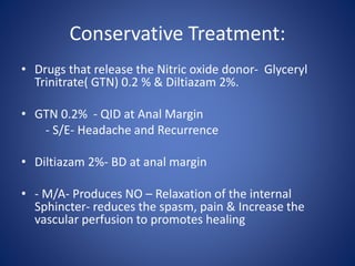
Conservative Treatment:
• Drugs that release the Nitric oxide donor- Glyceryl
Trinitrate( GTN) 0.2 % & Diltiazam 2%.
• GTN 0.2% - QID at Anal Margin
- S/E- Headache and Recurrence
• Diltiazam 2%- BD at anal margin
• - M/A- Produces NO – Relaxation of the internal
Sphincter- reduces the spasm, pain & Increase the
vascular perfusion to promotes healing
Conservative Treatment
• Botulinum toxin injection
• Site of Inj- Internal Sphincter
• M/A- Inhibits presynaptic release of Ach from
cholinergic nerve endings- Paresis of Striated
muscle and release the spasm .
• Other use- Achalasia cardia, Sphincter of Oddi
dysfunction, Frey Syndrome
Operative procedure for FIA.
• Anal Dilatation
• Posterior division of the exposed fibers of the
internal sphincter in the base of the fissure.
• Lateral Anal Sphincterotomy of Notaras
• Anal advancement Flap
Anal Dilatation: Lord’s Anal Dilatation
• Position- Lithotomy
• Under G/A
• Manual 4 to 8 finger sphincter dilatation
• Useful in Young men with very high sphincter
tone
• Risk: Incontinence.
Posterior division of the exposed
fibers of the internal sphincter in
the base of the fissure
• Indication – if fissure is associated with
INTERSPHINCTERIC FISTULA
• Disadvantage- Prolonged healing
- Passive anal leakage because of
resulting ‘ Keyhole gutter deformity’.
Lateral Anal Sphincterotomy:
• Position- Lithotomy
• Anesthesia- Regional or G.A
• Palpate the distal internal sphincter with the help
of bivalved speculum at the intersphincteric
groove.
• Give a small longitudinal incision in right or left
lateral position
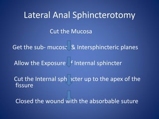
Lateral Anal Sphincterotomy
Cut the Mucosa
Get the sub- mucosal & Intersphincteric planes
Allow the Exposure of Internal sphincter
Cut the Internal sphincter up to the apex of the
fissure
Closed the wound with the absorbable suture
Complications of LAS:
• Hemorrhage
• Hematoma
• Bruising
• Perianal Abscess
• Fistula
• Incontinence.
Anal Advancement Flap:
• Very useful in women and those with Normal or Low
Resting Anal Pressures (persistent, chronic, non
healing fissure)
• Excised the edge as well as base of the fissure.
• Inverted house shaped flap of Perianal skin is
mobilized to cover the fissure.
• Post-op instruction- Stool softeners, Bulking agent &
Topical sphincter relaxants.
Fistula-in-ano:
• Chronic abnormal communication
• Between the Internal opening (anorectal lumen) &
External opening on the skin of the perineum or
buttock
• Lining is Granulation tissue.
• Commonest cause – Non-specific, Idiopathic & Crypto
glandular & Inter-Sphincteric anal gland infection.
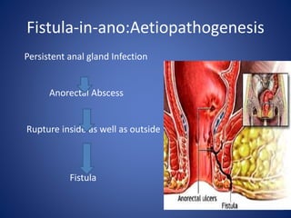
Fistula-in-ano:Aetiopathogenesis
Persistent anal gland Infection
Anorectal Abscess
Rupture inside as well as outside
Fistula
Fistula-in-ano:Underlying Condition –
CISTULA+ ARF
Carcinoma
Ileitis-Crohn’s
Schistosomiasis
Tuberculosis
Ulcerative colitis
L. Venereum
Anal Fissure Abscess
Actinomycosis
Rectal Duplication
Foreign Body
Fistula-in-ano:Clinical features
• Intermittent purulent discharge
• Pain
• External opening as sinus or Ulcer
• Bleeding/PR(sometimes)
Types of Fistula in ano:Standard
• Low type- Internal opening below the anorectal ring.
• High Type-Internal opening above the anorectal ring.
• Importance – Low type fistula- fistulotomy without
damage to sphincter
- High type fistula – Staged operation
Park’s Classification:
• Based on relationship of fistulous tract to the anal
sphincters- 4 types.
• Intersphincteric Fistula
In vast majority of Cases.
• Trans sphincteric Fistula
• Supra Sphincteric Fistula
• Extra Sphincteric Fistula
Park’s Classification
Intersphincteric Fistula:
• Most common type
• Incidence= 45%
• Don’t cross the external sphincter
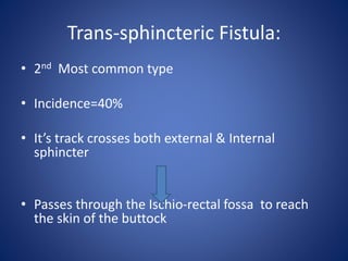
Trans-sphincteric Fistula:
• 2nd Most common type
• Incidence=40%
• It’s track crosses both external & Internal
sphincter
• Passes through the Ischio-rectal fossa to reach
the skin of the buttock
Supra-sphincteric Fistula:
• Very Rare
• Cause- Iatrogenic
• Very similar to high level
T-S Type.
Extra-sphincteric Fistula:
• Run without specific
relation to the sphincter
• Cause- Trauma or Pelvic
Disease.
• Originates in the rectal
Wall
• Tracks lateral to both
Sphincters
Clinical Assessment/Investigation:
A. Complete the General advise like
-Obstetric history
-Gastrointestinal history
-Surgical history
-Continence history
-Proctosigmoidoscopy examination
Clinical Assessment/Investigation
B.Important point about fistula
1. Site of the internal opening & External opening.
2. Course of the primary track
3. Presence of the secondary extension
4. Presence of other associated condition.
Goodsall’s Rule:
Clinical Assessment/Investigation
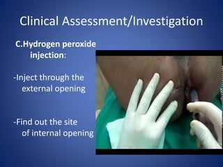
C.Hydrogen peroxide
injection:
-Inject through the
external opening
-Find out the site
of internal opening
Clinical Assessment/Investigation
D.Gentle use of Probe
Clinical Assessment/Investigation
E. Manometry:
- Resting anal tone
- Functional anal sphincter length
- Voluntary squeeze
F: Endoluminal USG: Sphincter
integrity, tract & anal canal.
MRI :
• Gold Standard
• Demonstrate the
secondary extension
Fistulography:
Demonstration of Fistula in Ano on CT
Management : Fistula in Ano:
• Fistulotomy
• Fistulectomy
• Setons- Loose & Tight Setons
• Biological Agent- Fibrin Glue
• Advancement Flap- To preserve both anatomy & Function .
• VAAFT: Video Assisted Anal Fistula Treatment.
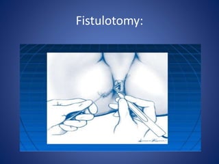
Fistulotomy
• Laid open the track( John of Arderne)
• Indication : Intersphincteric & Transsphincteric
Fistula.
• Steps:
1. - Position - Lithotomy
2. - Anesthesia - G/A.
3. -Identified the internal opening
Fistulotomy: Steps Continue
4. Pass the probe through E.O
to E.O to the I.O
5. The track is laid open over
the probe.
6. Curette the granulation
tissue and sent for HPE.
7.Wound edges are trimmed
E.O
I.O
Probe
Laid open
Fistulotomy:
FISTULECTOMY: Excision of whole
Fistulous tract:
Probe
Setons:Bristle material
• Thread
• Wire
• Proline
• Infant feeding tube
• Ksharsutra: kshar- corrosive & Sutra- Thread
Setons: Loose :
seton
Non-absorbable
Non-Degradable
Comfortable
No intent to cut
Ideal seton
No
tension
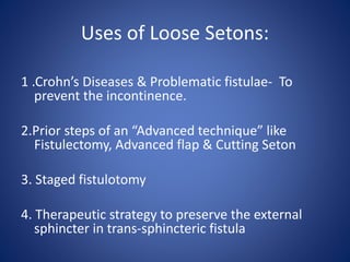
Uses of Loose Setons:
1 .Crohn’s Diseases & Problematic fistulae- To
prevent the incontinence.
2.Prior steps of an “Advanced technique” like
Fistulectomy, Advanced flap & Cutting Seton
3. Staged fistulotomy
4. Therapeutic strategy to preserve the external
sphincter in trans-sphincteric fistula
Purpose to use of Loose Setons:
Purpose:
- Eradicate the acute sepsis & Secondary
extension
- To simplify the fistula
- Allow fibrosis
Tight/Cutting Seton
• Placed with intention to cut the enclosed muscles.
• Also k/as “ Cheese Wiring through the ice”
• Fistulous tract is replaced by a thin line of fibrosis.
• Types- Elastic & Self cutting
- Non elastic & tightened
- Ksharsutra- most commonly used.
Tie the kharsutra to the eye of probe
E.O
Ksharsutra
Ksharsutra coming out through I.O
I.O
E.O
Cuting & healing simultaneously.
Biological Agent to fill the fistula.
Insertion of Fibrin Glue in the
fistula
VAAFT:Video Assisted Anal Fistula
Treatment• Visualization of the F.tract with the Fistuloscope
• Aim is to find the correct position of Internal Opening.
• A stapler to close the Internal opening.
• Fistuloscopy is done under irrigation & F.tract as well
as all granulation tissues are coagulated
• Total closure of the Internal opening with inserting the
Cyanoacrylate
Home message:Fissure:
• Post-midline is the commonest site for Fissure ( 90% )
• Main cause is Constipation – hard stool i.e trauma
• Pain during defecation is the commonest complaint.
• Clinical examination is sufficient to diagnose it
• GTN & Diltiazam 2% local application along with diet modification
have an excellent result as equivalent to LAS.
FISTULA IN ANO:
• Persistent anal gland infection is the commonest cause of Fistula in Ano
Home message:
• Goodsall’s rule is very useful in determining the site of external & internal
opening as well as about the fistulous tract.
• Intersphincteric type of fistula in Ano is the commonest type
( 45%)
• MRI is the gold standard for fistula imaging in complicated fistula
• Fistulotomy, Fistulectomy & Ksharsutra are common procedure to treat it.
• VAAFT is the recent advance in Fistula surgery
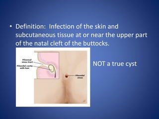
Pilonidal sinus
• Definition: Infection of the skin and
subcutaneous tissue at or near the upper part
of the natal cleft of the buttocks.
NOT a true cyst
History
• 1833- hair containing cyst located just below the
coccyx
Mayo
• 1880- Hodge coined the term “pilonidal”
Nest of hair
• In 19th and 20th century – considered to be
congenital
• In WW II
• Patey and Scarf – hypothesised origin of pilonidal
sinus acquired by penetration of hair into
subcutaneous tissue.
What causes pilonidal sinus???
• Midline holes – Hair follicles that have
enlarged
Pulling forces between sacrum
and skin
• Force concentrate on 1mm2 area where the
narrow gluteal crease comes in close contact
with the sharp angle of sacrum
• Weakest point of skin gives way first– Skin at
the bottom of the follicle.
• Primary cause – “Pit”
• Secondary casue – “ Hair follicles”
Cause of pilonidal sinus
• (1) Invader hair
• (2) Force causing hair penetration
• (3) Vulnerability of skin
Anatomy
 Intergluteal cleft: A groove between the buttocks that
extends from just below the sacrum to the perineum.
Anchoring of the deep layers of skin overlying the coccyx
to the anococcygeal raphe
Epidemiology
• Incidence : 26 per 100,000
• Mean age: 19 years for women and 21 years
for men
• Sex: M/F ratio – 2:1 to 4:1
• Equal incidence of acute:chronic
Risk factors
• Overweight/ obesity
• Local trauma or irritation
• Sedentary lifestyle/prolonged sitting
• Deep natal cleft
• Family history
Theory
• Acquired vs Congenital
• Tendency to recur following complete
excision.
• Tendency to occur in places other than natal
cleft.
Pathogenesis
• Hair and inflammation – inciting factors
• On sitting/bending natal cleft stretches-
breakage of follicles- opening of a pore/pit-
collection of debris - pilonidal sinus - abscess
• Proof??
• Pilonidal tract extends cephalad.
• Cavity contains hair, debris or granulation tissue.
Clinical manifestations
• Patient presentation:
- Acute onset mild to severe pain (sitting/bending)
- Intermittent mucoid/purulent/bloody discharge
- Recurrent / persisting pain
- Fever / malaise
Physical examination
• One/more pits in the natal cleft +/- painless
sinus opening cephalad and lateral to cleft
• Tender mass or sinus draining mucoid/bloody
or purulent fluid
Diagnosis
• Clinical
- Finding a pore/sinus in the natal cleft
- No imaging required
Differential diagnosis
• Perianal abscess/ fistula
• Hidradenitis suppurativa
• Perianal complications of Crohn’s disease
• Skin abscess/ furuncle/ carbuncle
• Folliculitis
Surgical treatment
• Drainage with/ without excision
• Marsupialisation
• Excision with primary closure
• Excision with grafting
• Sinus extraction
• Sclerosing injections
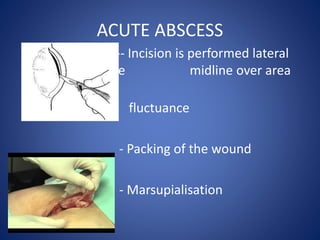
ACUTE ABSCESS
-- Incision is performed lateral
to midline midline over area
of maximum
fluctuance
- Packing of the wound
- Marsupialisation
Problems
• Recurrence rates are from 20 – 55 %
• During a 3 year period, 73 patients treated with I & D
for first episode of pilonidal abscess
• Healed : 42 patients (58%; 95% CI) within 10 weeks
• Recurrence : 9 patients (21%;95% CI)
• Follow up period : median of 60 months
• Constant cure rate : 76% (CI 95%) after 18 months
Prognosis after simple incision and drainage for a first-episode acute pilonidal abscess.
• Jensen SL, Harling H
• Br J Surg. 1988;75(1):60.
Chronic pilonidal sinus
Surgical approaches:
- Excision
- Wound closure
(1) Primary closure in midline/ off midline
> Z plasty
> V-Y advancement flap
> Rhomboid flap (limberg)
(2) Reconstruction using flaps
Karydakis surgery
• Karydakis believed that hair insertion is the cause
for pilonidal sinus
• Low recurrence rates due to:
- Wound placed away from midline
- Resulting new natal cleft was shallower
• Problems
- Sutured taken over the presacral fascia causing
pain
- Patients requiring GA
- Prolonged hospital stay