DISEASES OF THE ORBIT
BIPIN BISTA
OPHTHALMOLOGY
RESIDENT, NMCTH
DISCUSSION
Orbit
Content
Superior orbital fissure : structures
Inferior O F
Proptosis
Pre—septal cellulitis
Orbital cellulitis
Cavernous sinus thrombosis
Thyroid Ophthalmopathy
ORBIT
Quadrangular pyramids
situated between the
anterior cranial fossa and
the maxillary sinuses
below.
40 mm in height, width
and depth
Formed by 7 seven
bones : Frontal, Maxilla,
Zygomatic, Sphenoid,
Palatine, Ethmoid and
Lacrimal.
Has 4 walls – medial,
lateral, superior and
inferior.
WALLS OF ORBIT
Medial – 2 orbits are parallel , common for #.
Inferior – (floor) triangular , commonly involved in
blow—out #.
Lateral – triangular, covers posterior half.
Roof – formed by orbital plate of frontal bone.
Apex – posterior end where 4 walls converge , 2
orifices, the optic canal which transmits optic
nerve and ophthalmic artery and Superior orbital
fissure which transmits arteries, veins and nerves.
FASCIA BULBI
(TENON’S CAPSULE)
Envelops the globe from the margin of
cornea to the optic nerve.
It has thickened lower part to form a sling
where the globe rests ; ‘Suspensory
ligament of Lockwood’ .
CONTENTS OF ORBIT
Volume : 30 cc.
1/5th is occupied by eyeball.
Part of optic nerve, Extraocular muscles ,
lacrimal gland, lacrimal sac, ophthalmic artery
and its branches, 3rd, 4th and 6th cranial nerves
and ophthalmic & maxillary divisions of cranial
nerves , sympathetic nerve and fascia.
SUPERIOR ORBITAL
FISSURE
It is a foramen in skull lying between lesser
and greater wing of sphenoid.
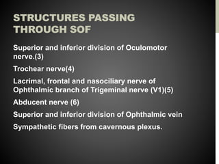
STRUCTURES PASSING
THROUGH SOF
Superior and inferior division of Oculomotor
nerve.(3)
Trochear nerve(4)
Lacrimal, frontal and nasociliary nerve of
Ophthalmic branch of Trigeminal nerve (V1)(5)
Abducent nerve (6)
Superior and inferior division of Ophthalmic vein
Sympathetic fibers from cavernous plexus.
INFERIOR ORBITAL
FISSURE
Zygomatic
branch of
trigeminal
nerve and
ascending
branch of
pterygopalatine
ganglion
PROPTOSIS
Forward displacement of eyeball beyond orbital
margins.
Classification :
1. Unilateral
2. Bilateral
3. Acute
4. Intermittent
5. Pulsating
UNILATERAL
PROPTOSIS
Congenital : dermoid, teratoma
Trauma : Orbital hemorrhage, FB, aneurysm,
emphysema
Inflammatory : cellulitis, thrombophlebitis, CST,
Panophthalmitis.
Circulatory : varix and aneurysms
Cysts : hydatid cyst, cysticercus
Tumours : primary, secondary or metastatic
Mucoceles of PNS : Common – frontal , ethmoidal
and maxillary.
BILATERAL
PROPTOSIS
• Developmental anomalies : Craniofacial
dysostosis
• Osteopathies : Rickets, acromegaly
• Inflammatory condition : Mikulicz’s
disease, CST
• Endocrinal exophthalmos : Thyroid ds
• Tumors : Lymphoma, lymphosarcoma
• Systemic disease :
Histiocytosis,amyloidosis
ACUTE PROPTOSIS
EXTREME RAPIDITY
• Orbital emphysema
• Orbital hemorrhage
• Rupture of ethmoidal mucocele
INTERMITTENT
PROPTOSIS
On/Off ,
• Orbital varix
• Periodic orbital oedema
• Recurrent orbital hemorrhage
• Vascular tumors
PULSATILE
PROPTOSIS
Caroticocavernous fistula
Saccular aneurysm of Ophthalmic artery
Deficient Orbital floor
INVESTIGATION
History
Local examination
• Inspection
• Palpation
• Auscultation
• Transillumination
• Visual acuity
• Pupil reaction
• Fundoscopy
• Ocular motility
• Exophthalmometry
• Perimetry
Systemic
examination
Laboratory
investigation
Imaging
Invasive procedure –
Orbital venography,
carotid angiography,
radioisotope
arteriography.
Histopathological
PRESEPTAL
CELLULITIS
Infection of subcutaneous tissues anterior
to the orbital septum.
Causes : Staphylococcus aureus or
Streptococcus pyogenes, occassionally
Haemophilus influenza.
MODES OF INFECTION
Exogenous
Endogenous
Neighbouring
structures
CLINICAL FEATURES
Inflammatory oedema of lids and periorbital skin sparing the
orbit.
Painful periorbital swelling
Erythema & hyperaemia of lids
Fever & leucocytosis
Proptosis – absent
Normal ocular movements
Conjunctiva- not congested
VA – Normal
TREATMENT
Systemic antibiotics
› Mild to Moderate : Oral co-amoxiclav 500/125 mg tds or
Clox 500 mg QID for about 10 days.
› Severe : IV Ceftriaxone 1-2 g/day in divided dose for 4-5
days.
› Systemic analgesics
› Warm compression
› Surgical exploration
ORBITAL CELLULITIS
Acute infection of orbital tissues of the orbit
behind the orbital septum.
May or may not develop to subperiosteal abscess
or orbital abscess.
 Pathology : similar to suppurative inflammation
of body in general.
• Infection establishes early d/t absence of
lymphatics
• Rapid spread with extensive necrosis
• Raised IOP d/t tight compartment.
CLINICAL FEATURES
• SYMPTOMS
Swelling & severe pain .
Associated general symptoms
Vision loss &/or diplopia
• SIGNS
Swelling of lids
Chemosis of conjunctiva
Axial proptosis
Restricted ocular movements
RAPD
Papillitis or Papilloedema
COMPLICATIONS
Ocular : Exposure keratopathy, optic neuritis and CRAO.
Orbital : Subperiosteal abscess &/or orbital abscess.
Temporal/parotid abscess
Intracranial complication : CST, Meningitis & Brain Abscess.
Septicaemia & pyaemia.
INVESTIGATIONS
Bacterial cultures
Complete haemogram
X-ray : PNS
Orbital USG
CT scan & MRI
TREATMENT
Intensive antibiotic therapy
Analgesic & anti-inflammatory
Topical antibiotics
Nasal decongestant
Revaluation
Surgical intervention : canthotomy/cantholysis
CAVERNOUS SINUS
THROMBOSIS
Septic thrombosis of cavernous sinus is a
disastrous sequela, resulting from spread
of sepsis travelling along the tributaries
from the infected sinuses, teeth, ears, nose,
and skin of the face.
COMMUNICATIONS
Anteriorly : superior and inferior
ophthalmic vein drain in the sinus.
Receive blood from face, nose, pns
& orbits.
Posteriorly : Superior and inferior
petrosal sinuses leave it to join
lateral sinus. Labyrinthine vein
opening into inferior petrosal
sinuses
Superiorly : veins of cerebrum and
may be infected from meningitis
and cerebral abscesses.
Inferiorly : pterygoid venous plexus
Medially : transverse sinus which
connects two cavernous sinuses.
CLINICAL FEATURES
Usually starts unilateral condition , which soon
becomes bilateral in more than 50% of cases d/t
intercavernous communication
General features : severe, high grade fever chills
and rigor , vomiting & headache.
OCULAR FEATURES
Severe pain in the eye and forehead on the affected side
Conjunctiva is swollen and congested.
Proptosis develops rapidly
Ipsilateral ophthalmoplegia – Diplopia caused d/t palsy of 3rd,
4th and 6th cranial nerves.
Ipsilateral ptosis, dilated pupil, and absence of direct and
consensual pupillary light reflex – 3rd nerve palsy.
Corneal anaesthesia
Oedema in the mastoid region.
Fundus : may be normal, retinal vein may be congested.
INVESTIGATIONS
Ct scan head
MR venography
Blood C/S
COMPLICATIONS
Hyperpyrexia and signs of meningitis or
pulmonary infarction.
TREATMENT
Antibiotics
Analgesics and anti-
inflammatory drugs
THYROID EYE
DISEASE
Also labelled as :
• Endocrine exophthalmos
• Malignant exophthalmos
• Dysthyroid ophthalmopathy
• Graves disease
• Thyroid associated ophthalmopathy
ETIOPATHOGENESIS
Hyperthyroidism (90%)
Hypothyroid (4%)
Euthyroidism (6%)
RISK FACTORS
Females (4-6 times) common than male.
Smoking
Middle age
Autoimmune thyroid disease
HLA-DR3 & HLA-B8
PATHOGENESIS
Autoimmune disease with orbital fibroblasts as the
primary target of inflammatory attack and EOM
being secondarily involved.
Target antigen is shared between the thyroid
follicular cells and orbital fibroblasts.
Activated T—cell act on fibroblast-adipocyte
lineage within the orbit and stimulate
adipogenesis, fibroblast proliferation and
glycosaminoglycan synthesis.
CLINICAL FEATURES
1. Lid signs
• Dalrymple’s sign :
retraction of the upper lids
• Von Graefe’s sign : when
globe is moved up, the
globe lags behind.
• Enroth’s sign : Fullness of
eyelid.
• Gifford’s sign: difficulty in
eversion of eyelid
• Stellwag’s sign : Infrequent
blinking
C/F (CONTINUED)
• Conjunctival signs : deep injection & chemosis.
• Pupillary signs : less important
– Ocular motility defects : Mobious sign – convergence
weakness to partial or complete immobility of one or all
EOM.
– Unilateral elevator palsy :d/t IR failure
– Failure of abduction due to MR involvement
• Exophthalmos
• Exposure keratitis
• Optic neuropathy
CLASSIFICATION
“NO SPECS”
Class 0:N: No signs and symptoms
Class 1:O: Only signs no symptoms(signs are limited to lid
retraction,with or without lid lag and mild proptosis)
Class 2:S: Soft tissue involvement with signs including
lacrimation,photophobia,lid or conjunctival swelling
Class 3:P: Proptosis is well established
Class 4:E: Extraocular muscle involvement
Class 5:C: Corneal involvement
Class 6:S: Sight loss due to optic nerve involvement
CLINICAL COURSE
• Self-limiting disease
• Lasts from 1-5 years
• Phases:
1. Congestive or active phase
2. Fibrotic or inactive phase
DIAGNOSIS
1. TFT
2. Thyroid Antibody Assay
3. USG
4. CT Scan
5. MRI
MANAGEMENT
1. Periodic clinical workup
2. Non-surgical measures
3. Surgical management
PERIODIC CLINICAL WORK UP
Pain 1.Retrobulbar pain
2.Pain on ocular movement
Redness 3.Redness of lids
4.rednesss of conjunctiva
Swelling 5.swelling of lids
6. swelling of conjunctiva
7. swelling of caruncle
8.proptosis(≥ 𝟐𝐦𝐦 𝐢𝐧𝐜𝐫𝐞𝐚𝐬𝐞 in proptosis over 1-3 months)
Loss of function 9.Decrease in eye movement by ±𝟓 𝐝𝐞𝐠𝐫𝐞𝐞 𝐨𝐯𝐞𝐫 𝟏 − 𝟑 𝐦𝐨𝐧𝐭𝐡𝐬
10.Decrease vision by ≥ 1 snellen line over 1-3 months
Ocular motility work up:Binocular,uniocular,VFA
NON-SURGICAL MANAGEMENT
1. Smoking cessation
2. Head elevation at night and cold compress in morning
3. Lubricating artificial tear drops
4. Eyelid taping
5. Guanethidine 5% eyedrops
6. Prisms
7. Systemic steroids
8. Radio therapy
9. Combined therapy
SURGICAL MANAGEMENT
1. Orbital decompression
2. Extra-ocular muscle surgery
3. Eyelid surgery
THANK YOU 