Cancer of the Vulva
www.aboutcancer.com
Vulvar cancer
accounts for
about 5% of
cancers of the
female genital
system in the
United States.
Median age 68
Possible signs of vulvar cancer include bleeding
or itching should lead to an examination by a
physician
Possible signs of vulvar cancer include bleeding
or itching.
-A lump or growth on the vulva.
-Changes in the vulvar skin, such as color
changes or growths that look like a wart
or ulcer.
-Itching in the vulvar area, that does not go
away.
-Bleeding
-Tenderness in the vulvar area.
Female Cancers in the US in 2014
Cancer Incidence Deaths
Breast 235,030 40,420
Uterus 52,630 8,590
Ovary 21,980 14,270
Cervix 12,360 4,020
Vulva 4,850 1,030
Vagina 3,170 880
What causes Vulva Cancer?
Two independent pathways of vulvar
carcinogenesis are felt to currently
exist, the first related to mucosal HPV
(Human Papilloma Virus) infection
second related to chronic
inflammatory (vulvar dystrophy) or
autoimmune processes
The risk of developing vulvar cancer
is increased by the following:
Older age
Precancerous changes (dysplasia) in vulvar
tissues
Lichen sclerosus, which causes persistent
itching and scarring of the vulva
Human papillomavirus (HPV) infection
Cancer of the vagina or cervix
Heavy cigarette smoking
Chronic granulomatous disease (a hereditary
disease that impairs the immune system)
Age Distribution NCDB
2000-2011
SEER rates for new vulvar cancer cases have been rising on average
0.5% each year over 2002-2011. Death rates have been rising on
average 0.5% each year over 2001-2010.
Histology and Prognosis
About 90% of vulvar carcinomas
are squamous cell cancers.
Survival is dependent on the
pathologic status of the inguinal
nodes and whether spread to
adjacent structures has
occurred.
Most Common Histologies
Squamous: >90%
Melanoma: 5-10%
Basal Cell: 2%
Sarcoma: 1-2%
Paget: <1%
Bartholin: rare
Other Histologies
Non-neoplastic epithelial disorders of skin and
mucosa
Lichen sclerosus (lichen sclerosus et atrophicus).
Squamous cell hyperplasia (formerly hyperplastic dystrophy).
Other dermatoses.
VIN vulvar intraepithelial neoplasias
Usual type (high-grade 2 and 3).
Differentiated type (high-grade 3).
Paget disease of the vulva
Characteristic large pale cells in the epithelium and skin adnexa.
Other histologies
Basal cell carcinoma.
Histiocytosis X.
Malignant melanoma.
Sarcoma.
Verrucous carcinoma.
Vulva Anatomy
The vulva is the
area
immediately
external to the
vagina, including
the mons pubis,
labia, clitoris,
Bartholin glands,
and perineum.
Vulva Anatomy The labia majora are
the most common site
of vulvar carcinoma
involvement and
account for about
50% of cases.
The labia minora
account for 15% to
20% of vulvar
carcinoma cases.
The clitoris and
Bartholin glands are
less frequently
involved.
Lesions are multifocal
in about 5% of cases.
uterus
nodes
nodes
vagina
nodes
Female Pelvic Lymph Nodes
Groin
(inguinal)
nodes
Para aortic
nodes
Pelvic
nodes
Female Pelvic Lymph Nodes
Para-aortic
Lateral External
Iliac
Deep
InguinalSuperficial
Inguinal
Obturator
Medial External
Iliac
Internal Iliac
Sacral
Highest Deep
Inguinal (Cloquet)
Sacral
Common Iliac
Pelvic Nodes
Pelvic Nodes
Odds of Lymph Node Spread
If the groin nodes are enlarged the
odds of finding cancer is 59% -
76%
If the groin nodes are not enlarged
the odds 25-35% (16-24%)
If there is cancer in the groin nodes
the odds of cancer in the pelvic
nodes is 28 - 30%
Odds of Lymph Node Spread
Depth of Tumor Positive Groin Nodes
<1mm 0%
1-2mm 7%
2-3mm 8%
3-4mm 22%
4-5mm 25%
Size of Tumor
<2cm 19%
> 2cm 42%
< 3cm 18-19%
> 3cm 29-72%
Extended beyond the
Vulva 54%
Stage I Tumor confined to the vulva.
IA Lesions ≤2 cm in size, confined to
the vulva or perineum and with
stromal invasion ≤1.0 mm, no nodal
metastasis.
IB Lesions >2 cm in size or with stromal
invasion >1.0 mm, confined to the
vulva or perineum, with negative
nodes.
Stage II Tumor of any size with extension to
adjacent perineal structures (1/3
lower urethra, 1/3 lower vagina,
Vulva Stage System
Stage I and II Vulva Cancer
Stage III Tumor of any size with or without
extension to adjacent perineal
structures (1/3 lower urethra, 1/3 lower
vagina, anus) with positive inguino-
femoral lymph nodes.
IIIA (i) With 1 lymph node metastasis (≥5
mm), or
(ii) 1–2 lymph node metastasis(es) (<5
mm).
IIIB (i) With 2 or more lymph node
metastases (≥5 mm), or
(ii) 3 or more lymph node metastases
(<5 mm).
IIIC With positive nodes with extracapsular
spread.
Stage III Vulva Cancer
PET-CT Scan Stage III Vulva Cancer
Stage IV Tumor invades other regional (2/3
upper urethra, 2/3 upper vagina), or
distant structures.
IVA Tumor invades any of the following:
(i) upper urethral and/or vaginal
mucosa, bladder mucosa, rectal
mucosa, or fixed to pelvic bone, or
(ii) fixed or ulcerated inguino-femoral
lymph nodes.
IVB Any distant metastasis including pelvic
lymph nodes.
Stage and Survival in the
US (2004-2010)
SEER Stage Incidence 5 Year Relative
Survival
Local 59% 86%
Regional 32% 54%
Distant 5% 16%
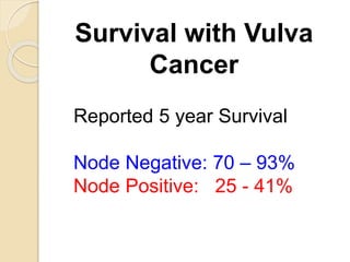
Survival with Vulva
Cancer
Reported 5 year Survival
Node Negative: 70 – 93%
Node Positive: 25 - 41%
Stage Distribution
NCDB 2000-2011
Stage Percent
Stage 0 29%
Stage I 29%
Stage II 14%
Stage III 12%
Stage IV 6%
Observed Survival
NCDB 2003-2006
Stage Percent
Stage 0 92%
Stage I 81%
Stage II 59%
Stage III 43%
Stage IV 21%
Five-Year Survival by Stage and Node Status
Clinical FIGO Stage
I 98%
II 85%
III 74%
IV 31%
Node Status
Groin Negative 91%
Groin Positive 52%
Pelvic Positive 11%
5 Year Survival
Stage Survival
I 79%
II 59%
III 43%
IV 13%
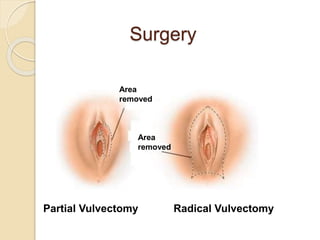
Surgery
Area
removed
Area
removed
Partial Vulvectomy Radical Vulvectomy
Surgery
Until the 1980’s, the standard
therapeutic approach was radical
surgery, including complete en bloc
resection of the vulva and regional
lymph nodes.
In tumors clinically confined to the vulva or perineum,
radical local excision with a margin of at least 1 cm has
generally replaced radical vulvectomy;
separate incision has replaced en bloc inguinal node
dissection;
ipsilateral inguinal node dissection has replaced
bilateral dissection for laterally localized tumors;
and femoral lymph node dissection has been omitted
in many cases.
Modern Treatment
Early Stage: Radical Local Excision
More Advanced: Modified Radical
Excision with Sentinel Node Biopsy
Advanced Stage: Radiation plus
Chemotherapy (chemoradiation)
possibly followed by limited surgery
Sentinel Node Biopsy
Node metastases were identified in 26% of sentinel
node procedures, and these patients went on to full
inguinofemoral lymphadenectomy. The patients with
negative sentinel nodes were followed with no further
therapy.
Side Effects Node Dissection Sentinel Nodes
wound breakdown 34% 11.7%
cellulitis 21% 4.5%
lymphedema 25% 2%
Frequency of Bilateral Nodes
Midline: 70%
Laterally ambiguous: 58%
Lateral position: 22%
Radiation Therapy
Radiation Instead of Surgery for Lymph Nodes
About 20-35% of patients will be found to
have spread to the groin lymph nodes
Small study compared surgery with radiation
to the groin and there were more relapse in
the radiation group (18% versus 0%) so the
study was discontinued
The radiation dose was very low in the study
so the results may not be valid
Radiation Instead of Surgery for Lymph Nodes
Women with positive groin nodes were
randomized between pelvic node surgery or
radiation.
Radiation was superior with better survival
(51%/6y versus 41%)
Lower vulva cancer mortality (29% versus
51%)
and less chronic lymphedema (16% s 22%)
PostOp Radiation
Lympho-vascular invasion
depth of invasion > 5mm
margins < 8mm
positive surgical margins
more than one + node
node with extracapsular invasion
Indications for
Chemoradiation
-Anorectal, urethral, or bladder
involvement (in an effort to avoid
colostomy and urostomy)
-Disease that is fixed to the bone
-Gross inguinal or femoral node
involvement (regardless of whether
a debulking lymphadenectomy was
performed)
Chemoradiation for Squamous
Cancer of the Vulva
Chemotherapy: 5FU plus cisplatin or
mitomycin
Radiation: 40 – 65Gy range
Cure Rate: 25-75% range
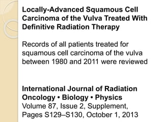
Locally-Advanced Squamous Cell
Carcinoma of the Vulva Treated With
Definitive Radiation Therapy
Records of all patients treated for
squamous cell carcinoma of the vulva
between 1980 and 2011 were reviewed
International Journal of Radiation
Oncology • Biology • Physics
Volume 87, Issue 2, Supplement,
Pages S129–S130, October 1, 2013
Eighty-eight patients were identified whose only
vulvar treatment was radiation therapy (RT) +/-
chemotherapy (CT) due primarily to
unresectable disease or co-morbidities
Median prescribed dose of RT to the vulva was
64 Gy
The median age 67 years
Clinical FIGO stages were T1 (10%), T2 (65%),
or T3 (25%);
70% had clinically positive inguinal nodes. The
Median tumor size was 5 cm.
Overall Survival rate for all patients
was 50% at 5 yrs.
Local Recurrence rate in the vulva
for all patients was 25% at 5 yrs.
Incidence of late grade 3 and 4
toxicities was 4% for gastrointestinal
and 10% for genitourinary.
CT scan is obtained at the time of
simulation
CT images are then imported
into the treatment planning
computer
In the simulation
process the CT
and PET scan
images are used
to create a
computer plan
Cross section anatomy of the female pelvis
CT Scans are used to identify the target
structures for radiation
bowel
sacrum
Iliac bone
Pelvic
nodes
Pre-sacral
nodes
CT Scans are used to identify the target
structures for radiation
bowel
nodes
vagina
bladder
rectum
hips
CT
Anatomy
Lymph
node
regions are
identified
IMRT (the colored areas are the
radiation zones)
Pelvic nodes Pelvic nodes Rectum
Groin nodes Vulva/Vagina Vulva
Computer reconstruction of radiation fields to
cover groin and pelvic lymph nodes but avoid
the bladder and rectum in vulva cancer patient
Tomotherapy fields used to hit the groin and
pelvic nodes but avoid the bladder and rectum
Side Effects of Pelvic Radiation
Side Effects of Vaginal/Vulvar
Radiation
•Vaginal and vulva
skin burning irritation
or discharge
•Urethra irritation or
frequency
•Anus burning
Cancer of the Vulva
www.aboutcancer.com