By Michael A. Medez
APOPTOSIS
• First described by Carl Vogt in 1842
• 1965, distinguished from necrosis
• Greek word “falling off”
• Programmed Cell Death
• Biochemical reactions lead to cell changes and
  death
• Around 50 to 70 billion cells die each day due to
  apoptosis
APOPTOSIS vs NECROSIS
• Apoptosis                          • Necrosis
    Natural Cell Death                  Traumatic Cell Death
    Stimulated by cell signals          Stimulated by factors
    Beneficial                           external to the cells
    Produces cell fragments that        Fatal
     are able to send signals that       Cannot send signals, leads to
     facilitate phagocytosis              build up of dead tissue and
                                          cell debris
PROCESS
• Controlled by a diverse range of cell signals
    Extracellular (Extrinsic Inducers)
    •   Toxins, hormones, cytokines
    •   Positive Induction or Negative Induction

   Intracellular (Intrinsic Inducers)
    •   Initiates in response to stress
         (heat, radiation, hypoxia, nutrient deprivation)
REGULATION
• Mitochondrial Regulation
   Mitochondria is essential to cell life
   Exploited by apoptotic pathways
   Cause mitochondrial swelling or may increase the
    permeability of the membrane

• Direct Signal Transduction
   TNF (Tumor Necrotic Factor) Pathway
   Fas Pathway
TNF and Fas
 TNF Pathway
                                      • Fas Pathway
    TNF is a cytokine produced
                                          Binds the Fas Ligand
     by macrophages
                                          Formation of Death
    Major extrinsic mediator of
                                           Inducing Signaling Complex
     apoptosis
                                           (DISC)
    Leads to caspase activation
                                          Caspase activation via the
     via membrane proteins
                                           FADD
     TRADD and FADD
    Indirectly leads to activation
     of transcription factors
     involved in cell survival and
     inflammatory response
COMPONENTS
• Following activation of TNF and Fas, a balance
  between proapoptotic and anti-apoptotic members
  of the Bcl-2 family is established
• Proportion of proapoptotic homodimers that form
  in the outer membrane of the mitochondria,
  required to make the membrane permeable for the
  release of caspase activators
CASPASES
• Cysteine-dependent aspartate-secific proteases
• Play a central role in transduction of apoptopic
  signals
• Highly conserved proteins
• Intiator Caspases
    Activates by binding to specific oligomeric
     adaptor protein
• Effector Caspases
    Activated by active initiator caspases through
     proteolytic cleavage
    Degrade intracellular proteins to carry out cell
     death program
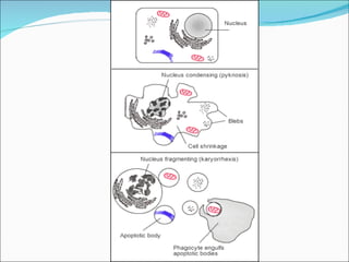
MECHANISM
1.   Cell receives stimulus
2.   Shrinks (breakdown of cytoskeleton)
3.   Dense cytoplasm, packing of organelles
4.   Condensation of chromatin (PYKNOSIS)
5.   Fragmentation of DNA, breaking of nucleus
6.   Formation of blebs
7.   Cell breaks, formation of apoptotic bodies
8.   Phagocytosis
REMOVAL OF DEAD CELLS
• Efferocytosis
• Dying cells display phagocytotic molecules,
  phosphatidylserine
• Mark the cell for phagocytosis
DEFECTS in APOPTOSIS
• p53
    Tumor suppressor protein
    Accumulates when DNA is damaged
    Prevents the cell fro replicating to give it time to
     repair
    Induce apoptosis if damage is extensive and repair
     efforts fail
    Any disruption to its regulation results in impaired
     apoptosis and possible formation of tumors
INHIBITION and HYPERACTIVITY
• INHIBITION            • HYPERACTIVITY
    Decrease in cell      Loss of control of cell
     death                  death
    Most common is        HIV
     CANCER