Presented by : Zinat Ashnagar, PT, PhD
Assistant Professor, Tehran University of Medical Sciences
https://orcid.org/0000-0001-5515-2130
Zinatashnagar@gmail.com
https://www.researchgate.net/profile/Zinat_Ashnagar
Kinesiology of the Ankle Joint
2
Ankle & Foot Complex 3
Deep plantar fascia
• The medial longitudinal arch and associated
connective tissues are the primary sources of
mechanical support for the foot during relatively
low stress or near static conditions (e.g. while
standing on ease).
Ankle & Foot Complex 4
• Without this arched configuration, the large and
rapidly produce forces applied against the foot
during running and marching, for examples,
would likely exceeds the physiologic weight-
bearing capacity of the bones.
Ankle & Foot Complex 5
ACCEPTING BODY WEIGHT DURING
STANDING
Ankle & Foot Complex 6
PES PLANUS – “ABNORMALLY DROPPED” MEDIAL
LONGITUDINAL ARCH
• Pes planus or “flatfoot” describes a chronically dropped
or abnormally low medial longitudinal arch.
• Often results from joint laxity and an overstretched or weak plantar
fascia.
• Flexible pes planus appears normal when unloaded, but
drops when loaded.
7 Ankle & Foot Complex
ACCEPTING BODY WEIGHT DURING
STANDING
Ankle & Foot Complex 8
PES CAVUS – ABNORMALLY RAISED MEDIAL LONGITUDINAL
ARCH
9 Ankle & Foot Complex
1/25/2020
Pes Cavus
• An abnormally high arch places the metatarsal bones
at a greater angle with a ground.
• When combined with the reduced area of plantar
contact, plantar pressures typically rise and shift
anteriorly over the forefoot.
• Furthermore, a foot with a chronically raised (and
relatively rigid) arch cannot, in theory, optimally
absorb the repeated impacts of walking and running.
• A person with significant pes cavus may therefore at
high higher risk of developing stress-related injuries
within the foot and lower limb.
• While walking, maximal dorsiflex occurs late in stance phase,
just before the heel rises off the ground (at about 40% of the
gait cycle).
• Realize that while in the stance phase of walking, the term
dorsiflex describes the position of leg relative to the foot.
14
Functional Considerations:
Most- and Least-Stable Positions of the Talocrural Joint
• At this point in the gait cycle, the ankle is most stable
because most of the collateral ligaments and all of
the plantar flexor muscles are stretched.
ROM
TALOCRURAL
JOINT
DURING GAIT
16 Ankle & Foot Complex
17
18
• The dorsiflexed ankle is further stabilized as the
wider, anterior part of the trochlea of the talus
become wedged into the mortise.
• For this reason the close-packed position of the ankle
is full dorsiflex.
19
• Such stability is necessary in late stance to prepare
for the action of the strongly activated plantar flexor
muscles during jumping or the push-off phase of
walking.
• The least stable position of the talocrural joint is full
plantar flex.
• Full plantar flex- loose-packed position of the joints-
slackens most of the collateral ligaments and all of the
plantar flexor muscles.
22
23
• The position of full plantar flex also causes the mortise
(distal tibia and fibula) to “loosen its grip” on the talus.
• This places the narrower width of the talus between the
malleoli, thereby releasing tension within the mortise.
• Weight bearing over a fully plantar flexed ankle,
therefore places the ankle at a relatively unstable
position.
24
25
FACTORS THAT INCREASE THE MECHANICAL STABILITY OF
DORSIFLEXED TALOCRURAL JOINT
Ankle & Foot Complex 26
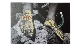
Subtalar joint:
Critical Kinematic Link Between the Leg and Foot
• Motion at the subtalar joint is commonly expressed in one of two
ways:
• When the calcaneus is free, such as during the swing phase, or
when it is in firm contact with the ground during the stance phase
of walking.
27
• While in the stance phase, the leg and talus moves as
one mechanical unit over the fixed calcaneus.
• Although the motion at the subtalar is small, it is
nevertheless important.
29
• The normal subtalar joint is well utilized during walking and
running, especially on unlevel terrain.
• For example, when standing on level ground, the leg and talus
are in relative alignment with the calcaneus (A).
30
• Consider what happens when the foot encounters uneven
ground (B).
• In this scenario, the calcaneus rotates, resulting in inversion of
the subtalar joint.
• This “righting” mechanism of the foot allows the leg to remain
vertical, even while standing or walking on uneven surfaces.
• If this motion is excessive, however, it may result in a sprain of
the lateral ligaments.
31
• In other circumstances, it may be necessary that the
calcaneus remains firmly planted on the ground,
while the leg and body cut in medial or lateral
directions (C).
• With the calcaneus well fixed, a medially directed
movement of the talus and leg can occur as subtalar
joint inversion.
32
• Realize that, although different bones are moving, in B and C,
the final position of the subtalar joint in both scenarios is the
same: “INVERSION”.
• Without the available motion provided by the subtalar joint,
walking on uneven surfaces would be extremely difficult and
would likely result in loss of balance or injury to the ankle and
foot.
CHANGE IN
HEIGHT IN THE
MEDIAL
LONGITUDINAL
ARCH
34 Ankle & Foot Complex
CHANGE IN HEIGHT IN THE MEDIAL LONGITUDINAL ARCH
• In the healthy foot, MLA (Medial Longitudinal Arch)
rises and lowers cyclically throughout the gait cycle.
• During most of the stance phase, the arch lowers
slightly in response to the progressive loading of body
weight.
36
• Structures that resists the lowering of the
arch absorb local stress as the foot is
progressively compressed by body
weight.
• This load attenuation mechanism offers
essential protection to the foot and lower
limb against stress-related, overuse
injury.
• During the first 30 to 35% of gait cycle, the subtalar
joint pronates (everts), adding an element of
flexibility to the midfoot.
• By late stance, the arch rises sharply as the now
supinating subtalar joint adds rigidity to the midfoot.
39
The rigidity prepares the foot to support the
large loads produced at the peak of the
push off phase.
• The ability of the foot to repeatedly transform from a
flexible and shock-absorbent structure to a more
rigid lever during each gait cycle is one of the most
important and clinically relevant actions of the foot.
• Immediately after the heel contact phase of gait, the
dorsiflexed talocrural joint and slightly supinated
subtalar joint rapidly plantar flex and pronate.
• The amount and the speed of the pronation
nevertheless influence the kinematics of the more
proximal joints of the lower extremity.
42
• These effects can be readily appreciated by
exaggerating and dramatically slowing the pronation
action of the rearfoot during the initial loading phase
of gait.
• While standing over a loaded and fixed foot, forcefully but
slowly internally rotate the lower limb and observe the
associated pronation at the rearfoot and lowering of the MLA.
• If sufficiently forceful, this action also tends to
internally rotate, slightly flex and adduct the hip and
create a valgus stress on the knee.
44
45
ACTIONS ASSOCIATED WITH EXAGERRATED PRONATION
OF THE SUBTALAR JOINT DURING WEIGHT BEARING
Joint of Region Action
Hip Internal rotation, flexion, and
adduction
Knee Increased valgus stress
Rearfoot Pronation (eversion) with a
lowering of medial longitudinal arch
Midfoot and Forefoot Supination (inversion)
46 Ankle & Foot Complex
47
1/25/2020
Basic Functions of Distal Intertarsal Joints
• As a group, the distal intertarsal joints:
1) Assist the transverse tarsal joint in pronating and
supinating the midfoot
2) Provide stability across the midfoot by forming the
transverse arch of the foot.
Cuneonavicular Joint
• The major function of Cuneonavicular joint is to help
transfer components of pronation and supination
distally from the talonavicular toward the forefoot.
1/25/2020
Cuboideonavicular Joint
• Synarthrodial (fibrous) or sometimes synovial
• Provides a relatively smooth contact point between the lateral
and medial longitudinal columns of the foot.
• Articular surfaces slide slightly against each other during
movement of the midfoot, most notably during inversion and
eversion.
Intercuneiform and Cuneocuboid joint Complex
• Consists of three articulation:
• Two between the set of three cuneiforms, and
• one between the lateral cuneiform and medial
surface of the cuboid.
1/25/2020
• Forms the transverse arch of the foot.
• This arch provides transverse stability to the midfoot.
• Under the load of body weight, the transverse arch
depresses slightly, allowing body weight to be shared
across all five metatarsal heads.
Intercuneiform and Cuneocuboid joint Complex
• The transverse arch receives support from intrinsic
muscles; extrinsic muscles, such as the tibialis
posterior and fibularis lungus; connective tissues;and
the keystone of the transverse arch:
the intermediate cuneiform
1/25/2020
1/25/2020
Tarsometatarsal joints
1/25/2020
Tarsometatarsal Joints
• The tarsometatarsal joints are the basilar joints of the
forefoot.
• Mobility is least at the second and third tarsometatarsal
joints, in part because of strong ligaments and the
wedged position of the base of the second ray between
the medial and lateral cuneiforms.
• The second and third rays produce an element of
longitudinal stability throughout the foot, similar to
the second and third rays in the hand.
• This stability is useful in late stance as the forefoot
prepares for the dynamics of push off.
The functional stability of the first tarsometatarsal
joint is considered an important mechanism that
assists the MLA in safely accepting and sharing the
loads incurred during walking.
• Most literature describes a natural mechanical
coupling of the kinematics at the first tarsometarsal
joint:
• Specifically, plantar flex occurs with slight eversion,
and dorsiflex with slight inversion.
OSTEOKINEMATICS OF FIRST TARSOMETATARSAL
JOINT
Ankle & Foot Complex 66
Combining plantar flex and eversion, for example, allows
the medial side of the foot to better conform around
irregular surfaces on the ground.
1/25/2020
• A pair of collateral ligaments spans each
metatarsophalangeal joint, blending with and
reinforcing the capsule.
• As in the hand, each collateral ligament courses
obliquely from a dorsal-proximal to a plantar-distal
direction, forming a thick cord portion and a fan-like
accessory portion.
METATARSOPHALANGEAL JOINT
71 Ankle & Foot Complex
• The accessory portion attaches to the thick, dense
plantar plate, located on the plantar side of the joint.
• Fibers from deep plantar fascia attach to the plantar
plates and sheaths of the flexor tendons.
• Movements at the MP joints occur in two degrees of
freedom:
• Extension (DF) and Flexion (PF) (Sagittal plane about
ML axis)
• Abduction and Adduction (horizontal plane about a
vertical axis)
Foot Flat (8% Point of the Gait Cycle)
• Foot flat is defined as the point at which the
entire plantar surface of the foot is in contact
with the ground.
• This event is often described as the loading
response phase.
Mid Stance (30% Point of the Gait Cycle)
• Mid stance occurs as the lower leg approaches
the vertical Position.
• The leg is in single-limb support, as the other limb
is freely swinging forward.
• The hip and the knee are in near extension, as the
ankle continues to move into greater dorsiflexion.
HALLUX VALGUS
Ankle & Foot Complex 77
1/25/2020
(1) adduction of the first metatarsal (toward the midline of
the body), evidenced by the increased angle between the
first and second metatarsal bones;
(2) lateral deviation of the proximal phalanx with
dislocation or subluxation of the first metatarsophalangeal
joint;
(3) displacement of the lateral sesamoid;
(4) rotation (eversion) of the phalanges of the
great toe; and
(5) exposed first metatarsal head, forming the so-
called “bunion.”
Windlass effect
1/25/2020
windlass effect
• The “windlass effect” of the plantar fascia is
demonstrated while a subject stands on tiptoes.
• A windlass is a lifting device consisting of a rope wound
around a cylinder that is turned by a crank.
• The rope is analogous to the plantar fascia, and the
cylinder is analogous to the metatarsophalangeal joint.
• In the normal foot, contraction of the extrinsic plantar
flexor muscles lifts the calcaneus, thereby transferring
body weight forward over the metatarsal heads.
• The resulting extension of the metatarsophalangeal
joints (shown collectively as white disk) stretches (winds
up) the plantar fascia within the medial longitudinal arch
(red spring).
Windlass effects
1/25/2020
• The increased tension from the stretch raises
the arch and strengthens the midfoot and
forefoot.
• Contraction of the intrinsic muscles provides
additional reinforcement to the arch.
• The foot with pes planus (flatfoot) typically has a poorly
supported medial longitudinal arch.
• During an attempt to stand up on tiptoes, the forefoot sags
under the load of body weight.
• The reduced extension of the metatarsophalangeal joints
limits the usefulness of the windlass effect.
• Even with strong activation of the intrinsic muscles, the
arch remains flattened and the midfoot and forefoot
unstable.
COMMON FIBULAR (PERONEAL) NERVE
88 Ankle & Foot Complex
TIBIAL NERVE
89 Ankle & Foot Complex
ACTIONS ACROSS TALOCRURAL AND SUBTALAR JOINTS
90 Ankle & Foot Complex
MUSCLES OF THE ANTERIOR COMPARTMENT OF THE LEG
(PRETIBIAL “DORSIFLEXORS”)
• Muscles
– Tibialis anterior
– Externsor digitorum longus
– Extensor hallucis longus
– Fibularis tertius
• Innervation
– Deep branch of the fibular nerve
91 Ankle & Foot Complex
PRETIBIAL MUSCLES
92 Ankle & Foot Complex
1/25/2020
LATERAL COMPARTMENT MUSCLES
94 Ankle & Foot Complex
1/25/2020
1/25/2020
LATERAL COMPARTMENT OF THE LEG (“EVERTORS”)
• Muscles
– Fibularis longus
– Fibularis brevis
• Innervation
– Superficial branch of the fibular nerve
97 Ankle & Foot Complex
• The line of force of several plantar flexor muscles
while the subject rises on the tip toes.
• Note that the fibularis longus and tibialis posterior
form a sling that supports the transverse and medial
longitudinal arches.
1/25/2020
1/25/2020
the pull of the
gastrocnemius and
tibialis posterior
muscles causes a
slight supination of
the rear foot, which
adds further stability
to the foot.