CELL
The human organism presents about 200 different cell
types, all derived from the zygote, the single cell formed
by fertilization of an oocyte with a spermatozoon.
 During their specialization process, called cell
differentiation, the cells synthesize specific proteins,
change their shape, and become very efficient in
specialized functions.
 The body's cells can experience both normal and
pathological conditions and the same cell type can
exhibit different characteristics and behaviors in different
regions and circumstances

CELLULAR FUNCTIONS IN SOME SPECIALIZED
CELLS.


The cell is composed of two basic parts:
Cytoplasm and
Nucleus.



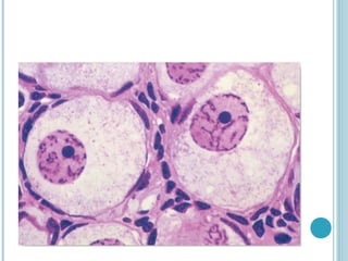
Individual cytoplasmic components are usually not clearly
distinguishable in common hematoxylin-and-eosin–stained
preparations.



The nucleus, appears intensely stained dark blue or black.



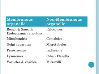
The cytoplasm is composed of a fluid component, or cytosol, which
containes metabolically active structures, the organelles, which can
be membranous (such as mitochondria) or non-membranous protein
complexes (such as ribosomes and proteasomes).
Membranous
organelle

Non-Membranous
organelle

Rough & Smooth
Endoplasmic reticulum

Ribosomes

Mitochondria

Centrioles

Golgi apparatus

Microtubules

Peroxisomes

Inclusions

Lysosomes

Cilia - Flagella

Vacuoles & vesicles

Microvilli
PLASMA MEMBRANE


The outermost component of the cell, separating the
cytoplasm from its extracellular environment, is the
Plasma membrane or Plasmalemma.



All eukaryotic cells are enveloped by a limiting
membrane composed of phospholipids, cholesterol,
proteins, and chains of oligosaccharides covalently
linked to phospholipid and protein molecules.



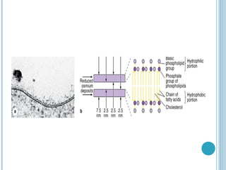
Membranes range from 7.5 to 10 nm in thickness and
visible only in the electron microscope.


With electron microscopy, the plasmalemma and, all other organellar
membranes appears a trilaminar structure after fixation in osmium
tetroxide.



Because all membranes have this appearance, the 3-layered structure was
designated the unit membrane.



Membrane phospholipids, such as phosphatidylcholine (lecithin),
consist of two non-polar (hydrophobic or water-repelling) long-chain
fatty acids linked to a charged polar (hydrophilic or water-attracting)
head group.



Membrane phospholipids are most stable when organized into a double
layer (bilayer) with their hydrophobic fatty acid chains directed toward
the middle away from water and their hydrophilic polar heads directed
outward to contact water on both sides.


Cholesterol molecules insert among the close packed the phospholipid
fatty acids nearly a 1:1 ratio , restricting their movement, and thus
modulate the fluidity and movement of all membrane components.



The lipid composition of each half of the bilayer is different.



In red blood cells phosphatidylcholine and sphingomyelin are more
abundant in the outer half of the membrane, whereas phosphatidylserine
and phosphatidylethanolamine are more concentrated in the inner half.



Some of the lipids, known as glycolipids, possess oligosaccharide chains
that extend outward from the surface of the cell membrane and thus
contribute to the lipid asymmetry.


Proteins, are major molecular constituent of membranes, can be
divided into two groups.

Integral proteins :
o Incorporated within the lipid bilayer.
o Some integral proteins span the membrane one or more times,
from one side to the other, they are called one-pass or
multipass transmembrane proteins.
Peripheral proteins :
exhibit a looser association with one of the two
membrane surfaces.








With freeze-fracture electron microscope studies of membranes
show that many integral proteins are only partially embedded in the
lipid bilayer and protrude from either the outer or inner surface.
Transmembrane proteins are large enough to extend across the two
lipid layers and may protrude from both membrane surfaces.
The carbohydrate moieties of the glycoproteins and glycolipids
project from the external surface of the plasma membrane; they are
important components of specific molecules called receptors that
participate in important interactions such as cell adhesion,
recognition, and response to protein hormones.
As with lipids, the distribution of membrane proteins is different in
the two surfaces of the cell membranes. Therefore, all membranes in
the cell are asymmetric.










Integration of the proteins within the lipid bilayer is mainly the
result of hydrophobic interactions between the lipids and nonpolar
amino acids present on the outer region of the proteins.
Some membrane proteins are not bound rigidly in place and are able
to move within the plane of the cell membrane.
unlike lipids, most membrane proteins are restricted in their lateral
diffusion by attachment to cytoskeletal components.
In most epithelial cells, tight junctions also restrict lateral diffusion
of unattached transmembrane proteins and outer layer lipids to
specific membrane domains.
The mosaic disposition of membrane proteins and the fluid nature of
the lipid bilayer and led to the well-established fluid mosaic model
for membrane structure.
Functions of plasmalemma :


A selective barrier.



To keep constant the ion content of cytoplasm.



Specific recognition and regulatory functions.
GLYCOCALYX










With the EM the external surface of the cell shows a fuzzy
carbohydrate-rich region called the glycocalyx.
This layer is made of carbohydrate chains linked to membrane
proteins and lipids and of cell-secreted glycoproteins and
proteoglycans.
The glycocalyx has a role in cell recognition and attachment to other
cells and to extracellular molecules.
Some ions, such as Na+, K+, and Ca2+, cross the cell membrane by
passing through integral membrane proteins.
This can involve passive diffusion through ion channels or active
transport via ion pumps using energy from the breakdown of
adenosine triphosphate (ATP).
ENDOCYTOSIS


Bulk uptake of material also occurs across the plasma membrane in
a general process called endocytosis, which involves folding and
fusion of this membrane to form vesicles which enclose the material
transported.



Cells show three general types of endocytosis.
1. Phagocytosis
2. Fluid-phase Endocytosis
3.Receptor-mediated Endocytosis
Phagocytosis :
o

o

o

Certain white blood cells, such as macrophages and neutrophils, are
specialized for engulfing and removing particulate matter such as
bacteria, protozoa, dead cells, and unneeded extracellular
constituents.
When a bacterium becomes bound to the surface of a neutrophil,
cytoplasmic processes of the cell are extended and ultimately
surround the bacterium.
The membranes of these processes meet and fuse, enclosing the
bacterium in an intracellular vacuole, a phagosome.
PHAGOCYTOSIS
Fluid-phase Endocytosis :


In fluid-phase pinocytosis , smaller invaginations of the cell
membrane form and entrap extracellular fluid and anything it has in
solution.



Pinocytotic vesicles (about 80 nm in diameter) pinch off inwardly
from the cell surface.



In most cells such vesicles usually fuse with lysosomes.
PINOCYTOSIS
Receptor-mediated Endocytosis :
 Receptors for many substances, such as low-density
lipoproteins and protein hormones, are integral proteins of the
cell membrane.
 Binding of the ligand (a molecule with high affinity for a
receptor) to its receptor causes widely dispersed receptors to
aggregate in special membrane regions called coated pits.
 The electron-dense coating on the cytoplasmic surface of the
membrane is composed of several polypeptides, the major one
being Clathrin.
 In a developing coated pit clathrin molecules, forming that
region of cell membrane into a cage-like invagination that is
pinched off into the cytoplasm, forming a coated vesicle
carrying the ligand and its receptor.
RECEPTOR-MEDIATED ENDOCYTOSIS
In endocytotic processes, the vesicles or vacuoles
produced quickly enter and fuse with the endosomal
compartment, a dynamic system of membranous
vesicles and tubules located in the cytoplasm near the
cell surface (early endosomes) or deeper in the
cytoplasm (late endosomes).
 The clathrin molecules separated from the coated
vesicles recycle to the cell membrane to participate in
the formation of new coated pits.
 The membrane of endosomes contains ATP-driven H+
pumps that acidify their interior.

While phagosomes and pinocytotic vesicles soon fuse with
lysosomes, molecules penetrating the endosomal
compartment after receptor-mediated endocytosis may take
more than one pathway.
 The acidic pH of early endosomes causes many ligands to
uncouple from their receptors, after which the two
molecules are sorted into separate vesicles.
 The receptors may be returned to the cell membrane to be
reused.
 The ligands typically are transferred to late endosomes.
 Some ligands are returned to the extracellular milieu with
their receptors and both are used again.
 Late endosomes most commonly fuse with lysosomes for
degradation of their contents.

EXOCYTOSIS
In exocytosis a membrane-limited cytoplasmic vesicle
fuses with the plasma membrane, resulting in the release
of its contents into the extracellular space without
compromising the integrity of the plasma membrane.
 Often exocytosis of stored products from epithelial cells
occurs specifically at the apical domains of cells, such as
in the exocrine pancreas and the salivary glands.
 The fusion of membranes during exocytosis is a highly
regulated process involving interactions between several
specific membrane proteins.
 Exocytosis is triggered in many cells by transient
increase in cytosolic Ca2+.



During endocytosis, portions of the cell membrane
become endocytotic vesicles; during exocytosis, the
membrane is returned to the cell surface.



This process of membrane movement and recycling is
called membrane trafficking.



Trafficking and sorting of membrane components occur
continuously in most cells and are not only crucial for
cell maintenance but also physiologically important in
processes such as reducing blood lipid levels.
SIGNAL RECEPTION AND
TRANSDUCTION


Cells in a multicellular organism need to communicate with one
another to regulate their development into tissues, to control
their growth and division, and to coordinate their functions.



Soluble extracellular signaling molecules bind receptor proteins
only found on their target cells.



Each cell type in the body contains a distinctive set of receptor
proteins that enable it to respond to a complementary set of
signaling molecules in a specific, programmed way.
Signaling can take different routes:
Endocrine signaling:
 the signal molecules (called hormones) are carried in the blood to target
cells throughout the body.
Paracrine signaling :
 the chemical mediators are rapidly metabolized so that they act only on
local cells very close to the source.
Synaptic signaling :
 a special kind of paracrine interaction, neurotransmitters act only on
adjacent cells through special contact areas called synapses.
Autocrine signaling :
 signals bind receptors on the same cell type that produced the messenger
molecule.










Hydrophilic signaling molecules :
including most hormones, local chemical mediators (paracrine
signals), and neurotransmitters activate receptor proteins on the surface
of target cells.
These receptors, often transmembrane proteins, relay information to a
series of intracellular intermediaries that ultimately pass the signal (first
messenger) to its final destination in either the cytoplasm or the nucleus
in a process called Signal transduction.
One of the intermediary proteins, the G proteins, binds guanine
nucleotides and acts on other membrane-bound intermediaries called
effectors which propagate the signal further into the cell.
Effector proteins are usually ion channels or enzymes that generate large
quantities of small second messenger molecules, such as 1,2diacyglycerol (DAG), cyclic adenosine monophosphate (cAMP), and
inositol 1,4,5-triphosphate (IP3).
The ions or second messengers diffuse through the cytoplasm,
amplifying the first signal and triggering a cascade of molecular
reactions that lead to changes in gene expression or cell behavior.
MEDICAL APPLICATION
Several diseases have been caused by defective receptors.
 Pseudohypoparathyroidism and a type of Dwarfism are
caused by nonfunctioning parathyroid and growth hormone
receptors.
 In these two conditions the glands produce the respective
hormones, but the target cells do not respond because they
lack normal receptors.

Hydrophobic signaling molecules :
such as small steroid and thyroid hormones, bind
reversibly to carrier proteins in the plasma for transport
through the body.
 Such hormones are lipophilic and once released from their
carrier proteins, they diffuse directly through the plasma
membrane lipid bilayer of the target cell and bind to specific
intracellular receptor proteins.
 With many steroid hormones, receptor binding activates that
protein, enabling the complex to move into the nucleus and
bind with high affinity to specific DNA sequences.
 This generally increases the level of transcription from
specific genes.

NUCLEUS
The nucleus contains a blueprint for all cell structures
and activities encoded in the DNA of the chromosomes .
 Nucleus is the site of deoxyribonucleic acid (DNA)
replication and transcription of DNA into precursor
ribonucleic acid (RNA)molecules.
 It contains all of the enzymes required for replication
and repair of newly synthesized DNA, as well as for
transcription and processing of precursor RNA
molecules.
 It is enclosed by the nuclear envelope and contains the
nuclear lamina, nucleolus, and chromatin

Nuclear envelope :


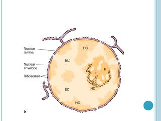
The nuclear envelope is a double membrane
containing pores that are approximately 90 nm in
diameter.



The outer nuclear membrane is continuous with
the endoplasmic reticulum.
Nuclear Lamina :


The nuclear lamina is a latticelike network of
proteins that include lamins.



Lamins attach chromatin to the inner membrane
of the nuclear envelope and participate in the
breakdown and reformation of the nuclear
envelope during the cell cycle.



Phosphorylation of the lamins, (by lamin kinase)
during prophase of mitosis initiates nuclear
disassembly into small vesicles.
Nuclear Lamina
Functions:
Maintenance of nuclear shape
Spatial organization of nuclear pores within nuclear membrane
Regulation of transcription
Anchoring of interphase heterochromatin
DNA replication.
Lamina

bio.winona.msus.edu/.../ Lec-note/16-new.htm
Nucleolus :


The nucleolus is responsible for ribosomal RNA
(rRNA) synthesis and ribosome assembly.



It contains three morphologically distinct zones:

• Granular zone—found at the periphery; contains
ribosomal precursor particles in various stages of
assembly.
• Fibrillar zone—centrally located; contains ribonuclear
protein fibrils.
• Fibrillar center—contains DNA that is not being
transcribed.
Nucleolus :


The nucleolus is responsible for ribosomal RNA
(rRNA) synthesis and ribosome assembly.



It contains three morphologically distinct zones:

• Granular zone—found at the periphery; contains
ribosomal precursor particles in various stages of
assembly.
• Fibrillar zone—centrally located; contains ribonuclear
protein fibrils.
• Fibrillar center—contains DNA that is not being
transcribed.
Chromatin :
Chromatin is a complex of DNA, histone proteins, and nonhistone
proteins.
• DNA—a double-stranded helical molecule that carries the genetic
information of the cell.
 It exists in three conformations: B DNA, Z DNA, and A DNA.
• Histone proteins—positively charged proteins enriched with lysine
and arginine residues.
 They are important in forming two types of structures in chromatin:
nucleosomes and solenoid fibers.
 The nucleosomes are the basic repeating units of the chromatin
fiber, having a diameter of approximately 10 nm.
• Nonhistone proteins—include enzymes involved in nuclear functions
such as replication, transcription, DNA repair, and regulation of
chromatin function.
 They are acidic or neutral proteins.
Chromatin

6 nucleosomes
become coiled
around an axis,
forming a solenoid.
Nucleosome, Solenoid model of chromatin, and chromosome

Nucleosome
Forms of Chromatin :
• Heterochromatin :
highly condensed and transcriptionally inactive.
 In a typical eukaryotic cell, approximately 10% of the
chromatin is heterochromatin.
• Euchromatin :
a more extended form of DNA, which is
potentially transcriptionally active.
 In a typical cell, euchromatin accounts for approximate
90%of the total chromatin, although only about 10% is
being actively transcribed.
CYTOPLASM
RIBOSOMES
Ribosomes are composed of rRNA and protein. They
consist of large (60S) and small (40S) subunits.
 Ribosomes are assembled in the nucleus and transported
to the cytoplasm through the nuclear pores.
 The large ribosomal subunits are synthesized in the
nucleolus, whereas the small subunits are synthesized in
the nucleus.
Polysomes :
 Ribosomes often form polysomes, which consist of a
single messenger RNA (mRNA) that is being translated
by several ribosomes at the same time.
 The ribosomes move on the mRNA from the 5' end
towards 3' end.
 The two ribosomal subunits associate on the mRNA,
with the small subunit binding first

Forms of Ribosomes :
Ribosomes exist in two forms:
• Free polysomes : are the site of synthesis for
proteins destined for the nucleus, peroxisomes, or
mitochondria.
• Membrane-associated polysomes : are the site of
synthesis of secretory proteins, membrane
proteins, and lysosomal enzymes.
ENDOPLASMIC RETICULUM
The endoplasmic reticulum exists in two forms, rough
endoplasmic reticulum (RER) and smooth endoplasmic
reticulum (SER).
 Rough Endoplasmic Reticulum :
 RER is a single, lipid bilayer continuous with the outer
nuclear membrane.
 It is organized into stacks of large flattened sacs called
cisternae that are studded with ribosomes on the cytoplasmic
side.
 RER snthesize poteins that are destined for the Golgi
apparatus, secretion, the plasma membrane, and lysosomes.
 RER is very prominent in cells that are specialized in the
synthesis of proteins destined for secretion
(eg : pancreatic acinar cells).

ENDOPLASMIC RETICULUM
SMOOTH ENDOPLASMIC RETICULUM
Smooth Endoplasmic Reticulum :


SER is a network of membranous sacs, vesicles, and
tubules continuous with the RER, but lacking ribosomes.



SER contains enzymes involved in the synthesis of
phospholipids, triglycerides, and sterols
Functions of SER :
o

o
o
o

Detoxification Reactions :
Hydroxylation.
Conjugation.
Glycogen Degradation and Gluconeogenesis
Reactions in Lipid Metabolism
Sequestration and Release of Calcium Ions
GOLGI APPARATUS
GOLGI APPARATUS


The Golgi apparatus consists of disc-shaped smooth
cisternae that are assembled in stacks (dictyosomes), and
associated with numerous small membrane-bound vesicles.



The Golgi apparatus has two distinct faces:
• The cis (forming) face is associated with the RER.
• The trans (maturing) face is often oriented toward the
plasma membrane.



Important in glycosylation, phosphorylation, sulfation, etc.

Takes part in synthesis, concentration & storage of
secretory products.

Functions of the Golgi Apparatus :
Proteins and Lipids
 The Golgi apparatus is the site of post trauslational
modification and sorting of newly synthesized proteins
and lipids.
Glycoproteins
 Further modification of the carbohydrate moiety of
glycoproteins produces complex and hybrid
oligosaccharide chains.
 This determines which proteins remain in the Golgi
apparatus or leave the Golgi apparatus to become
secretory proteins, lysosomal proteins, or part of the
plasma membrane.
 Two diseases are caused by a breakdown in this process,
I-cell disease and hyperproinsulinemia
MEDICAL APPLICATION
Hyperprolnsulinemia :
Hyperprolnsulinemia is characterized by elevated levels of
proinsulin in the serum resulting from the failure of a
peptidase to cleave proinsulin to insulin and C-peptide in the
golgi apparatus.
I-Cell Disease :
Phosphorylation of mannose in glycoproteins targets proteins
to lysosomes.
 Phosphate is added in a two-step sequence of reactions that
are catalyzed by N-acetylglucosamine-phosphotransferase
and N-acetylglucosaminidases.
 A deficiency in N-acetylglucosamine-phosphotransferase
results In I-cell dieease .
LYSOSOMES


Lysosomes are spherical membrane-enclosed organelles
that are contain enzymes required for intracellular
digestion.

Lysosomes consist of two forms :
•Primary lysosomes have not yet acquired the materials to
be digested.
 They are formed by budding from the trans side of the
Golgi apparatus.
•Secondary lysosomes are formed by the fusion of the
primary lysosome with the substrate to be degraded and
have contents that are in various stages of degradation



•

Lysosomes contain approximately 60 hydrolytic
enzymes.
All lysosomal enzymes are acid hvdrolases, with
optimal activity at a pH of approximately 5.0.

The synthesis of the lysosomal hydrolases occurs
in RER.
 All hydrolasea are transferred to the Golgi
apparatus where they are modified and packaged
into lysosomes.

MEDICAL APPLICATION
Glycogen-Storage Disease Type II (Pompe Disease) :
 an autosomal recessive disorder that results from the deficiency
of acid alpha-glucosidase, a lysosomal hydrolase , is required
for the degradation of a small percentage (1-3%) of cellular
glycogen.
 Because the main pathway for glycogen degradation is not
deficient in glycogen-storage disease type II disease, energy
production is not impaired, and hypoglycemia does not occur.
 However, the deficiency of this enzymatic activity results in the
accumulation of structurally normal glycogen in lysosomes and
cytoplasm in affected individuals.
 Excessive glycogen storage within lysosomes may interrupt
normal functioning of other organelles and leads to cellular
injury. In turn, this leads to enlargement and dysfunction of the
entire organ involved (eg, cardiomyopathy).
RESIDUAL BODIES
•Lysosomes containing
indigestible compounds
are called residual
bodies.
•The indigestible
compounds are usually
exocytosed.
•The unreleased
indigestible compounds
in long-living cells
appear as lipofuscins or
aging pigments.
RESIDUAL BODIES IN LUNG CELLS
AUTOPHAGOSOMES
Primary Liposomes fuse
with membrane-bound
organelles or a portion
of cytoplasm to form
autophagosomes.


Autolysis occurs when
lysosomes rupture and
destroy the cell itself.

PEROXISOMES


Peroxisomes are a heterogeneous group of small,
spherical organelles with a single membrane.

Functions:
Synthesis and degradation of hydrogen peroxide.
 Oxidation of very long chain fatty acids (> C24).
 Phospholipid exchange.
 Bile acid synthesis.

PEROXISOMES (MICROBODIES )
MEDICAL APPLICATION
Peroxisome Deficiency :
Several genetic diseases are associated with the impairment
or absence of peroxisomes.
 These patients fail to oxidize very long chain fatty acids and
accumulate bile acid precursors.




The four most common disorders are:
• Zellweger (cerebrohepatorenal) syndrome
• Neonatal adrenoleukodystrophy
• Infantile Refsum disease
• Hyperlipopecolaternia
MITOCHONDRIA
MITOCHONDRIA
Mitochondria have two membranes , They synthesize
adenosine triphosphate (ATP), contain their own doublestranded circular DNA, and make some of their own
proteins.
 Mitochondria have several compartments :
Outer Membrane
 The outer membrane is smooth, continuous, and
highly permeable.
 It contains an abundance of porins, an integral
membrane protein that forms channels in the
outer membrane through which molecules of less
than 10 kD can pass.

Inner Membrane
 The inner membrane is inpermiable to most small ions (Na,
K*, H*) and small molecules (ATP, adenosine diphosphate,
pyruvate).
 The impermeability is likely related to the high content of
the lipid cardiolipin.
 The inner membrane has numerous infoldings, called
cristae.
 The cristae greatly increase the total surface area. They
contain, enzymes for electron transport and oxidative
phosphorylation.
 The number of mitochondria arid the number of cristae per
mitochondrion are proportional to the metabolic activity of
the cells in which they reside.
Mitochondria

Two types of cristae: tubular-like and plate-like.
Most cells contain mitochondria with plate-like cristae. Steroid
secreting cells (eg. Adrenal, gonadal cells) have tubular cristae
Mitochondria

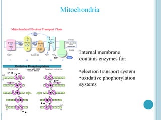
Internal membrane
contains enzymes for:
Oxidative Phosphorylation

•electron transport system
•oxidative phophorylation
systems
Intermembrane Compartment :
 The intermembrane compartment is the space between
the inner and outermembranes.
 It contains enzymes that use ATP to phosphorylate other
nucleotides (creatine phosphokinase and adenylate kinase).
Matrix :
 The matrix is enclosed by the inner membrane and
contains:
Dehydrogenases : oxidize many of the substrates in the
cell (pyruvate, amino acids, fatty acids), generating
reduced nicotinamide adenine dinucleotide (NADH) and
reduced flavin adenine dinucleotide (FADH,) for use by
the electron transport chain and energy generation.
Mitochondria
Citric acid cycle
Matrix contains enzymes for:
•Citric acid cycle for generation
of ATP
•Dehydrogenases
A double-stranded circular DNA genome—encodes a
few of the mitochondrial proteins.
 Mitochondrial DNA is always inherited from the
mother, resulting in transmission of diseases of enery
metabolism.
 RNA, proteins and ribosomes—although there is some
protein synthesis, most mitochondrial proteins are
synthesized in the cytoplasm and are transferred into the
mitochondria.
Intramitochondrial granule : contain calcium and
magnesium. Their function is not known, but it is
believed that they may represent a storage site for
Calcium.
